Cell structure
Organisms are made up of cells. Most organisms are multicellular and have cells that are specialised to do a particular job. Microscopes are needed to study cells in detail.
Cell measurement
Light microscopes
Cells range in size:
- animal cells are between 0.01 mm – 0.05 mm
- plant cells are between 0.01 mm – 0.10 mm
The human eye can see objects as small as around 0.05 mm. A microscope is required to see cells in any detail.
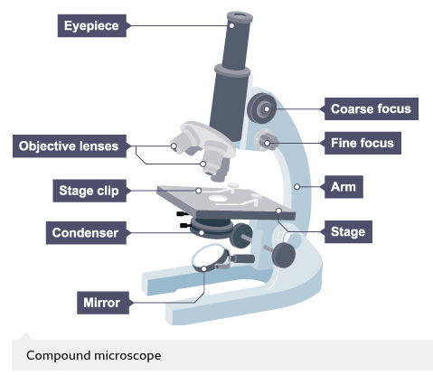
Microscopes magnify the image of a biological specimen so that it appears larger. The type of microscope used in a school laboratory is a compound microscope.
Calculating the magnification of the microscope
The compound microscope uses two lenses to magnify the specimen – the eyepiece and an objective lens.
In most microscopes, there is a choice of objective lenses to use. Magnification can be varied according to the size of the specimen and the level of detail required.
The magnification of a lens is shown by a multiplication sign followed by the amount the lens magnifies, eg ×10.
Magnification of the microscope = magnification of eyepiece × magnification of objective
So, if the magnification of an eyepiece is ×10 and the objective is ×4, the magnification of the microscope is:
magnification of eyepiece × magnification of objective
= 10 × 4
= 40
Calculating the magnification of an image
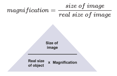
Microscopes use lenses to magnify the image of a biological specimen so that it appears larger.
The formula to calculate magnification is:
Cell size
Most animal and plant cells are 0.01 – 0.10 mm in size. The smallest thing seen with the naked eye is about 0.05 mm.
For all cells we need a microscope to see them in any detail.
The best unit to measure most cells is the micrometre, symbol μm.
For some sub-cellular structures, for instance ribosomes, or organisms such as viruses, it’s best to use a smaller unit – the nanometre, symbol nm.
One metre can be broken down into the following measurements:
| Millimetre, mm | Micrometre, μm | Nanometre, nm | |
|---|---|---|---|
| 1/1000 metre | 1/1000 millimetre | 1/1000 micrometre | |
| Division of a metre as a fraction | 1/1000 metre | 1/1,000,000 metre | 1/1,000,000,000 metre |
| Division of a metre in standard form | 1 × 10^-3 m | 1 × 10^-6 m | 1 × 10^-9 m |
Standard form
When writing and working with very large or very small numbers, we use standard form.
Standard form shows the size of numbers as powers of ten.
Standard form numbers are written as:
- A × 10^n
- A is a number greater than one but less than 10
- ^n is the index or power, always in powers of 10
Using standard form for large numbers
- A population of 120 000 000 microorganisms could be written as 1.2 × 10^8.
- This number can be written as 120 000 000.0.
- If the decimal place is moved eight spaces to the left we get 1.2.
- So we put x 10^8 after 1.2 to show this.
- Because the original number is greater than one metre the minus sign before the 8 is not needed.
- It makes a very large number easier to write down.
Using standard form for small numbers
- A red blood cell's diameter of 7 μm or 0.000007 m could be written as 7 × 10^-6 m.
- This number can be written as 0.000007.
- If we move the decimal place six spaces to the right we get 7.0
- So we put x 10^-6 after 7 to show this.
- Because the original number is less than one metre we put a minus sign before the 6.
- It makes a very small number easier to write down.
Calculating the magnification of a cell
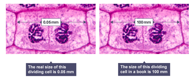
It’s important to work in the same units when calculating magnification. Sizes of most cells are given in micrometres, symbol μm.
Preparing biological samples for examination
Aims of the experiment
- To stain cells for examination with a light microscope.
- To examine a range of cells and other structures with a microscope to understand their basic structure.
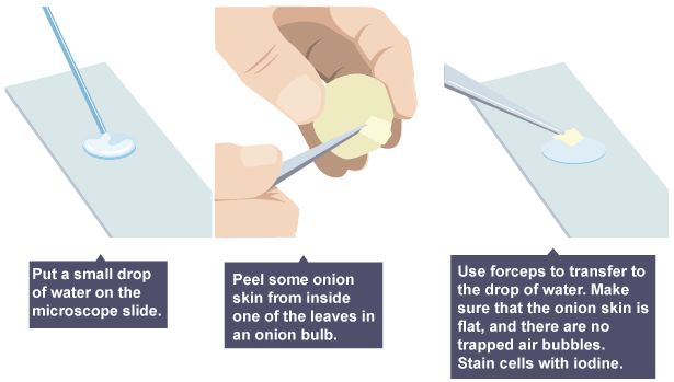
Plant cells
Onion epidermal cells
Animal cells
Cheek cells

Other biological structures
Human hair or animal fur
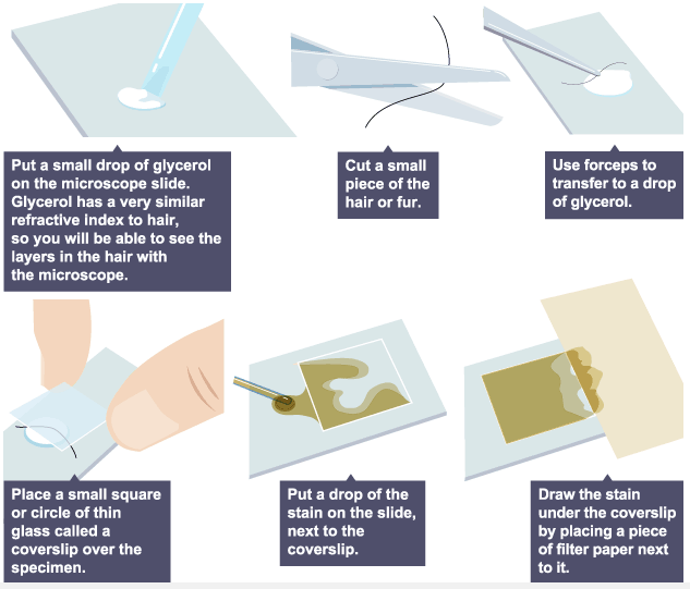
When viewing any slide with a microscope, a small square or circle of thin glass called a coverslip is placed over the specimen. It protects the microscope and prevents the slide from drying out when it’s being examined.
The coverslip is lowered gently onto the specimen. A mounted needle can be used to hold the specimen in place as the coverslip is lowered. It is important that no air bubbles are trapped underneath. Most cells are colourless. Stains are used to add contrast. Certain stains are also used to stain specific cell structures or cell products.
Risks
- Care must be taken when looking down the microscope if the illumination is too bright.
- Care when using microscope stains.
- Care when handling coverslips, microscope slides and mounted needles.
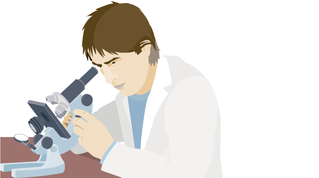
Investigating cells with a light microscope
Once slides have been prepared, they can be examined under a microscope.
Aims of the experiment
- To use a light microscope to examine animal or plant cells.
- To make observations and draw scale diagrams of cells.
Method
Rotate
Rotate the objective lenses so that the low power, eg x10, is in line with the stage.

Focus
Turn the coarse focus so that the stage is as close to the objective lens as possible. You should not look through the microscope to do this.

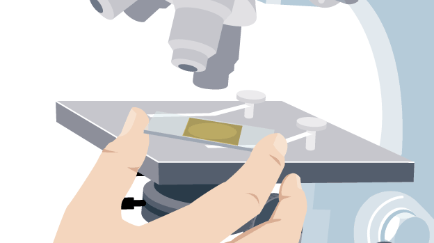
Place the slide
Place the microscope slide – either one you have prepared, or a permanent slide – on the stage. Line it up so that the specimen – if you can see it – is in the centre of the stage, where the light passes through.
Focus
Focus the slide towards you by turning the coarse focus adjustment.
Record an image
Draw a low power image or record a digital image of what you see. Then, rotate the objectives so that the high power objective, eg x40, is in line with the stage.

Re-focus
Bring the slide back into focus using the fine focus adjustment. If you do not succeed, go back to low power and re-focus, then try again.

Risks
- Care must be taken when looking down the microscope if the illumination is too bright.
- Care must be taken when using microscope stains.
- Care must be taken when handling coverslips and microscope slides.
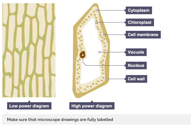
Drawing the image
Record the microscope images using labelled diagrams or produce digital images.
When first examining cells or tissues with low power, draw an image at this stage, even if going on to examine the slide with high power.
A low power diagram is used:
- as a plan to show the arrangement of any distinct regions of the tissue, for example the tissues in a plant root
- to show the outline of individual cells that make up the tissue, if the tissue is uniform
A high power diagram is then produced – a detailed image of a part of the slide. It is usually drawn to show a single cell, eg of a single cheek cell or onion cell.
Microscopes
How have light microscopes developed?
No-one knows who first invented the microscope, but there have been key stages in their development:
1590s - Dutch spectacle makers Janssen experimented with putting lenses in tubes. They made the first compound microscope. None of their microscopes have survived, but they are thought to have magnified from ×3 to ×9.
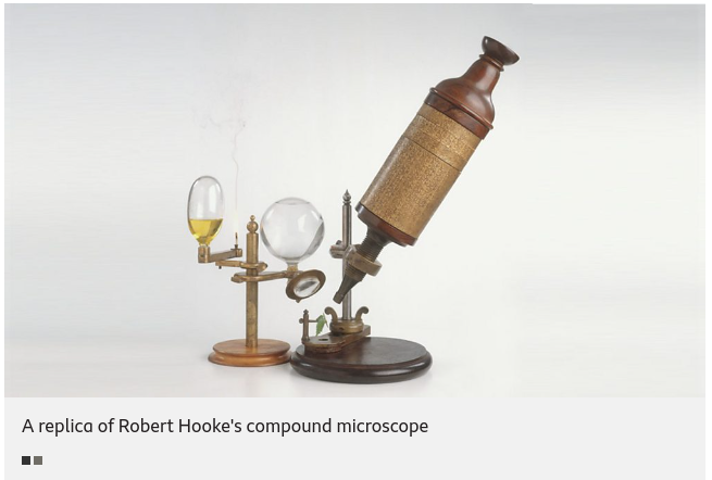
1650 - British scientist, Robert Hooke 1650 – also famous for his law of elasticity in Physics – observed and drew cells using a compound microscope.
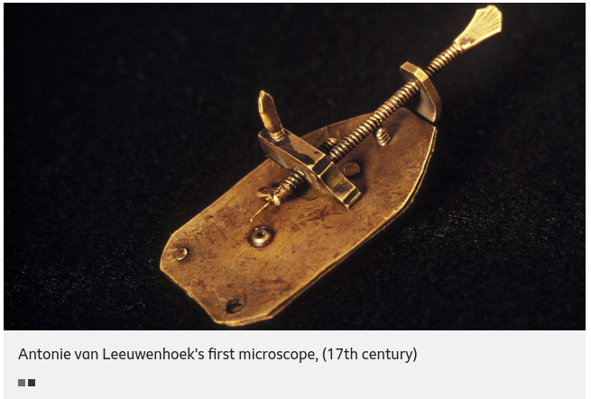
Late 1600s – Dutch scientist Antonie van Leeuwenhoek constructed a microscope with a single spherical lens. It magnified up to ×275.
1800s - the optical quality of lenses increased and the microscopes are similar to the ones we use today.
Throughout their development, the magnification of light microscopes has increased, but very high magnifications are not possible. The maximum magnification with a light microscope is around ×1500.
The limits of the light microscope
The magnification of a microscope is not the only factor that’s important when viewing cells. The detail that can be seen is also important.
The ability to see greater detail in an image depends on the resolution or resolving power. This is the ability to see two points as two points, rather than merged into one.
Think about a digital photo. It can be enlarged, but over a certain size, you won’t be able to see any more detail. It will just become blurry.
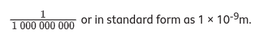
The resolution of a light microscope is around 0.2 μm, or 200 nm. This means that it cannot distinguish two points closer than 200 nm. One nm, or nanometre, is one billionth of a metre. This is written as:
The electron microscope
Electron microscopes use a beam of electrons instead of light rays.
There are two types of electron microscope:
- The scanning electron microscope (SEM) has a large field of view so can be used to examine the surface structure of specimens. SEMs are often used at lower magnifications.
- The transmission electron microscope (TEM) is used to examine thin slices or sections of cells or tissues.
TEMs have a maximum magnification of around ×1 000 000, but images can be enlarged beyond that photographically. The limit of resolution of the transmission electron microscope is now less than 1 nm.
The TEM has revealed structures in cells that are not visible with the light microscope.
Animal cells
Almost all animals and plants are made up of cells.
Animal cells have a basic structure. Below the basic structure is shown in the same animal cell, on the left viewed with the light microscope, and on the right with the transmission electron microscope.
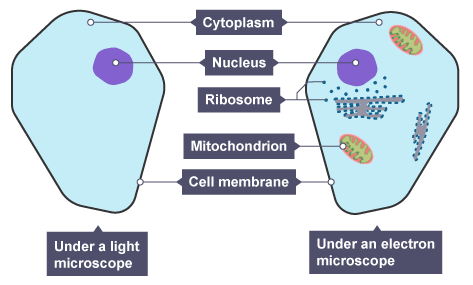
Mitochondria are visible with the light microscope but can’t be seen in detail.Ribosomes are only visible with the electron microscope.
Cell structures and their functions
| \n | Function |
|---|---|
| Cytoplasm | A jelly-like material that contains dissolved nutrients and salts and structures called organelles. It is where many of the chemical reactions happen. |
| Nucleus | Contains genetic material, including DNA, which controls the cell’s activities. |
| Cell membrane | Its structure is permeable to some substances but not to others. It therefore controls the movement of substances in and out of the cell. |
| Mitochondria | Organelles that contain the enzymes for respiration, and where most energy is released in respiration. |
| Ribosomes | Tiny structures where protein synthesis occurs. |
Most cells are specialised and are adapted for their function. Animals and plants therefore consist of many different types of cell working together.
Plant cells
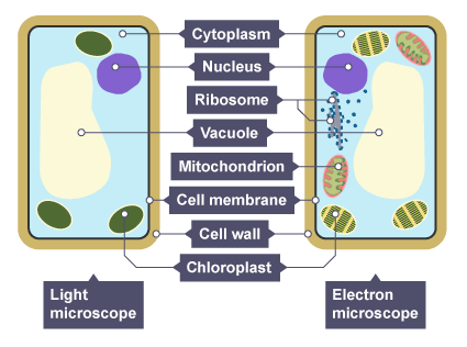
This basic structure of a plant cell is shown below – the same plant cell, as viewed with the light microscope, and with the transmission electron microscope.
Animal and plant cells have certain structures in common.
| Function | |
|---|---|
| Cytoplasm | A jelly-like material that contains dissolved nutrients and salts and structures called organelles. It is where many of the chemical reactions happen. |
| Nucleus | Contains genetic material, including DNA, which controls the cell’s activities. |
| Cell membrane | Its structure is permeable to some substances but not to others. It therefore controls the movement of substances in and out of the cell. |
| Mitochondria | Organelles that contain the enzymes for respiration, and where most energy is released in respiration. |
| Ribosomes | A tiny organelle where protein synthesis occurs. |
Plant cells also have additional structures:
| Function | |
|---|---|
| Chloroplast | Organelles that contains the green pigment, chlorophyll, which absorbs light energy for photosynthesis. Contains the enzymes needed for photosynthesis. |
| Cell wall | Made from cellulose fibres and strengthens the cell and supports the plant. |
| Permanent vacuole | Filled with cell sap to help keep the cell turgid. |
Animal cells may also have vacuoles, but these are small and temporary. In animals, they are commonly used to store or transport substances.
Measuring cell size
Cell size can be measured using an eyepiece graticule. The graticule has a scale ruled on it.
You must find out the distance measured for each division of the graticule. You can then use the graticule to measure cells. The distance will be different for each objective.
To do this, you will use a stage micrometer. You will use this to calibrate the eyepiece graticule. Once it's calibrated, you can use the eyepiece graticule every time you use the microscope.
Place a stage micrometer on the stage of the microscope.
Line up one of the divisions on the eyepiece graticule with a fixed point on the stage micrometer.
Count the number of divisions on the eyepiece graticule that correspond with a set measurement on the stage micrometer.
Calculate the distance in micrometres of one division on the eyepiece graticule.
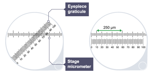 The distance of 250 μm on the stage micrometer lines up against two divisions at 10 and 61 on the eyepiece graticule.
61 − 10 = 51 divisions on the eyepiece graticule are equivalent to 250 μm on the stage micrometer.
One division on the eyepiece graticule is equivalent to μ25051μm on the stage micrometer: = 4.9 μm (to two significant figures).
Therefore one division is equal to 4.9 μm.
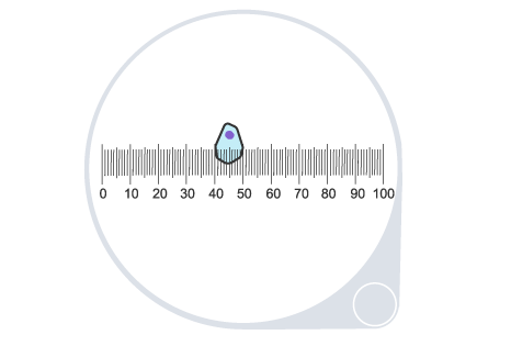
Using the same calibrated eyepiece graticule to measure a cell:
The width of the cell highlighted = 52 – 40 = 12 eyepiece graticule divisions.
The real width of the cell is 12 × 4.9 μm = 59 μm (to two significant figures).
Comparing sizes
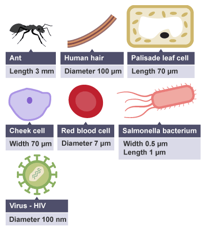
The diagram shows the size of three organisms, different cells and other structures.
Sizes can be compared using a straightforward calculation.
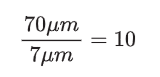
For instance, the length of the leaf cell above is ten times the diameter of a red blood cell.
The calculation would be:
When comparing the size of a bacterium with the Human Immunodeficiency Virus (HIV) different units have to be converted to be consistent.
The length of the bacterium = 1 μm = 1000 nm
The diameter of the HIV = 100 nm
Therefore the length of the bacterium is 1000/100 = 10 times that of the bacterium.
Order of magnitude
Differences in size are often described as differences in order of magnitude. That's the difference calculated in factors of 10.
Notation used to write numbers in multiples and sub-multiples of 10
| 1000 | = 10 × 10 × 10 | = 10^3 |
|---|---|---|
| 100 | = 10 × 10 | = 10^2 |
| 10 | =10 | =10^1 |
| 1 | =1 | =10^0 |
| 0.1 | =1/10 | =10^-1 |
| 0.01 | =1/100 | =10^-2 |
| 0.001 | =1/1000 | =10^-3 |
If you increase a number by one order of magnitude, you are multiplying the number by 10.
If you decrease a number by one order of magnitude, you are dividing the number by 10, which is equivalent to multiplying by 0.1.
For instance, there is a one order of magnitude difference between a person 2 m tall, and an oak tree, 20 m tall.
The person's height = 2 m = 2 × 10^0
The oak tree's height = 20 m = 2 × 10^1
Meaning there is one order of magnitude between the height of a human being (2m) and the height of an oak tree (20 m).
Notice that when dividing numbers in standard form, we subtract the powers.
When comparing orders of magnitude, actual distances can be approximated. It’s the relative difference that is important.
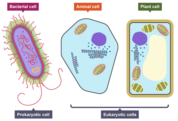
Eukaryotes and prokaryotes
Bacteria are amongst the simplest of organisms – they are made of single cells. Their cell structure is simpler than the cells of animals, plants and fungi.
Cells of bacteria are called prokaryotic cells.
Cells of animals, plants and fungi are called eukaryotic cells.
Comparing cell types
| Eukaryotic cell | Prokaryotic cell | |
|---|---|---|
| Size | Most are 5 μm – 100 μm | Most are 0.2 μm – 2.0 μm |
| Outer layers of cell | Cell membrane - surrounded by cell wall in plants and fungi | Cell membrane - surrounded by cell wall |
| Cell contents | Cytoplasm, cell organelles include mitochondria, chloroplasts in plants and ribosomes | Cytoplasm, ribosomes, no mitochondria or chloroplasts |
| Genetic material | DNA in a nucleus - plasmids are found in a few simple eukaryotic organisms | DNA is a single molecule, found free in the cytoplasm - additional DNA is found on one or more rings called plasmids |
| Type of cell division | Mitosis | Binary fission |
A group of organisms called Archaea are also prokaryotic.
Plant and animal cells
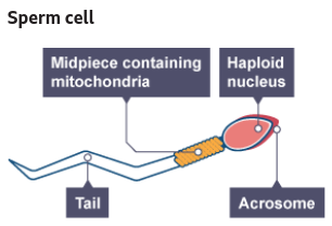
How different types of animal cell are adapted to carry out their function
There are many different types of cells in animals. Each type is specialised for a particular role. These ensure that the organism functions as a whole.
The head of the sperm contains the genetic material for fertilisation. The acrosome in the head contains enzymes so that the sperm can penetrate an egg. The middle piece is packed with mitochondria to release energy needed to swim and fertilise the egg. The tail enables the sperm to swim.
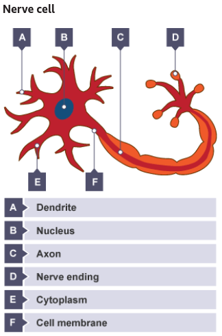
The nerve cell is extended, so that nerves can run to and from different parts of the body to the central nervous system. The cell has extensions and branches, so that it can communicate with other nerve cells, muscles and glands. The nerve cell is covered with a fatty sheath, which insulates the nerve cell and speeds up the nerve impulse.
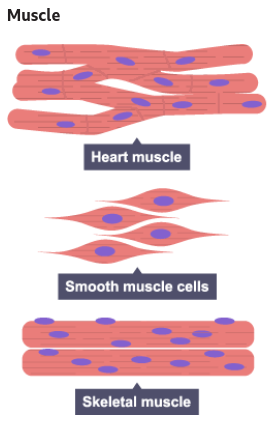
Muscle cells contain filaments of protein that slide over each other to cause muscle contraction. The arrangement of these filaments causes the banded appearance of heart muscle and skeletal muscle. They contain many well-developed mitochondria to provide the energy for muscle contraction. In skeletal muscle, the cells merge so that the muscle fibres contract in unison.
How different types of plant cells are adapted to their function
There are many different types of cells in plants. Each type is specialised to do a particular role and ensures that the organism functions as a whole.
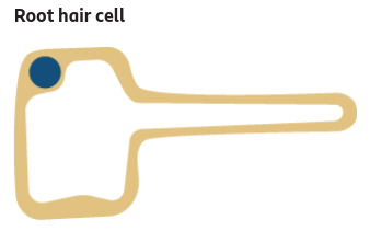
The root hair cell has a large surface area to provide contact with soil water. It has thin walls so as not to restrict the movement of water.
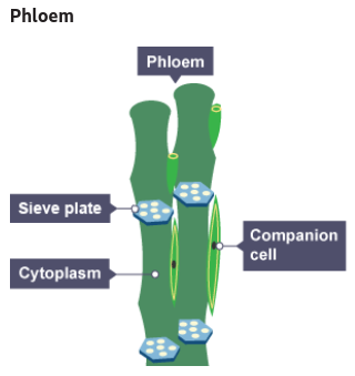
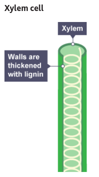
There are no top and bottom walls between xylem vessels, so there is a continuous column of water running through them. Their walls become thickened and woody. They therefore support the plant.
Dissolved sugars and amino acids can be transported both up and down the stem. Companion cells, adjacent to the sieve tubes provide energy required to transport substances in the phloem.