Chromosomes and Karyotypes
REVIEW:
Not all chromosomes are in ‘x’ shape. Humans have 46 chromosomes.
Bacteria: Circular shape chromosome
Centromere is not always in the center of the chromosome.
There are still 46 chromosomes when it’s duplicated, its just the chromatids that are duplicated.
Karyotype: Image of all your chromosomes, from a cell in mitosis stage typically metaphase
Anaphase And Telophase: NOT the phase to do Karyotype
Metaphase: Has two sister chromatids
Homologous pairs:
22 of the pairs are autosomes.
Last two are called sex chromosomes: 23 chromosomes from fathers (x and y), 23 chromosomes from mothers ( x )
CHROMOSOMES AND KARYOTYPES
Chromosomes condense during mitosis and become visible.
We take a photo of the chromosomes and then cut and paste them until the chromosomes are paired by length and banding.
This picture is called a karyotype.

Karyotype
Karyotypes show the number and types of chromosomes in a eukaryotic cell.
The chromosomes are stained and photographed to generate a visual profile
The chromosomes of an organism are arranged into homologous pairs according to size (with sex chromosomes shown last)

Karyotype Notations
It has to be written in format of:
# of chromosomes, gender, and location of the abnormality if any

Nondisjunction
This is caused when the chromosomes do not separate properly during meiosis, forming a monosomy or trisomy.
When a sex cell with an extra or one less chromosome gets fertilized, the zygote will have an extra or one less chromosome.
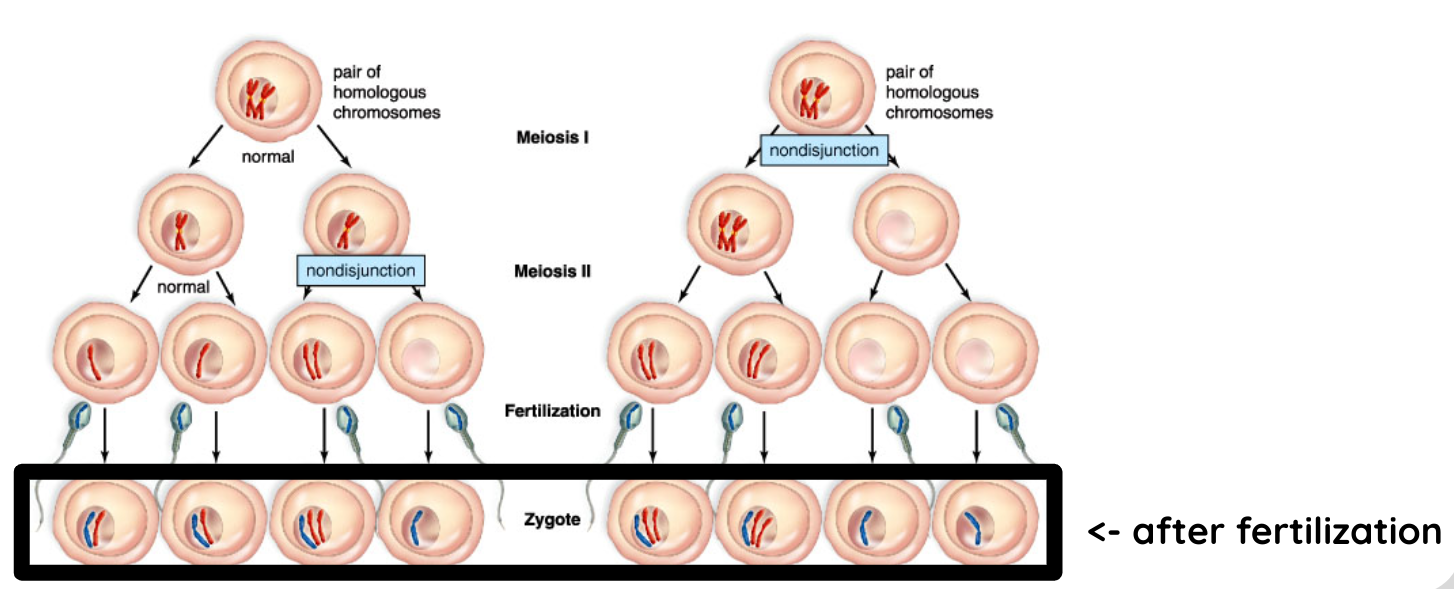

Trisomy VS Monosomy
When there is an extra chromosome in a pair = trisomy

Examples of trisomy disorders:
Down syndrome
Patau syndrome
Klinefelter syndrome
When there is only one chromosome in a pair = monosomy

Examples of monosomy disorders:
Turner syndrome
Cri du Chat Syndrome