Thorax and Lungs
Thorax extends from the base of the next to the diaphragm
Lower Respiratory System:
lungs, distal portion of the trachea, bronchi, bronchioles, alveoli
supposed by the diaphragm and rib cage
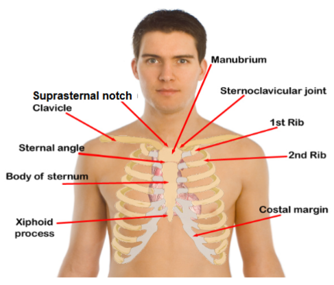
Thoracic cage:
outer structure of the thorax
provides support and protection for organs - heart, liver, spleen, kidneys
Thoracic cavity:
contains respiratory components -mediastinum and lungs lined by pleural membranes
mediastinum - central compartment of the thoracic (chest) cavity located between the two lungs
Lungs
Structure: cope-shaped, elastic structures.
apex of each lung extends slightly above the clavicle, base at level of the diaphragm
Lung lobes: sizes are not identical but don’t differ much but have different lobe distributions
D. Right lung has three lobes
C. Left lung has two lobes
the lower lobes of both lungs primarily located toward posterior surface of the chest wall
fissures are separate lobes
lobes appear as diagonal sloping segments
laterally, lung tissue reaches eight rib level
posteriorly, base lies at about tenth rib
Mechanisms of breathing
inspiration = contraction of diaphragm & expansion of the chest creating a negative pressure within the lungs
expiration = usually passive relaxation of the diaphragm & the intercostal muscles
breathing patterns change in response to cellular demands
Control of respirations:
breathing controlled by brainstem (involuntary)
medulla oblangata and pons and cerebral cortex (voluntary)
medulla oblangata = control center, adjusts rate and depth of breathing based on chemical signals (PH, CO2 and O2 levels) from the body
Respiratory drives:
hypercapnia = increase in CO2. normal stimulus to breath
hypoxemia = decrease in O2. also increases respirations, but less effective than hypercapnia
COPD = chronic state of hypercapnia, relies solely on hypoxic drive to breath rather than high CO2. thus give controlled 02
When could a disease process or medical condition affect the acid base-balance enough to change a person’s breathing pattern?
Hypoventilation
not breathing enough CO2 accumulates
patient breathes too slowly or too shallowly to remove enough CO2 carbon dioxide builds up in the blood which can cause respiratory acidosis
chronic obstructive pulmonary disease, barbiturate overdose and protected vomiting can all lead to hypoventilation
Hyperventilation
breathing more than the body needs - too much CO2 is removed
patient removes CO2 faster than the body produces it. CO2 decreases in the blood which can cause respiratory alkalosis
aspirin overdose, panic and diabetes complicated with ketoacidosis can all lead to hyperventilation


Subjective data collection for lung assessment:
cough = acute or chronic
shortness of breath = activity, position, time of day, season
chest pain with breathing = location and characteristics
history of respiratory illnesses = asthma, TB, bronchitis
smoking history = how many packets/days, years
environmental exposure = asbestos, factory, coal mine, farming, high traffic, pets, chemical plant
patient centered care = ask about last TB test, chest x-ray, pneumonia, vaccine, flu immunization
Objective data collection for lung assessment: inspection, palpation, percussion, auscultation, evaluate functioning of lungs
Inspection
begins with observation of breathing difficulties
RR and patter, ease of breathing
inspect for nasal flaring and pursed lip breathing
pallor (abnormal paleness, grayish, ashen, or yellowish cast of skin) or cyanosis (bluish, gray, ashen, or dusky discoloration)
observe color of face, lips, chest
inspect color and shape of nails
inspect configuration - symmetry and shape of thoracic cage
observe retraction, use of accessory muscles and intercostal spaces
inspect the client’s positioning
airway obstruction or atelectasis - observing the respiratory effort of a client you note that the client’s intercostal spaces are retracting on inspiration
Respiratory patterns:
normal = 10-20 breaths per minute, even
tachypnea = >20/25 breaths per minute, shallow
abnormally rapid and shallow
pleurisy/pluritis —> creates difficulty in getting enough oxygen and the body responds by increasing the respiratory effort in attempt to compensate
more examples: pneumonia, TB, COVID 19, pulmonary imbolism
bradypnea = <10/12 breaths per minute
rate less than 10/min and can be associated with medicated induced depression of the respiratory center, diabetic coma, or neurologic damage
hyperventilation = increased rate and depth
hypoventilation = shallow, slow, irregular pattern
cheyne-stokes = unusual pattern associated with serious problems, breaths become shallow, then stop for a period of time before restarting.


Inspecting face, lips, and chest:
red blotchy spots may be seen in clients with COPD or congestive heart failure as a result of polycythemia (overproduction of RBCs)
Inspecting face, lips, fingers, nails and chest:
cyanosis may be seen if client is cold or hypoxic
pale or cyanotic nails may indicate hypoxia. early clubbing (180-degree angle) and late clubbing (180-degree angle) can occur from hypoxia
variation information cyanosis makes white skin appear blue tinged especially in the perioral, nail bed, and conjunctival areas. cyanosis makes dark skin appear blue and dull in the same areas

Normal (elliptical) vs barrel chest:
normal = wider side to side (transverse diameter) vs narrower front to back (anterior posterior diameter 2:1 ratio
barrel chest = hyperinflation of the lungs and often seen in emphysema because of hyperinflation of the lungs. 1:1 ratio

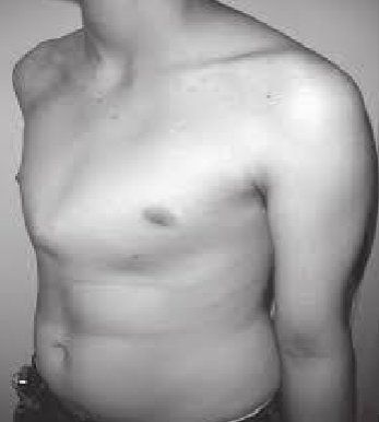
Pectus excavatum or funnel chest:
congenital malformation
breastbone (sternum) and ribs grow inward, creating a sunken or caved in chest appearance
affecting 1 in 400-1000 births
often becomes more pronounced during adolescent growth spurts, potentially causing exercise intolerance, fatigue, chest pain and significant body image issues

Pectus carinatum or pigeon chest:
congenital malformation
affecting 1 in 1200 to 1500 births
sternum protrudes forward. increased AP diamete
severe cases can restrict heart and lung function, leading to decreased stamina, shortness of breath, chest pain and tenderness and palpitation
Tripod position and use of accessory muscles: client leaning forward with their arms supporting their body weight and lift chest to increase breathing capacity
often seen in COPD - emphysema

Orthopneic position: a client with heart failure would most likely assume an to ease any breathing difficulties


Kyphosis = increased curve of the thoracic spine. may be a normal finding in older clients. it results from a loss of lung resiliency and loss of skeletal muscle
Palpating the thorax:
confirm symmetric chest expansion
palpate for tactile fremitis
detect lumps, masses, tenderness
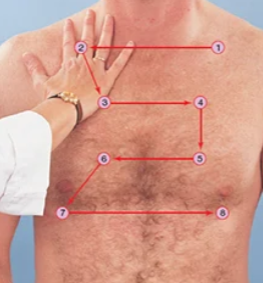
Palpate for symmetric chest expansion: palpate for symmetric chest expansion
chest expansion assessed by measuring the distance the examiner’s thumbs move when the client takes a deep breath.

Palpate for tactile fremitus:
assess vibrations of chest wall when a client speaks - ask client to say “ninety-nine” as the nurse palpates the thorax
nurse uses ball or ulnar edge of one hand to assess vibrations of air and the bronchial tubes transmitted to the chest wall
use ball or ulnar of the hand for best assessing
Increased tactile fremitus: denser lung tissue such as pneumonia, pulmonary fibrosis, atelectasis, or lung tumor
Decreased tactile fremitus: conditions that add air or fluid between the lung and chest wall, such as pleural effusion, pneumothorax, emphysema, or a thick or obese chest wall

Palpate for crepitus:
respiratory crepitus - cracking, popping, or grating sound/sensation (subcutaneous emphysema or subcutaneous crepitus) caused by air trapped in subcutaneous tissues, usually around the chest or neck. it indicates an underlying, sometimes serious, issue like pneumothorax, injury, or severe infection
Percussion of thorax and lungs:
percuss the lung fields
tone over lung fields should be resonant/resonance is the normal finding on lung percussion
hyperresonance - emphysema, pneumothorax due to air trapping
tone over bone is typically flat like the scapula ribs
tone over organs is dull. dullness is noted with fluid or solid tissue replacing air in the lung pleural effusion, pneumonia, tumor
tone over stomach is tympany. tympany would be noted over areas of air, such as a gastric bubble in the stomach
Auscultation of the lungs
1. BRONCHIAL SOUNDS OVER THE TRACHEA ARE HIGH PITCHED &
HARSH ,SHORT DURING INSPIRATION AND LONG DURING EXPIRATION
BRONCHOVESICULAR SOUNDS OVER MAJOR BRONCHI ARE
MODERATE IN PITCH, EQUAL DURING INSPIRATION AND EXPIRATION
VESICULAR SOUNDS OVER PERIPHERAL LUNG FIELDS ARE LOW
PITCHED, SOFT AND RUSTLING LIKE THE “WIND IN THE
TREES”/BREEZY
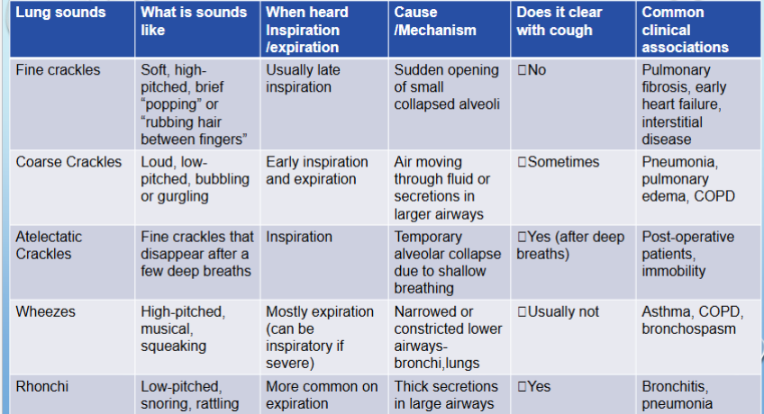
Adventitious sounds of the lungs:
crackles = fluid or alveoli popping open
wheezes = airway narrowing (think asthma)
rhonchi = secretions in large airways
pleural rub = inflamed pleura rubbing
stridor = upper airway obstruction (emergency)

Developmental changes reflected in the lungs
newborn
lungs are fluid filled at birth and should clear with the first few breaths
adequate surfactant (develops after 32 weeks gestation) is essential for alveolar expansion
immature lungs more vulnerable to infections and environmental exposures
pregnancy
breathing becomes deeper due to increased oxygen demands from growing fetus
enlarging uterus pushes diaphragm upward, limiting downward lung expansion
estrogen causes relaxation of thoracic ligaments, allowing more horizontal chest expansion
aging adults
thoracic cage becomes calcified and less flexible
kyphosis is common in older clients
respiratory muscles weaken
lungs lose elasticity
fewer alveoli —> reduced surface area for gas exchange
increased risk for pneumonia and postoperative complications
deep breathing may be especially difficult for the older adult client therefore, the nurse will offer rest as needed
How do these changes affect the older adult’s ability to tolerate anesthesia or respiratory infections?
decreased lung elasticity and fewer functional alveoli
reduce gas exchange efficiency, making it harder for older adults to clear anesthetic gases or recover from hypoventilation
weakened respiratory muscles and a less effective cough reflex
impair the ability to clear secretions, increasing the risk for atelectasis (collapses alveoli) and pneumonia
calcified rib cage
limits chest expansion, further reducing ventilation
these changes collectively make older adults more sensitive to anesthesia, slower to recover post-op, and more vulnerable to respiratory infections
nursing considerations
close post-op monitoring - assess for shallow respirations, oxygen desaturation, or ineffective cough
encourage early mobilization and deep breathing exercises as tolerated
Why are older adults more prone to developing pneumonia after surgery or prolonged bed rest?

What preventive nursing interventions can help maintain lung functions in aging adults?

What happens if a newborn does not have enough surfactant at birth?

Why might a premature infant have more respiratory problems than a full term infant?

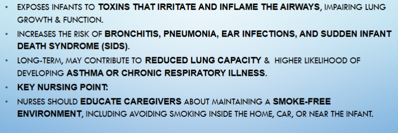
How could exposure to secondhand smoke affect an infants developing lungs?