genetics m2
Cell Division: Mitosis & Meiosis
The cell cycle multiplies cells
> The cell cycle is an ordered sequence of events
for cell division
It consists of two stages:
Interphase: duplication of cell contents
- G 1—growth, increase in cytoplasm
- S—duplication of chromosomes
- G 2—growth, preparation for division
Mitotic phase: division
– Mitosis—division of the nucleus
– Cytokinesis—division of cytoplasm
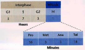
Time Duration in the Cell Cycle
The large, complex chromosomes of eukaryotes duplicate with each cell division
Eukaryotic chromosomes are composed of chromatin
– Chromatin = DNA+proteins
– Chromatins are highly compact
– Chromosomes are visible under a microscope
– Chromosomes are made of two sister chromatids joined together by a centromere
PARTS OF A METAPHASE CHROMOSOMES
1. Chromatin
< Heterochromatin- where the DNA is more condensed, and usually there is not much transcriptional activity. Some heterochromatin will remain condensed throughout the cycle.
< Euchromatin- this is where “active” genes are- usually this region is much less condensed.
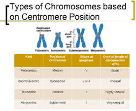
2. Centromere
- during mitosis, spindle fibers attach to the centromere via kinetochore – a highly complex multiprotein structure that is responsible for the actual events of chromosome segregation (anaphase)
3. Chromosome arms
< P arm
< Q arm
> Secondary constriction
- Site of nucleolus formation
> DNA Satellites
- Rounded or elongated (has extra nucleotides)
- Also called trabants
> Telomeres
- Located at the tip of linear chromosomes
- Maintains chromosomal stability
- Shortens as cells divide (helps determine age)
- Prevents adhesion between chromosomes
- Adheres to the inner side of the nuclear envelope
Polytene Chromosome
> Occurs when duplication happens repeatedly without segregation
> Found in salivary glands of larval Drosophila
> Characterized by having alternating light and dark bands
○ Dark bands = inactive areas
○ Light bands= transcriptionally active
Lampbrush Chromosome
> Found in the growing oocytes (immature eggs
Of most animals, except mammals.
> Organized into a series of chromomeres with large chromatin loops extended laterally giving a hairy “lampbrush” appearance.
> Present during Prophase I
Cell division is a continuum of dynamic changes
> Mitosis progresses through a series of stages
– Prophase
– Metaphase
– Anaphase
– Telophase
> Cytokinesis often overlaps telophase
> A mitotic spindle is required to divide the chromosomes
> The mitotic spindle is composed of microtubules
> It is produced by centrosomes
-Organizes individual tubulin into microtubules
– Contain a pair of centrioles in animal cells
– The role of centrioles in cell division is unclear
-- Centrioles are absent in plant cells
STAGES OF MITOSIS
< Interphase
< Prophase
< Metaphase
< Anaphase
< Telophase
< Cytokinesis
Allium cepa Chromosome Abberations Test for Genotoxicity Analysis
> Chromosomal aberrations
- Changes in chromosome structure or number
> Two kinds of chromosomal aberrations
- Bridges - chromatids that didn’t separate
- Fragments - pieces of chromosome that broke off
> Colchicine-like mitosis (c-mitoses)
- Colchicine is a chemical that disrupts spindle formation
- Full c-mitosis = no spindle (cell death)
- Vagrants = weak spindle (aneuploidy)
- Multipolar anaphases = partly disrupted spindle
(aneuploidy)
F: fragment
s: sticky chromosomes
v: vagrants
b: bridges
c: c-mitosis
Asynthetic Fission:
A new type of cell division
STAGES OF MEIOSIS:
< Meiosis l
< Meiosis ll
Prophase l
● Leptotene
- Chromosomes are in diploid number
● Zygotene
- Homologous chromosomes begin to pair
● Pachytene
- Crossing over between homologous pairs take place
- Each chromosome has doubled and is consist of 4 strands
● Diplotene
- Each pair of synapsed chromosomes begin to uncoil
- Each pair has 4 chromatids (2 per chromosome or bivalents)
● Diakinesis
- Bivalents disperse in the nucleus
- Chromosomes start to assume different shapes due to repulsions and chiasmata attachments
Homologous chromosomes wrap around each other during meiosis, as seen in this micrograph of a meiotic cell of a lily. The points at
which the homologous
chromosomes are crossed are termed chiasmata (arrows).
Gametogenesis
< Is the process of diploid or haploid cells entering cellular division and undergoing differentiation to mature haploid gametes.
Spermatogenesis
< process of making sperm cells
NOTES:
Meiosis I: Separate the Homologues.
Meiosis ll: Separate the sister chromatids (by mitosis)
Normal Human Karyotype
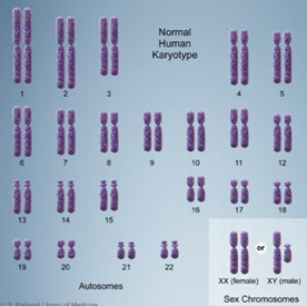
44 Autosomes, 2 Chromosomes
(syndromes are seen when there’s an extra chromosome in the said number)
Down Syndrome / Trisomy 21
< Trisomy is the presence of
an extra chromosome in cells. Down syndrome is an
example of a condition
caused by trisomy,
Patau Syndrome / Trisomy 13
< The baby has no eyes, no nose opening, and an elongated bulb hanging from forehead.
Edward Syndrome / Trisomy 18
< The baby has elongated skull, short neck, short breastbone, malformed ears and mentally deficient
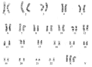
Karyotype of Metafemale (47-XXX)
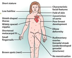
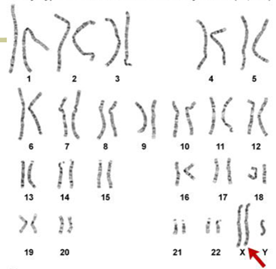
Turner Syndrome
Klinefelter Syndrome
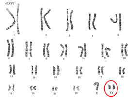
Jacob Syndrome (44-XXY)
Richard Speck: A Wrong Case of Jacob Syndrome
< Speck systematically killed eight student nurses from South Chicago Community Hospital in Chicago, Illinois on July 14, 1966.
Chromosomal Mosaicism
< When an individual has two or more cell populations with a different chromosomal
makeup, this situation is called chromosomal mosaicism.
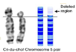
Cri-du-chat Syndrome
Philadelphia chromosome or Philadelphia translocation
< Associated with chronic myelogenous leukemia (cancer of the WBC)
< Reciprocal translocation between chromosome 9 and 22
Robertsonian Translocation
< Chromosome 21 breaks off and attaches to chromosome 14.
<The person in which this happens (called translocation carrier) is normal because, he or she only has the material of two chromosome 21s, but part of one of them just happens to be attached to chromosome 14.
< However, their child can inherit three “doses” of chromosome 21 material because the child might receive both a normal chromosome 21 and the chromosome 14 that has the extra chromosome 21 material, then the child would have down syndrome. About 3-5% of down syndrome cases occur this way.
< For many parents who have had a child with down syndrome due to a translocation, there is a 3-12% chance of that couple having another child with down syndrome because the parent could pass on both a normal chromosome and the chromosome 14 that has the extra chromosome 21 material to any child of theirs.
Cancer
< When normal cells are damaged beyond repair, they are eliminated by apoptosis. Cancer cells avoid apoptosis and continue to multiply in un unregulated manner.