Unit 3
Vision, Hearing & Equilibrium (Ch 10)
1. Vision
Know the function of external anatomy of the eye.
-Lacrimal gland secretes tears
-Muscles attached to the external surface of eye movement
-The orbit is a bony cavity that protects the eye
-Nasolacrimal duct drains tears into nasal cavity
Know the pathway of light to the photoreceptor.
Light reflected off objects --> enters cornea --> pupil
-->lens --> vitreous humor --> arrives at retina
Understand the role of the cornea and lens with relation to focus on the fovea.
-cornea: focuses light
-lens: bends light to focus it on the retina
-retina: layer that contains photoreceptors
-Fovea: region of sharpest vision
Understand different hypothesis explaining why the iris is pigmented.
-controls the diameter of pupil and is a ring of pigmented muscle
-two pigmented layers absorb light
-blue and green mostly caused by changed amount or type of melanin in outer layer (stroma)
-sexual selection? Is the reason for eye color variation
Understand explanation for the human blind spot.
-Optic disk (blind spot): region where optic nerve and blood vessels leave the eye
Know common definitions of vision disorders.
Myopia:
trouble focusing on distance (near sightness) and main issue is with how long eyes are
Hyperopia:
trouble focusing on near objects (far sightness) and main issue is with how short eyes are
Presbyopia:
degenerative loss of ability to focus on near objects (aging is the main cause). Thickening and loss of flexibility of lens, less elastic ciliary muscles
Astigmatism:
blurred vision caused by curvy cornea (multiple focus points). Lack of + converging on a single spotDifferentiate sensitivity and distribution of rods v cones.
Understand cellular physiology of phototransduction in rods.
Light arrives at retina
-->protein (a pigment) in rods/ cones changes shape
--> signal is produced
-->permeability of membrane to ions change --> action potential --> transduction thru optic nerve to brain
What differentiates ON vs. OFF bipolar cells? (hint: they have different types of glutamate receptors).
ON (in light): trans retinal in rhodopsin--> no neurotransmitter released --> action potential
OFF (in dark): cis retinal in rhodopsin --> neurotransmitter released --> no action potential
What are some basic properties which contribute to visual processing? (convergence, integration, specialization)?
Convergence: turn two eyes inward toward each other to look at a close object
What are “visual fields”?
Total area in which objects can be seen in the side (peripheral) vision as you focus you eyes on a central point)
What are examples of top-down versus bottom-up visual processing?
bottom-up
detection of contrast, color, movement
what pathway: recognize and identify objects
where pathway: object movement and location
controls where your eyes are “attending”
ex: hermann grid
top-down
experience and focus combine to alter our perception by feeding back and influencing “lower-level” signaling
controls which input you consciously pay “attention”
ex: the basketball and monkey suit; charlie chaplin mask
2. Hearing and Equilibrium
Describe how the functional anatomy of the outer, middle, and inner ear provide our sense of hearing.
Outer:
sound waves strike the tympanic membrane and become vibrations
Middle:
The sound wave energy is transferred to three bones to vibrate. The vibrations of the oval window create fluid waves within the cochlea.
Inner:
The fluid waves push on membranes of cochlear duct. Hear cells bend and ion channels open, creating an electrical signal that alerts neurotransmitter release.
What type of receptor is the inner ear hair cell?
neurons
What is one important reason that auditory information from the left and right ears “crosses over” on its way to the auditory cortexes?
allows for sound regulation
Understand and be able to differentiate among the three type of hearing loss?
sensorineural
loss of receptors
conductive:
breaking the connection - loss of transmission through ear
central:
nerve or brain damage
What type of receptors are used for the sense of equilibrium? (mechanical proprioceptors)
vestibular apparatus
Distinguish between the arrangement of cells that provide a sense of linear vs. rotational acceleration.
cristea - rotational acceleration
macula and otoliths - linear acceleration
otoliths are like lil crystals
What is the difference in response of a tonic vs. phasic receptor in terms of their frequency of action potentials? Be able to give examples of senses that use tonic v phasic receptors.
-Tonic: slowly adapting receptors that respond for the duration of the stimulus (nociceptors, and proprioceptors- pain)
-Phasic: rapidly adapt to a constant stimulus and turn off (olfactory)
Somatic Nervous System & Muscle Physiology (Ch 11-12)
1. Somatic nervous system and skeletal muscle
What is the somatic nervous system? Compare and contrast with the autonomic nervous system.
Somatic controls voluntary movements and
autonomic controls involuntary responses.
Describe three features of a somatic motor pathway.
-consist of one neuron
-originates in the CNS (brain or spinal cord)
-myelinated, very long, always excitatory--> ON switch and triggering contraction
-terminal branches close to target and each terminal innervates a single skeletal muscle fiber
What are the three types of muscles? Compare and contrast their basic appearance and control.
-Skeletal
striated and voluntary
Cardiac
striated involuntary
Smooth
spindle like and involuntary (organs)
Describe flexor-extensor pairs (antagonistic muscle groups).
flexor
brings bones together
extensor
moves bones away
pairs form antagonistic muscle groups
move bones in opposite directions
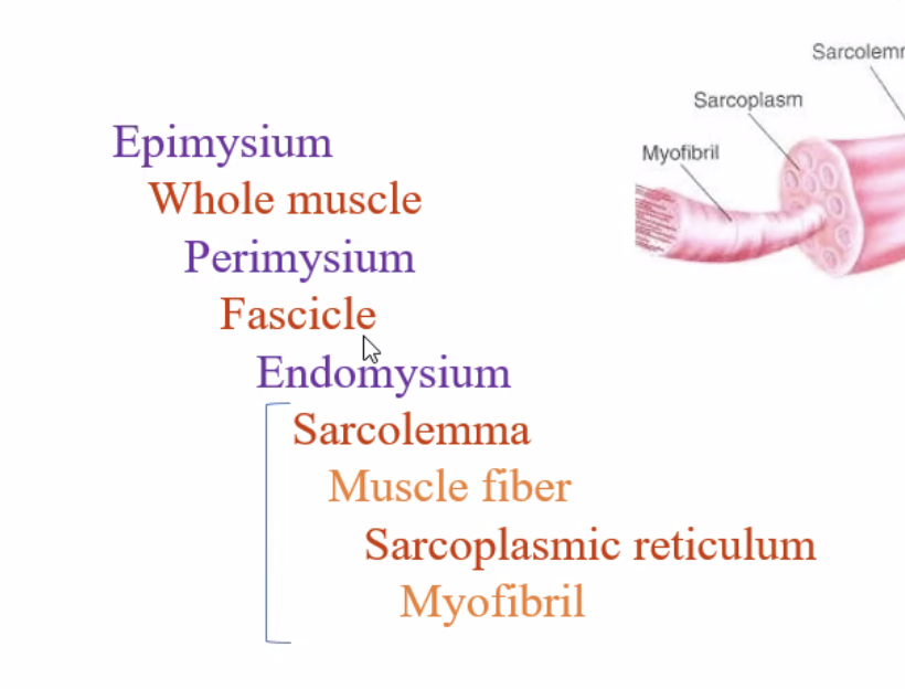
Understand skeletal muscle terminology and basic organization (Table 12.1).
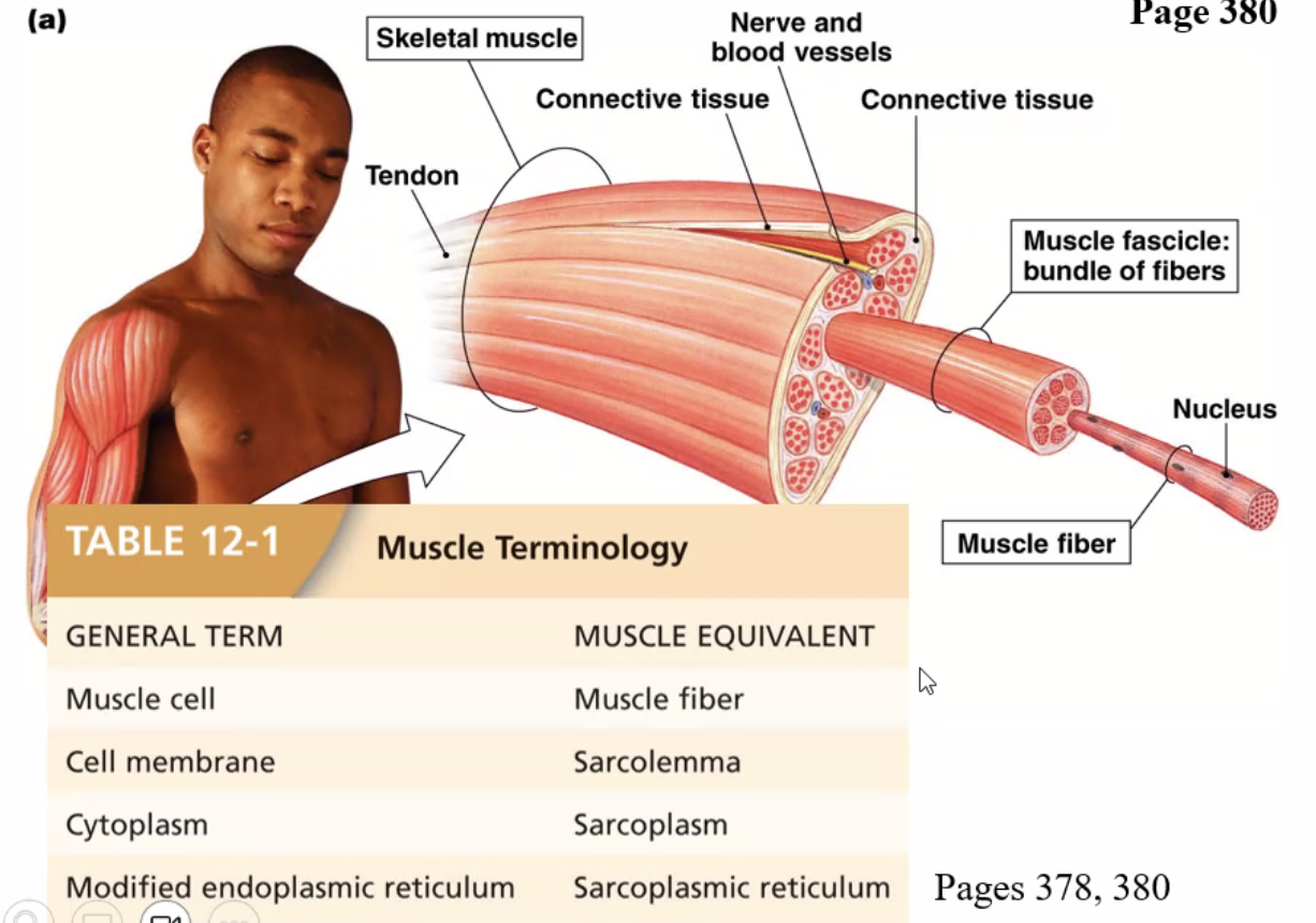
What are the unique and important features of a skeletal muscle cell?
Muscle fiber:
muscle cell
Endomysium:
surrounds each muscle fiber
Fascicle:
bundle of fibers
Perimysium:
surrounds a fascicle
Muscle:
collection of muscle fibers
Epimysium:
surrounds the whole muscle
Distinguish thick vs thin filaments and be able to label the diagram of a sarcomere.
thick = myosin = motor protein
actin = all have myosi binding sites
tropomyosin = blocks myosin binding site
troponin = hold tropo. in place, has Ca+ binding sites
Understand the role of troponin and tropomyosin in regulating actin.
actin = all have myosi binding sites
tropomyosin = blocks myosin binding site
troponin = hold tropo. in place, has Ca+ binding sites
Understand skeletal muscle contraction from (1) events at neuromuscular junction, (2) excitation-contraction coupling involving calcium signaling, and (3) the contraction-relaxation cycle as it relates to cross-bridge cycling and the sliding filament theory.
1.voltage gates Ca+ channels open.
2. Excitation-contraction coupling involving Ca signaling, Ach is released into synapse.
3. T-tubule depolarization triggers the opening of voltage gated Ca +2 channels in the SR membrane.
Name 3 roles/uses of ATP in skeletal muscle cells.
unbinding of myosin from actin and energizing myosin for power stroke. -
reuptake of Ca +2 into sarcoplasmic reticulum via Ca +2 pumps.
Na/K+ pump activity to help maintain proper concentration gradients
Describe the cellular mechanisms that allow a skeletal muscle cell to relax.
Ca+2 pumped back into SR
2. Skeletal muscle and Exercise
Differentiate between isotonic & isometric contractions
isometric= create force, no movement
same length
isotonic = contractions move loads
same tone
Understand motor units and how muscle innervation can provide sustained contraction.
motor unit - motor neuron and all innervated muscle fibers
Asynchronous recruitment: if skeletal muscle needs to sustain contraction, the CNS recruits motor units asynchronously
one muscle may have many motor units of different types
Understand the basics of fuel use during exercise, including the role of creatine phosphate as an intermediate energy store in metabolically demanding tissues.
Short term: ATP, creatine
Medium term: anaerobic glycolysis "lactic fermentation"
long term - mitochondrial respiration
resting muscle stores energy from ATP in the high-energy bonds of phosphocreatine. working muscle them uses that as stored energy
Distinguish among different muscle types with special emphasis on variation in cellular metabolism might determine difference in their performance.
Slow twitch oxidative muscle fibers:
darker color due to myoglobin and more red blood cells as it needs more oxygen
marathon runners, less likely to fatigue
fast-twitch oxidative-glycolytic (type 2A)
shorter thicker muscles
sprinter
low mitochondrial content
fast-twitch glycolytic (type2X)
Fast-twitch glycolytic muscle fibers: pale
3. Smooth Muscle
Where is smooth muscle found?
Involuntary muscle found in layers of organs
vascular (blood vessels)
gastrointestinal
urinary
respiratory
reproductive
ocular
How is smooth muscle organized at the tissue level? (single- vs. multi-unit)
Location: ^
by contraction pattern:
phasic smooth muscles
tonic smooth muscles
esophageal and urinary bladder sphincters
Communication with neighboring cells
Single-unit smooth muscle, or urinary smooth muscles, or visceral smooth muscles
Multi-unit smooth muscle
Multiunit smooth muscle is primarily under neural control. This is characteristic of vascular smooth muscle and ciliary muscles and iris of the eye.
Single-unit smooth muscle is usually made up of cells that are coupled by gap junctions, allowing ions and action potentials to pass between adjacent cells.
How is smooth muscle organized at the cellular level compared to skeletal muscle? How might the cellular organization of myofilaments explain differences in appearance of skeletal vs muscle under the microscope?
action potential not required
no t-tubules and no troponin
skeletal muscle fibers have a highly ordered arrangement of actin and myosin filaments in sarcomeres, which gives rise to the characteristic striated appearance under the microscope
Compare and contrast mechanisms of contraction and relaxation in smooth muscle versus skeletal muscle.
Skeletal: strong contraction for a short period of time
Smooth: weaker contraction for a longer period of time
Name four different mechanisms that can increase cytosolic Ca2+ and trigger contraction of smooth muscle.
chemically-gates channels (ligands)
Mechanically-gates channels (stretch)
Second messenger systems (IP3)
Intracellular regulatory signals (store operated channels)
Neural Reflexes (Ch 13)
Define neural reflex.
the integration of sensory info an involuntary response neural pathway link sensory (afferent) to autonomic or somatic (efferent). Can be innate or learned
What types of efferent pathways can a neural reflex use?
reflex
synapse
neurotransmitters
spinal cord
neurons
Give an example of an innate reflex and a learned reflex.
innate: plantar response
learned: potty training bladder
What can distinguish autonomic vs. skeletal reflexes at the anatomical level?
Autonomic: presynaptic axon (myelinated), central neuron synapses with ganglionic neuron, autonomic post synaptic axon (unmyelinated)
skeletal: Somatic motor neuron (myelinated), central fiber proteins to the target effector, Target effector: skeletal muscle, and axons ends at the synapse
Define and distinguish monosynaptic vs. polysynaptic reflexes.
Monosynaptic: reflex has a single synapse between the afferent and efferent neuron (skeletal muscles)
Polysynaptic: reflexes have two or more synapses. This somatic motor reflex has both synapses in the CNS. (divergence)
Can autonomic reflexes by influenced by higher CNS input?
These reflexes are subject to influence from higher nervous centers, they may occur without input from the brain
Describe an example of a simple skeletal reflex, reciprocal inhibition, and a withdrawal reflex (crossed extensor).
Simple skeletal reflex: the addition of a load stretches the muscle and spindles, creating a flex contraction
Reciprocal inhibition: antagonist muscle groups (patellar tendon- knee jerk)
Withdrawal reflex (crossed extensor): A flexion of one limb causes extension in the opposite limb. The coordination of reflexes with postural adjustments is essential for maintaining balance.
What is a “feedforward reflex” as it relates to voluntary movement
Causing changes in anticipation in preparation for movement.
Cardiovascular Physiology & Blood (Ch 14-16)
1. Cardiovascular System
Describe the gross anatomical features of the heart.
arteries - carry blood away from heart (anatomical left)
veins carry blood to heart (anatomical right)
oxygen from lungs to all cells
Understand the path of blood flow through the heart. Be able to differentiate pulmonary vs. systemic circulation.
BLUE/deoxygenated
1)enter via vena cava
2) right atrium
3) right AV valve
4) right ventricle
5) pulmonary semilunar valve
6) out thru pulmonary arteries towards lungs
RED/oxygenated
7) enter thru pulmonary veins
8) enter left atrium
9) left AV (mitral) valve
10) left ventricle
11) aortic semilunar valve
12) out thru aorta to body (systemic)
What are key differences in tissue composition and blood pressure gradient between arterial and venous side of circulation?
arterial circulation has higher blood pressure
What are exceptions to the rules in terms of arteries carrying oxygenated blood and veins carrying deoxygenated blood?
pulmonary arteries carry deoxygenated blood to the lungs whereas pulmonary veins carry oxygenated blood to the heart.
Distinguish diastole vs systole.
diastole - relaxation of the muscle to allow the chambers of the heart to fill with blood.
systole - contraction of the muscle pushes out ~60% of the blood from the chamber.
2. Cardiac muscle contraction
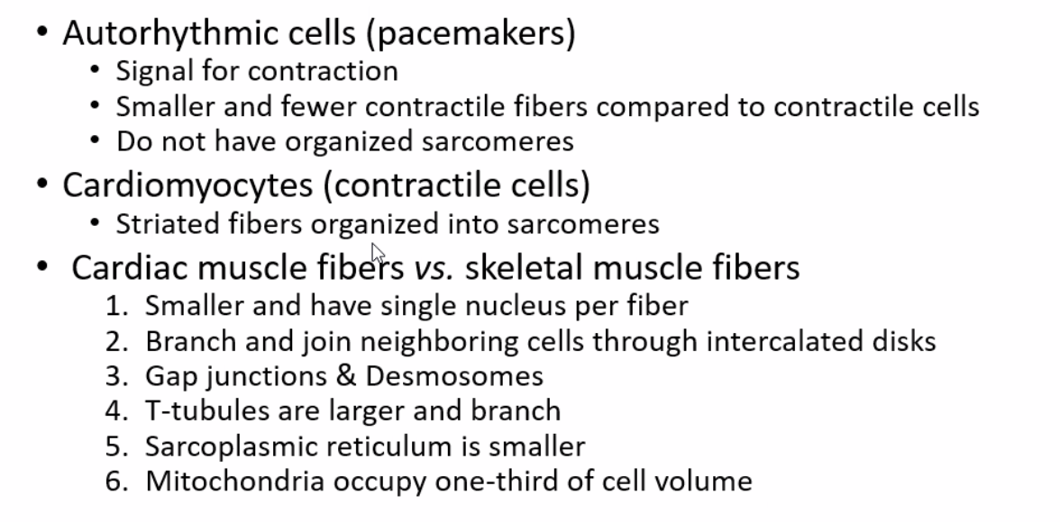
Name four physical features of myocardial cells.
striated
only have one nucleus per cell
branched
connected by intercalated disks → desmosomes and gap junction
Describe the functional components of intercalated discs. What do they do?
Intercalated discs are composed of desmosomes and gap junctions.
Desmosomes provide structure and adhesion
gap junctions provide rapid cell-cell communication
Are gap junctions formed between all cells of the heart? Why or not?
no, because the signal is passed by special cells
Gap junctions between auto-rhythmic cells and contraction cells to pass action potential
Describe general differences between pacemaker vs contractile cardiac cells.
Pacemakers:
-signal for contraction
-smaller/fewer contractile fibers compared to contractile cells
-do not have organized sarcomeres
contractile:
-striated fibers organized into sarcomeres
How does the anatomy of the cardiac conduction system contribute to the pathway of electrical signaling and resulting coordination of blood pumping?
1)SA node spontaneously APs
2) AP spreads through both atria, forcing blood into ventricles
3) AP does not immediately spread to ventricles
4) AP reaches AV node
5) AV node passes AP down muscles called bundle of his in septum
6) AP spreads into bundle branches and to smaller parking e fibers
7) ventricle walls contract from apex up, forcing blood out
What is the main cellular feature that makes pacemaker signals automatically fire action potentials?
Ion funny channels that let both Na+ and K+ through-
Are there other features of the pacemaker potential that differ from a neuronal action potential?
different starting potential
What about the action potential of cardiac contractile cells? Describe the cellular mechanism causing the “charge plateau” AND how one component of this mechanism couples the AP to contraction (i.e. sliding filaments).
1) resting membrane potential
2) action potential-> Na+ channels open
3) Na+ channels close and fast K+ channels open
4) ca2+ channels open and fast K+ channels close, creating a charge plateau during which the cell cannot depolarize
5)ca2+ close and slow K+ open
6)return to resting
4) calcium rushes in, binding to troponin moving off tropomyosin, allowing actin and myosin to bind causing contraction-
What physiological events are recorded in each phase of an ECG?
P wave: depolarization of atria
QRS complex: wave of ventricular depolarization, atrial repolarization
T wave: repolarization of the ventricle
Compare and contrast cardiac muscle cells, skeletal muscle cells, and smooth muscle cells (Table 12.4).
-only skeletal and cardiac are striated with sarcomeres
-skeletal: longer, more nuclei
-smooth: longer myofibrils
-smooth and cardiac: can have gap junctions, cardiac only have intercalated discs
-smooth and cardiac: pacemaker potential
3. Blood
What type of muscles help control blood flow thru blood vessels?
smooth muscle
Understand the what, where, and how of the “blood pressure reservoir”, “variable resistance”, and the “blood volume reservoir”.
Aorta/arteries: pressure reservoir, elastic, maintains blood flow during ventricular relaxation
arterioles: variable resistance through adjustable screws
Veins: blood volume reservoir, expandable, thinner walled
Be able to classify different blood vessels based on their basic physical and functional properties.
Arteries: lots of elastic/fibrous CT, smooth muscle, withstands high bp
arterioles: large amount of smooth, little CT, body temp homeostasis and O2 regulation
capillaries: exchange here! Either continuous or fenestrated, most present in muscle and gland
venules: little CT and smooth
veins: large lumen, less CT and smooth than arteries, lower bp than arteries-
What do you know about autonomic signaling and muscle tone of vascular smooth muscle?
Most vascular smooth muscle controlled by the sympathetic branch of the autonomic nervous system
increase in norepinephrine causes constriction (excitatory)
tonic control (so usually on)
Do capillaries vasodialate?
no because they do not have smooth muscle
What is the most important function of capillaries and what are some cellular features that can aid this function?
Site of exchange
either continuous or fenestrated (large pores)
simple structure allows exchange of materials between cells and blood
What is the role of the lymphatic system with respect to capillary exchange?
any excess is picked up by the lymphatic muscles
Capillaries lose fluid→ lymphatic system gets fluid back to circulatory system
What is a skeletal muscle pump and what function does it serve?
when they compress the veins, they force blood toward the heart by putting pressure on the valve to open it
it helps open the valves that are closed. they are closed to prevent backflow
Describe the cellular and non-cellular elements of blood.
blood is 55% plasma, the plasma is 92% water
Cellular elements:
-erythrocytes (hemoglobin)
-leukocytes (involved in immune response
-platelets: blood clot formation, formed by fragmented megakaryocytes
-45% erythrocytes, <1% Buffy coat with leukocytes and platelets
non-cellular:
-plasma (55% of blood)
What is special about erythrocytes?
made from fragmenting megakaryocytes which have multiple copies of DNA in the nucleus, which fragment off into platelets and once activated they form a sticky outer surface
4. Hemostasis
What is special about platelets? How are they made?
-made from fragmenting megakaryocytes which have multiple copies of DNA in the nucleus, which fragment off into platelets and once activated they form a sticky outer surface
Describe the three (or 4) steps of hemostasis (i.e. blood clot formation).
hemostasis is also thrombus (blood clot) formation
1) vasoconstriction: vessel constricts due to paracrine release
2a) platelet plug formation: platelets activated by exposed collagen , forming sticky clog in vessel wall
2b) coagulation: fibrinogen->fibrin via thrombin
3) vessel repair: clot dissolves via plasmin