3.1 - Intro to Skeletal Cartilages
Components and Functions of the Skeletal System
The skeletal system is a complex structural framework that encompasses more than just the hard bones of the skeleton. It consists of:
Bones
Cartilages
Ligaments
Other supporting connective tissues
Functional Role: These components work in transition to provide structural integrity to the body. Specifically, cartilages, ligaments, and other connective tissues are essential for:
Holding bones together securely.
Facilitating and allowing the movement of bones at various joints.
Basic Structure and Chemical Composition of Skeletal Cartilages
General Tissue Classification: Cartilage is classified as a type of connective tissue defined by a flexible matrix.
Proteoglycan Formation: The unique microscopic appearance of cartilage is due to the presence of specific polysaccharides known as chondroitin sulfates. These chondroitin sulfates bind with ground substance proteins to create complex molecules called proteoglycans.
Cellular Composition:
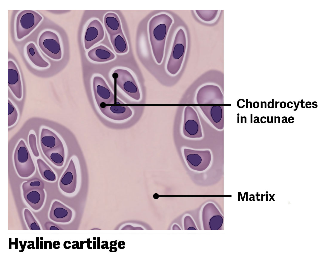
Chondrocytes: These are the specialized cells that make up cartilage.
Lacunae: Chondrocytes are not scattered loosely; they are located within small, dedicated spaces or cavities in the matrix called lacunae (singular: lacuna).
The Perichondrium: This is a layer of dense irregular connective tissue that encapsulates the cartilage structure.
Physiological Characteristics: Avascularity and Diffusion
Avascular Nature: Unlike most other connective tissues, cartilage is avascular, which means it contains no blood vessels.
Nutrient and Waste Management: Because there are no blood vessels to provide direct transport, all essential functions—such as the delivery of nutrients to cells and the removal of metabolic wastes—must be carried out via diffusion through the cartilage matrix.
Clinical Implications: The lack of a direct blood supply has significant consequences for healing. Damaged cartilage does not repair itself as readily or as quickly as most other tissues that possess a vascular supply.
Comparative Analysis of the Three Main Types of Cartilage
Hyaline Cartilage:
Prevalence: This is the most common type of cartilage found in the human body.
Anatomic Locations: It is found in structures such as the nose and the rib cage.
Articular Cartilage Subtype: This is a specific form of hyaline cartilage found at joint articulations. It acts as a cushion to help joints move freely. While it shares the basic structure of hyaline cartilage, it is unique because it has no perichondrium surrounding it.
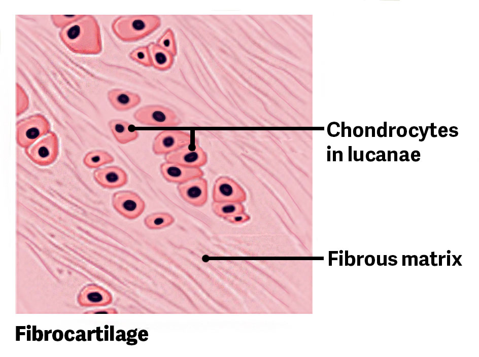
Fibrocartilage:
Structural Integrity: Fibrocartilage is characterized by being exceptionally tough.
Matrix Composition: It contains thick bundles of collagen fibers dispersed throughout the matrix, providing high tensile strength.
Anatomic Locations:
Menisci of the knee joint.
Intervertebral discs of the spinal column.
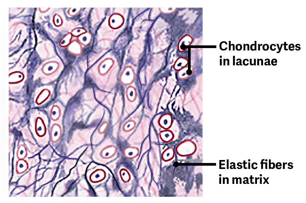
Elastic Cartilage:
Matrix composition: In addition to collagen and proteoglycans, this tissue contains a high concentration of elastic fibers.
Functional Properties: It provides rigid support while maintaining a high degree of elasticity.
Anatomic Example: The external ear. When a person gently tugs at an ear lobe, it returns to its initial shape because of the elastic cartilage within the structure.