[Answer Key] Unit E Review Package Answer Key
Anatomy & Physiology 12 Name: _________________________
Unit E (The Digestive System): Review Package
Key Words To Know and Understand:
Unit E1:
| Unit E2:
Unit E3:
|
|---|
Unit E1 Practice Questions:
- What are the three processes of digestion? Provide a description of what occurs during each process.
Process of Digestion | Description |
|---|---|
Digestion | the mechanical and chemical breaking down of ingested food into particles, then into molecules small enough to move through epithelial cells and into the internal environment. |
Absorption | the passage of digested nutrients from the gut lumen into the blood or lymph, which distributes them through the body. |
Elimination | the expulsion of indigestible residues from the body. |
- Identify the four major biomolecules, and identify what each biomolecule is broken down into during digestion:
Biomolecule | Broken down into…? |
|---|---|
Proteins | Amino Acids |
Carbohydrates | Glucose |
Fats (Lipids) | Glycerol and Fatty Acids |
Nucleic Acids | Nucleotides |
- Describe the structure of the mouth.
The mouth is divided into an anterior (“near the front”) hard palate, and a posterior (“further to the back”) soft palate.
The hard palate gets its name because it contains several bones, while the soft palate is mainly composed of muscle tissue.
The thing that hangs down in the back of your throat is the uvula, and forms the end of the soft palate.
The tonsils are found on the sides of the throat. They function in filtering out bacteria and other germs that can otherwise cause infections in the body.
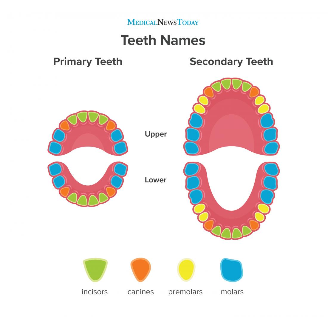
- Identify the four different types of teeth, and describe each tooth type’s method of mechanical digestion.
Teeth Type | Method of Mechanical Digestion |
|---|---|
Incisors | Biting |
Canines | Tearing |
Premolars | Grinding |
Molars | Crushing |
- In the given diagram, identify where each of the four types of teeth are found.
- Describe the following maladies of the teeth:
Malady | Description |
|---|---|
cavities | Caused by bacteria in the mouth that feed on foods (like sugars) and give off acids that corrode the tooth. |
gum disease | Gum disease (inflammation of the gums, or “gingivitis”) is the most common disease in the world! |
periodontitis | If gum disease spreads to the periodontal membrane (the lining of the tooth socket), it can cause bone loss in the socket and loosening of the teeth (“periodontitis”) |
- Identify the three sets of salivary glands, describe where they can be found, and explain how the duct openings can be located.
- parotid (below the ears)
- sublingual (below the tongue)
- submandibular (under the lower jaw)
You can locate the duct opening of the salivary glands with your tongue: parotid by the second upper molar, sublingual and submandibular flaps are under the tongue.
- What is the purpose of saliva? What does saliva contain?
When you chew food, you moisten and lubricate it with saliva. Saliva contains water, mucus, and salivary amylase (a hydrolytic enzyme that breaks down starch in the presence of water). Starch is broken down into maltose (a disaccharide of glucose).
- Describe what occurs during the process of swallowing.
Swallowing is a reflex action, meaning that it requires no conscious effort. Note that it is impossible to breathe and swallow.
When you swallow, the following happens in order to block air passages:
- The soft palate moves back to cover the openings to the nose (nasopharyngeal openings).
- The trachea (or windpipe) moves up under a flap of tissue called the epiglottis.
- The opening to the larynx (or “voice box”) is called the glottis. This opening is covered when the trachea moves up. It gets covered by the epiglottis.
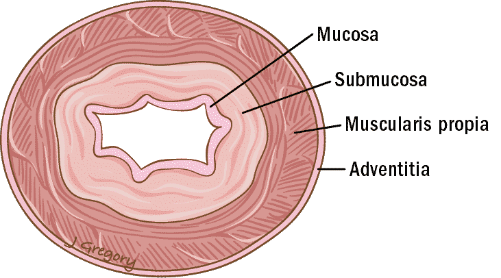
- In the given diagram, identify the names of the various linings and layers of the esophagus.
- Describe how peristalsis occurs in the esophagus.
Food moves down the esophagus through peristalsis, which are rhythmical contractions of the esophageal muscles.
Through peristalsis, the food bolus reaches the end of the esophagus and arrives at the cardiac sphincter, which connects to the stomach.
Sphincters are made of muscles that encircle tubes, and function like valves.
Unit E2 Practice Questions:
- Describe why the stomach does not digest itself.
The stomach has an inner wall that produces a thick layer of mucus, which is secreted by mucosal cells.
If hydrochloric acid does penetrate through, pepsin will start to digest the stomach lining, which will form an ulcer (an open sore on the wall of the stomach).
- What are ulcers, and what are they caused by?
Ulcers can be caused by many factors, including too much gastric juice or too much nervous exertion or stimulation (i.e., stress), which can also cause over-secretion of gastric juices.
However, the #1 cause of ulcers is actually bacterial infections (i.e., Helicobacter pylori), which impair the cell’s ability to produce mucus.
Most ulcers can be cured with antibiotics.
- What are the three zones of the small intestine?
- the duodenum
- the jejunum
- the ilium
- Describe the two types of secretions that are sent to the duodenum. Where do they come from? What is each type of secretion made up of, and what is their function?
Secretion Type | Place of Origin | Contents of Secretion | Function |
|---|---|---|---|
Pancreatic Juice | Pancreas | Sodium bicarbonate, pancreatic enzymes (such as pancreatic amylase, trypsin, and lipase) | Neutralize the acid chyme, digest starch to maltose, digest proteins to peptides, digest fat droplets into glycerol and fatty acids |
Bile | Liver | Bile salts | Break down fats into fat droplets |
- Describe how the small intestine interacts with fats, sugars, and amino acids. Ensure that your descriptions include how the villi and/or microvilli aid in these interactions.
Both the pancreas and the liver send secretions into the first part of the duodenum. These secretions contain a variety of agents and enzymes that break down fats and peptides. The walls of the duodenum and the small intestine are lined with millions of interstitial glands that produce juices containing enzymes that finish the digestion of protein and starch.
The lining of the small intestine is actually not smooth; it is long and convoluted.
The lining itself, under closer examination, is shown to consist of millions of finger- like projections called villi (sing. = villus).
The lining of each villus is made of columnar epithelial cells which have microvilli (folds of cell membrane) across which nutrients are absorbed.
Fatty acids and glycerol, which were broken down by the bile from the liver, are absorbed across the villi. Then, the fatty acids and glycerol are recombined into fat molecules in the epithelial cells of the villus. The fats then move into the lacteal of each villus and enter the lymphatic system.
Sugars and amino acids will enter the blood through the capillary network.
The blood vessels from the villi in the small intestine merge to form the hepatic portal vein, which leads to the liver.
- List the ten functions of the liver.
- maintains blood concentrations of nutrients, hormones, etc., and ensures that they remain constant
- interconversions of nutrients (e.g., carbohydrates to fats, amino acids to carbohydrates and fats)
- removes toxins from the blood (or detoxifies), and removes unwanted particulate matter from the blood.
- produces up to 1.5 litres of bile per day.
- destroys old red blood cells.
- in embryos (of vertebrates), also makes new red blood cells.
- manufactures plasma proteins, such as fibrinogen and albumin.
- produces urea (deamination of amino acids and excretion of resulting ammonia as urea, uric acid, etc.).
- manufactures cholesterol.
- stores iron and vitamins.
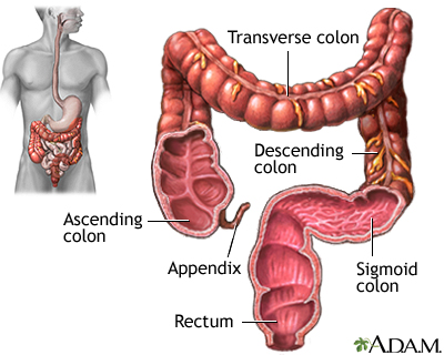
- In the given diagram, identify the various segments of the large intestine.
Unit E3 Practice Questions:
- Identify the four different hormones at play in our digestive system, and give a detailed description of each hormone.
Hormone | Released by what part? | Released in response to what? | Acts on what part? | Function |
|---|---|---|---|---|
Gastrin | Upper part of the stomach | Protein in the stomach | Secreting cells at the top of the stomach | Causes the secretion of gastric juices |
Secretin | Small intestine | Influx of acid chyme into the small intestine (duodenum) from the stomach | Pancreas | Causes the pancreas to release sodium bicarbonate (NaHCO3) and pancreatic enzymes |
CCK | Small intestine | Influx of acid chyme into the small intestine (duodenum) from the stomach | Pancreas and liver | Causes the liver to secrete bile and the pancreas to secrete pancreatic juices |
GIP | Small intestine | Influx of acid chyme into the duodenum that is rich in fats | Stomach | Inhibits stomach peristalsis and acid secretion (opposes gastrin) |
- Provide a complete description of how the secretion of digestive hormones are controlled.
- When food is eaten, sensory cells in the stomach detect the presence of peptides. Other sensory receptors detect that the stomach is distending (i.e., stretching). This causes other stomach cells to release gastrin into the blood.
- Gastrin travels through the blood and finally reaches other cells in the stomach that produce gastric juices (which takes about 1 minute), and stimulates its release.
- Most digestion of food occurs in the duodenum. The acid chyme seeps in from the stomach and is first neutralized. Secretin mediates the neutralization by stimulating the release of sodium bicarbonate by the pancreas.
- The presence of amino acids or fatty acids in the duodenum also triggers the release of cholecystokinin (CCK) which stimulates the release of digestive enzymes by the pancreas and bile by the gallbladder.
- A fourth hormone, enterogastrone (or GIP), slows the process of digestion by inhibiting stomach peristalsis and acid secretion when acid chyme that is rich in fats (which require additional digestion time) enters the duodenum.
- Identify four important vitamins that are essential to the body, describe how they can be acquired, and explain what a deficiency in each vitamin would look like.
Vitamin | How are they acquired? | What does a deficiency in the vitamin look like? |
|---|---|---|
D | Manufactured naturally by the skin upon exposure to sunlight | Rickets: bowing of the legs |
C | Found in various fruits and vegetables, especially citrus fruits | Scurvy |
Riboflavin (B2) | Found in plant- and animal-based foods, including meat, eggs and milk | Cheilosis: fissures of the lips |
Niacin (B3) | Found in various meats and grains, including red meat, poultry, fish, and brown rice | Pellagra: dermatitis of areas of skin exposed to light |
- Why should fat-soluble vitamins never be taken in large “mega-doses”? (hint: watch the video included in the Unit E3 slide deck)
They can build up in the body, especially in the liver.
- Describe the differences between minerals that are macronutrients vs. micronutrients.
Macronutrient minerals | Micronutrient minerals |
|---|---|
|
|