Nervous System Cells
Nervous System Cells (online)
Anatomy and Physiology
- Made up of brain, spinal cord, and nerves
- Central nervous system: brain and spinal cord
- Somatic nervous system
- Autonomic nervous system
- peripheral nervous system: cranial nerves and spinal nerve
- Purpose is to communicate between different parts of body
- Detects changes in internal and external environment, then initiates a response
Functional Classification of Neurons
Afferent (sensory): conduct impulses from sensory receptors to CNS
Interneuron: process information from afferent neuron, then sends out response
Efferent (motor): conducts outgoing impulses from the interneuron toward effectors (muscles/glands)
Terms
CNS: brain and spinal cord
PNS: nerves in outer regions, spinal and cranial nerves
Afferent nerves: sensory, collect from periphery to send to CNS
Efferent nerves: motor, send information from CNS to periphery
Somatic Nervous system: body and skeletal muscles, carries information to and from voluntary skeletal muscles
Autonomic nervous system: controls involuntary functions, sends out response to visceral (organs) effectors, eg. muscles, glands
Sympathetic nervous system:
- prepares body for threats, sends out appropriate response, eg. breathing rate increases
Parasympathetic nervous system:
- rest and repair system, keeping them in balance
Cells of Nervous system
Neurons
- Excitable cells that initiate impulses in nervous system
- Has dendrites, cell body, axon
Structure
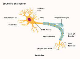
Dendrites: receiving information from sensory receptors or other nerves, and then information sent to cell body
Nucleus: instructions to govern cell function
Mitochondria: atp for neuron
RER and ribosomes: protein synthesis of neurotransmitters
Cell body(perikaryon or soma): synthesizes material, protein and mitochondria, and material to maintain and regenerate nerve fibers
Axon: nerve fiber/process, takes information away from cell body, sometimes covered in myelin sheath, varies in size
Synaptic Knob: far end of neuron(distal), communicate with gland, muscle, or another nerve
Synapse: area where there is communication between synaptic knob and other effector
Nodes of renvier: in between myelin where axon is exposed, AP jumps from node to node to allow faster transmission
Axon Hillock: information gathers here and then gets sent along axon
Axon collateral: side branches of an axon
Cytoskeleton:
- Microtubules, mibrofilaments, neurofibrils(bundles of intermediate neurofilaments
- Structural support, allows for axon transport, can transport substances from cell body can travel along axon to specific areas
Axonal Transport
- Mitochondria and vesicles with neurotransmittles can travel through the cell from the cell body, they do this buy travelling down microtubules
- Motor molecules take the vesicles, and the materials are transported to end of axon, motor molecules use microtubules as a railway and walk it down the axon
- Neurotransmitters are released into the synapse at the end of axon, some return back to terminate action
- Reverse of this transport returns neurotransmitters and vesicle membrane back up where they are recycled or degraded
Functional Region of Neuron
Input zone: receives the input, includes dendrites and cell body
Summation zone: inputs are combined which generates impulse, includes axon hillock, many voltage gated sodium and potassium channels
Conduction zone: impulse conducts along axon away from cell body, includes axon
Output zone: neurotransmitters are released into a synapse to communicate, include telodendria and synaptic knobs of axon, has many voltage gated calcium channels
Glia Cells in CNS (Neuroglia)
- Support cells of nervous system
- Support neurons, promotes their efficiency
- Can retain capacity for cell division, but makes them susceptible to uncontrolled cell division, eg. cancer
Microglia
- Small, stationary, can become enlarged when brain tissues are inflamed
- Undergoes phagocytosis, engulfs and destroys microorganisms and cellular debris during inflammation
Ependymal Cells
- Thin sheets of epithelia cells, line fluid filled cavities in CNS
- Produce cerebroapinal fluid (CSF)
- Others cells have motile cilia to keep CSF moving
Oligodendrocytes
- Smaller than astrocytes, have fewer processes, specific to CNS
- Holds nerve fibers together and produce myelin sheaths (myelination)
Astrocytes
- Largest and most numerous of gliio, star shapes, have cell extensions that connect neurons and capillaries through brain tissue
- Transports snutrients from blood to neurons, helps restore ion gradients in extracellular fluid
- Intense neuron activity can lead to excess in electrolytes, and astrocytes help return to normal state
- Has feet that attache to basement membrane of brains blood vessels/capillaries to form transport barrier called blood brain barrier, barrier prevents certain substances from entering brain tissue
Glia Cells in PNS- Shwann cells
- Only in PNS
- Functional equivolent to oligodendrocytes
- Wrap around nereves, form sthick myelin sheath around peripheral nerve fiber, myelinated fiber
- In between where schwann cells have formed myelin there are gaps; nodes of envier
- Bundle of nerve fibers wrapped by a schwann cell, unmyelinated nerve fiber
- Satellite cells: schwann cells that cover cell bodies in ganglion in PNS
- Ganglion: collection of cell bodies of neurons in PNS
- Nuclei: collection of cell bodies of neurons in CNS
Myelinated axon appearance in PNS
Myelin: plasma membrane of shwann cells, made of white fatty substance (phospholipid), forms sheath around nerve axon
Myelin Sheath: the inner core made of many layers of myelin
Nodes of Ranvier: gaps in myelin sheath, faster conduction
Neurilemma: where the nucleus and cytoplasm of shwann cell is located at perimeter, essential for growth
Neurolemmocyte: another name for shwann cell
Neuronal sheath: both the myelin sheath and neurilemma surrounding nerve axon/fiber
Nerves and Tracts
CNS
- Bundles of nerve fibers/axons referred to as tracts
- Grey matter called nuclei
- White matter made of myelinated nerves, myelinated tracts are NOT held together by connective tissue layers
- Individual fibers can extend through a tract
PNS
- Bundles of nerve fibers/axons referred to as nerves
- Grey matter called ganglia
- Whit matter made of myelinated nerves, Myelinated nerves ARE held together by connective tissue layers
- We have nevers so that neurons can be packagedd together neurons that have common characteristics
- Individual fibers can exten through a nerve
Gray matter
- Made up of collections of cell bodies and unmyelinated fibers
Note
- Motor nerves: motor neurons,
- Sensory nerves: sensory neurons
- Mixed nerves: contain sensory and motor neurons
Connective tissue coverings of nerves in PNS
Endoneurium
- Each axon/nerve fiber wrapped by endoneurium,
- Fibrous connective tissue inner covering
Perineurium
- A group of axons/nerve fibers forming a fasicle that is held together by perineurium
- Connective tissue middle covering
Epineurium
- Grouping all of the fasicles together is an outer covering epineurium
- Has superficial and more deep layer, fibrous connective tissue
Repair of Nerve Fibers
- Mature neurons cannot go through cell division
- Damage to nervous tissue can be permanent
- Sometimes neurons can be prepared
- Damage cannot be extensive
- Cell body and neurilemma must be intact
- Scarring has not occurred