immunology
Sompayrac, 7th e.d.
Lecture 1
innate immune system: second defense barrier below the skin, in all animals
macrophage:
defender cell and professional phagocyte, has receptors on surface which recognise characteristics of common microbial invaders (e.g. membranes of bacteria being made up of certain fats and carbohydrates not normally found in body)
also eats almost anything
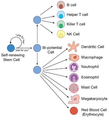
is (along with all other blood cells) a descendant of self-renewing blood stem cells→ensures that there are allways blood stem cells (that reside in bone marrow) in reserve to carry out the process of making mature blood cells
monocytes: the cells that mature in macrophages first, exit the bone marrow and enter the blood stream; can remain the blood avg 3 days; travel to capillaries looking for a crack between the endothelial cells that line inside of capillaries
phagocytosis:
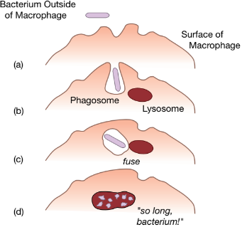
1. macrophage engults bacterium in a vesicle called phagosome
2. vesicle containing vacterium in talen inside the macrophage
3. it fuses with another vesicle: lysosome
lysosomes have chemicals and enzykmes that destroy bacteria→hence inside a vesicle to protect macrophage
cytokines: proteins secreted by macrophages during battle with bacteria, hormone-like messengers that facilate communication between cells of immune system
some alert monocytes and other immune systems in nearby capillaries→encourages these cells to exit the blood to fight the bacteria→triggers inflammatory response
innate system: professional phagocytes (neutrophilis, macrophage, monocytes, etc; eat invaders) + complement proteins (punch holes in bacteria) + natural killer cells (destroy bacteria, parasites, virus-infected cells, some cancer cells)
adaptive immune system: third line of defense, adapt to protect against any invader
antibodies: proteins that circulate in blood of immunized ppl (agent that caused them to made: antigen)
5 classes of antibodies: IgA, IgD, IgE, IgG, IgM (Ig = Immunoglobulin); all produced by B cells (WBC born in bone marrow and can mature to be antibody factories called plasma B cells)
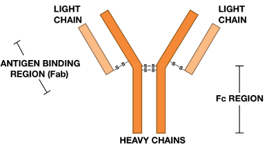
IgG antibody molecule (Immunoglobulin G): ~ 75% of antibodies in blood, made up of two pairs of two diff. proteins: Hc (heavy chain) + Lc (light chain)→due to this stucture, has two identical hands (Fab regions) that bind to antigens
Fc (constant region): an antibody also has Fc “tail” that can bind to Fc receptors on the surface of cells i.e. macrophages; special structure of the Fc region determines its class, which immune system cells it will bind to, and how it will function
modular design + junctional diversity:
hands of each antibody bind to a specific antigen (e.g., protein on surface of smallpox virus), so many diff. antibody molecules are needed to bind to many diff. antigens→100 mil. are needed (10,000 Hc’s x 10,000 Lc’s would be needed..)
Hc assembly: in every B cell, chromosones that encode the antibody Hc are multiple copies of DNA modules (gene segments): V, D, J, C; B cell chooses at random one of each kind of gene segment:
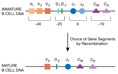
Lc assembly: DNA that encodes Lc is also assembled by picking gene segments and pasting them tgt.
junctional diversity (full 100 mil.): additional DNA bases are added or deleted when the gene segments are joined tgt
modular design + junctional diversity to create 100 mil. B cells (with each the ability to make diff. antibody) with small amount of genetic info.:
mature antibodies are made my modular design (creates 10 mil. diff. antibodies):
principal of clonal selection:
BCR (B cell receptors, tester antibodies): 100,000 of these are attached to each B cell with antigen-binding regions facing out
all BCRs on a given B cell recognize the same antigen, and their Fab regions have the shape to grasp their cognate antigen
vast majority of B cells never find their match
proliferation: once B cell’s receptors bind to its cognate antigen, it’s triggered to 2x in size and divide into 2 daughter cells, so on so forth
each cycle of cell growth and cell division→12 h
period of proliferatoin→~ a week
by the end, roughly 20,000 identical B cells will have been produced, all have receptors that can recognize the same antigen
after the selected B cells proliferate, most of them begin to make antibodies, and their is no “anchor” to attach the antibody molecules on the B cell’s surface→antibodies are transported out of B cell and into blood
at full capacity, a working B cell can pump ~ 2,000 antibody molecules / sec
after this, most of these B cells die, only working for about a week
what antibodies do: opsonization→“tagging” the bacteria and viruses (most common in developted countries) + neutralizing viruses
antibodies opsonize bacteria / viruses→bind to the invader with Fab regions
this leaves Fc tails to bind to Fc receptors of cells such as macrophages→forms a bridge between invader and phagocyte, preparing invader for phagocytosis
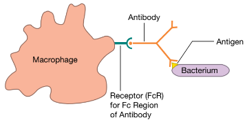
when phagocyte’s Fc receptors bind to opsonizing antibodies, ↑ appetite of phagocyte→ability of antibodies to form a bridge allows macrophage to ↑ catalog of enemies to include any invader→antibodies focus a macrophage’s attention on invaders, the uncommon ones a macrophage would otherwise ignore
neutralizing antibodies: some antibodies can attach to part of the virus that normally would plug into its cellular receptor→prevents the virus from attaching itself to surface of a cell because:
viruses enter cells by binding to certain receptor molecules on cell’s surface (e.g. Fc receptors)→virus then uses cell’s machinery to make copies of itself→(sometimes kills cell) and then infects neighbouring cells, so the neutralizing antibodies render the virus useless
T cells