Lesson 12C: Ear and Audition
Ear and Audition
The ideas below are not module objectives, but rather these are concepts that you will hopefully feel more comfortable with after reviewing this lesson:
Describe the structures of the external, middle, and inner ear.
Explain how the transmission of sound waves leads to hearing.
Structures of the Ear and Hearing
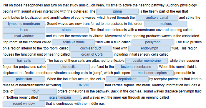
Hearing, or audition, is the transduction of sound waves into a neural signal that is made possible by the structures of the ear (Figure 12C-1). The large, fleshy external structure on the lateral aspect of the head is known as the auricle. Some sources will also refer to this structure as the pinna, though that term is more appropriate for a structure that can be moved, such as the external ear of a cat. The C-shaped curves of the auricle direct sound waves toward the auditory (ear) canal. The canal enters the skull through the external auditory meatus of the temporal bone. At the end of the auditory canal is the tympanic membrane, or ear drum, which vibrates after it is struck by sound waves. The auricle, ear canal, and tympanic membrane are often referred to as the external ear.
The middle ear consists of a space spanned by three small bones called the ossicles. The three ossicles are the malleus, incus, and stapes, which are Latin names that roughly translate to hammer, anvil, and stirrup. The malleus is attached to the tympanic membrane and articulates with the incus. The incus, in turn, articulates with the stapes. The stapes is then attached to the inner ear, where the sound waves will be transduced into a neural signal. The middle ear is connected to the pharynx through the Eustachian tube (also called the auditory tube), which helps equilibrate air pressure across the tympanic membrane. The tube is normally closed but will pop open when the muscles of the pharynx contract during swallowing or yawning, such as you might do when trying to "clear" your ears during an ascent on an airplane or a mountain road.

Figure 12C-1: The external ear contains the auricle, auditory (ear) canal, and tympanic membrane. The middle ear contains the ossicles and is connected to the pharynx by the Eustachian tube. The inner ear contains the cochlea and vestibule, which are responsible for audition and equilibrium, respectively. (Image courtesy of OpenStax.)
The inner ear is highly protected as it sit deep in the temporal lobe where a bony labyrinth is hollowed through the bone forming a series of canals that embed the sensory structures. The inner ear has three regions, the cochlea, the vestibule and the semicircular canals; the cochlea is responsible for hearing and the vestibule and semicircular canals sense different forms of equilibrium. The neural signals from these regions are relayed to the brain stem through separate fiber bundles. However, these distinct bundles travel together from the inner ear to the brainstem as the vestibulocochlear nerve. Sound is transduced into neural signals within the cochlear region of the inner ear, which contains the sensory neurons of the spiral ganglia. These ganglia are located within the spiral-shaped cochlea of the inner ear. The cochlea is attached to the stapes through the oval window.
The oval window is located at the beginning of a fluid-filled tube within the cochlea called the scala vestibuli. The scala vestibuli extends from the oval window, traveling above the cochlear duct, which is the central cavity of the cochlea that contains the sound-transducing neurons. At the uppermost tip of the cochlea, the scala vestibuli curves over the top of the cochlear duct. The fluid-filled tube, now called the scala tympani, returns to the base of the cochlea, this time traveling under the cochlear duct. The scala tympani ends at the round window, which is covered by a membrane that contains the fluid within the scala. As vibrations of the ossicles travel through the oval window, the fluid of the scala vestibuli and scala tympani moves in a wave-like motion. The frequency of the fluid waves match the frequencies of the sound waves (Figure 12C-2). The membrane covering the round window will bulge out or pucker in with the movement of the fluid within the scala tympani.

Figure 12C-2: A sound wave causes the tympanic membrane to vibrate. This vibration is amplified as it moves across the malleus, incus, and stapes. The amplified vibration is picked up by the oval window causing pressure waves in the fluid of the scala vestibuli and scala tympani. The complexity of the pressure waves is determined by the changes in amplitude and frequency of the sound waves entering the ear. (Image courtesy of OpenStax.)
A cross-sectional view of the cochlea shows that the scala vestibuli and scala tympani run along both sides of the cochlear duct (Figure 12C-3). The cochlear duct contains several organs of Corti, which transduce the wave motion of the two scala into neural signals. The organs of Corti lie on top of the basilar membrane, which is the side of the cochlear duct located between the organs of Corti and the scala tympani. As the fluid waves move through the scala vestibuli and scala tympani, the basilar membrane moves at a specific spot, depending on the frequency of the waves. Higher frequency waves move the region of the basilar membrane that is close to the base of the cochlea. Lower frequency waves move the region of the basilar membrane that is near the tip of the cochlea.

Figure 12C-3: The three major spaces within the cochlea are highlighted. The scala tympani and scala vestibuli lie on either side of the cochlear duct. The organ of Corti, containing the mechanoreceptor hair cells, is adjacent to the scala tympani, where it sits atop the basilar membrane. (Image courtesy of OpenStax.)

Figure 12C-4: The hair cell is a mechanoreceptor with an array of stereocilia emerging from its apical surface. The stereocilia are tethered together by proteins that open ion channels when the array is bent toward the tallest member of their array, and closed when the array is bent toward the shortest member of their array. (Image courtesy of OpenStax.)
The organs of Corti contain hair cells, which are named for the hair-like stereocilia extending from the cell's apical surfaces (Figure 12C-4). When the stereocilia bend toward the tallest member of their array, tension in the protein tethers opens mechano-gated K+ ion channels in the hair cell membrane. Endolymph surround the hair cell is high in K+. When ion channels open, K+ moves into the hair cell mediating an excitatory event. This will depolarize the hair cell membrane, triggering neurotransmitter release from these anaxonic neurons. When the stereocilia bend toward the shortest member of their array, the tension on the tethers slackens and the ion channels close. When no sound is present, and the stereocilia are standing straight, a small amount of tension still exists on the tethers, keeping the membrane potential of the hair cell slightly depolarized.
Hair cells communicate with fibers of CN VIII, which synapses in the pons twice. Information is then sent to the midbrain's inferior colliculus, which transmits signals to the thalamus that relays auditory input to the temporal lobe cortex. In total, there are four orders of sensory neurons in the pathway.
Summary of the process of hearing (from auricle to brain):
Sound waves amplified by outer ear
Sound waves strike tympanic membrane
Sound waves travel through ossicles (malleus, incus, stapes)
Sound waves reach oval window that vibrates
Waves produced in perilymph of scala vestibuli
Endolymph in cochlear duct displaced (also perilymph of scala tympani displaced as waves exit the cochlea at round window)
Basilar membrane vibrates
Stereocilia of hairs cell bends, K+ influx depolarizes the cell and it releases neurotransmitter
Postsynaptic sensory neuron (branches of CN VIII) sends information to CNS
Four orders of sensory neurons carry auditory to the temporal lobe (two stops in the pons, one in the midbrain and one in the thalamus)
Videos
The following Crash Course A & P YouTube video looks at how your sense of hearing works, and also introduces the concept of equilibrium, which we will cover in the next lesson. It follows sounds as they work there way into the ear where they are registered and transformed into action potentials. This mechanism not only helps you hear but also helps maintain your equilibrium. Note: scala media = cochlear duct. Be sure that you have your audio turned on. Closed captioning is available.
Hearing & Balance: Crash Course Anatomy & Physiology #17 [10:39]
Please also view the following Dr. Bruce Forciea YouTube video. It reviews the anatomy of the ear and hearing, and also introduces the concept of equilibrium. Note: scala media = cochlear duct. Be sure that you have your audio turned on. Closed captioning is available.
Anatomy of the Ear (v2.0) [5:33]