CHAPTER 5: Biochemistry Notes: Protein-Ligand Binding and Hemoglobin
Foundations of Biochemistry: Binding Protein Concepts
5.1 Binding Protein Concepts
Reversible Binding of Ligands: This process is fundamental to protein function.
Specificity: Ligands and their binding sites exhibit high specificity, meaning only certain ligands will bind to particular sites.
Conformational Changes (Induced Fit): Ligand binding often causes significant shape changes in the protein. This idea, called "Induced Fit" (Daniel Koshland, 1958), helps proteins bind more tightly and with high affinity to various ligands. It can make predicting binding partners challenging.
Cooperativity: In multi-subunit proteins, conformational changes in one subunit can influence the others. This phenomenon is known as cooperativity.
Regulation: Protein-ligand interactions are subject to various regulatory mechanisms.
Illustrative Examples: These concepts are well illustrated by Hemoglobin/Myoglobin interactions with oxygen. (Antibodies and muscle contraction are mentioned but will be skipped as per the original content's emphasis).
Functions of Globular Proteins
Globular proteins perform a diverse range of functions, including:
Storage of ions and molecules:
Examples: myoglobin (oxygen storage), ferritin (iron storage).
Transport of ions and molecules:
Examples: hemoglobin (oxygen transport), serotonin transporter.
Defense against pathogens:
Examples: antibodies, cytokines.
Muscle contraction: (Often involve enzymes that bind, but also binding proteins themselves)
Examples: actin, myosin.
Biological catalysis (enzymes):
Examples: chymotrypsin, lysozyme.
Binding Proteins (Not Enzymes)
Reversible, Transient Process: Ligand binding to a protein is viewed as a reversible and transient chemical equilibrium:
represents the protein, represents the ligand, and represents the protein-ligand complex.
Ligand: A molecule that binds to a protein. Typically, ligands are small molecules or peptides.
Binding Site: A specific region within the protein where the ligand binds.
Noncovalent Forces: Ligands bind to proteins via the same noncovalent forces (e.g., hydrogen bonds, ionic interactions, hydrophobic interactions, van der Waals forces) that dictate protein structure (as discussed in Chapter 4). These transient noncovalent interactions are crucial for enabling the reversibility of binding.
Binding: Thermodynamic Connections
Interaction Strength Expression: The strength of protein-ligand interactions can be quantified using several thermodynamic parameters:
Association (Binding) Constant ():
Units:
Definition:
Dissociation Constant ():
Units:
Definition:
Relationship:
Interaction (Binding) Free Energy ():
Units:
Relationship to enthalpy () and entropy ():
Relationships for :
Where is the gas constant ( ) and is the absolute temperature in Kelvin.
At (), .
Magnitudes of Binding Strength:
Strong binding: Typically indicated by a dissociation constant .
Weak binding: Typically indicated by a dissociation constant K_d > 10 \text{ \mu M}.
Specificity: Lock-and-Key Model
High Specificity: Proteins generally exhibit high specificity, meaning only particular ligands can bind to them.
Complementarity: This high specificity is explained by the complementary nature of the binding site and the ligand. Complementarity extends to:
Size
Shape
Charge distribution
Hydrophobic/hydrophilic character
"Lock and Key" Model (Emil Fischer, 1894): This classic model postulates that the complementary surfaces of the protein and ligand are preformed, fitting perfectly together like a lock and key upon encounter.
Specificity: Induced Fit
Conformational Changes upon Ligand Binding (Daniel Koshland, 1958): In contrast to the rigid "Lock and Key" model, the Induced Fit model proposes that conformational changes can occur in both the protein and the ligand upon binding.
Adaptation: This mutual adaptation is termed "induced fit."
Benefits of Induced Fit:
Tighter Binding: Allows for a more precise and tighter fit of the ligand to the binding site.
High Affinity for Different Ligands: Can enable a single protein to bind a variety of ligands with high affinity, adapting its binding site to each.
Prediction Difficulty: The dynamic nature of induced fit can make it challenging to predict which ligands might bind based solely on static protein structure.
Globins are Oxygen-Binding Proteins
Limitations of Protein Side Chains: Protein amino acid side chains generally lack sufficient affinity for to effectively bind and transport it.
Problems with Free Transition Metals: Some transition metals (e.g., iron in heme) bind well. However, if left free in solution, they would generate harmful free radicals. Furthermore, free in heme can be easily oxidized to , which cannot bind oxygen.
Solution: Protein-Bound Heme: The biological solution involves capturing the oxygen molecule with a heme group that is securely bound within a protein structure. This arrangement prevents both free radical generation and oxidation of the iron.
Myoglobin (Mb): The primary oxygen storage protein, found predominantly in muscle tissue.
Hemoglobin (Hb): A circulating oxygen-binding protein responsible for oxygen transport in the blood.
How Can Affinity to Oxygen Change?
For a protein to alter its affinity for oxygen, it must possess specific characteristics:
Multiple Binding Sites: The protein must have more than one site where oxygen can bind.
Interacting Binding Sites: These binding sites must be able to communicate with each other, meaning that a binding event at one site affects the properties of another site.
Cooperativity: This phenomenon, where binding at one site influences binding at other sites, is fundamental to variable oxygen affinity.
Positive Cooperativity: The initial binding event of a ligand increases the affinity of the remaining sites for that ligand. This is characterized by a sigmoidal (S-shaped) binding curve when plotting fractional saturation against ligand concentration.
Negative Cooperativity: The initial binding event reduces the affinity of the remaining sites. Examples include alcohol dehydrogenase and GAP dehydrogenase. The advantage of negative cooperativity is less clear, with sites often appearing to "fire" alternately.
Applicability: Both binding proteins (like hemoglobin) and enzymes can exhibit cooperativity in their function.
Model of Positive Cooperativity
Empty Protein: Consider an empty protein with, for example, two binding sites.
First Ligand Binding: The binding of a ligand at one site induces a conformational change. This change might make the other site "less wiggly" or stabilize it in a conformation more receptive to ligand binding.
Second Ligand Binding: Consequently, the second site becomes better able to bind a second ligand, reflecting an increased affinity.
The Hill Plot of Cooperativity
The Hill plot is used to analyze cooperativity in ligand binding.
It plots vs , with the slope of the linear region being the Hill coefficient ().
At low oxygen concentrations: Hemoglobin (Hb) behaves as if it were composed of individual subunits, binding oxygen weakly ().
At medium oxygen concentrations: Hb undergoes a transition from a low-affinity state to a high-affinity state, exhibiting strong cooperativity (, typically around for Hb, reflecting its four subunits).
At high oxygen concentrations: Hb becomes saturated with oxygen; it again behaves as a molecule with individual, tightly bound sites, nearing saturation ().
Hill Coefficient (): represents the apparent cooperativity and is always less than or equal to the total number of binding sites (n \le \text{# of binding sites}).
Cooperativity is a Special Case of Allosteric Regulation
Allosteric Protein: An allosteric protein is one in which the binding of a ligand to one site (the allosteric site) affects the binding properties of a different site (the active or primary binding site) on the same protein.
Types of Allosteric Regulation:
Positive Allosteric Regulation: Binding of a regulator increases the affinity of the primary binding site.
Negative Allosteric Regulation: Binding of a regulator decreases the affinity of the primary binding site.
Types of Allosteric Regulators:
Homotropic Regulator: The normal ligand of the protein itself acts as the allosteric regulator (e.g., binding to hemoglobin). Cooperativity is a classic example of positive homotropic regulation.
Heterotropic Regulator: A different ligand (distinct from the protein's normal ligand) affects the binding of the normal ligand.
Hemoglobin Binds Oxygen Cooperatively
Hemoglobin (Hb) Structure: Hb is a tetramer composed of two identical alpha subunits and two identical beta subunits, often denoted as (alpha2\beta2).
Subunit Similarity to Myoglobin: Each of the four subunits (two and two ) in hemoglobin is structurally and functionally similar to myoglobin, containing its own heme group that can bind oxygen.
R and T States of Hemoglobin
Hemoglobin exists in two primary conformational states, which dictate its oxygen affinity:
T = Tense State (Deoxyhemoglobin):
Characterized by more numerous and stronger interactions (salt bridges and hydrogen bonds) between subunits, making the structure more rigid.
Exhibits a lower affinity for .
This state is favored in tissues where oxygen needs to be released.
R = Relaxed State (Oxyhemoglobin):
Characterized by fewer interactions between subunits, making the structure more flexible.
Exhibits a higher affinity for .
This state is favored in the lungs where oxygen needs to be picked up.
Binding Trigger: The binding of oxygen to one Hb subunit triggers a conformational change from the T state to the R state across the entire tetramer.
"Communication" Between Subunits: This crucial conformational change involves the breaking of specific ion pairs (salt bridges) located particularly at the alpha1 - beta2 interface, facilitating the transition and signaling between subunits.
Spectroscopic Detection of Oxygen Binding to Myoglobin
The heme group in myoglobin/hemoglobin is a chromophore, meaning it strongly absorbs light in UV and visible ranges. -
An important absorption band, the Soret band changes when oxygen binds to the heme iron.
When oxygen is absent ( in deoxy-myoglobin/hemoglobin), the Soret band is at . - When oxygen binds, the Soret band shifts to . This shift can be measured using UV-Vis spectrophotometry to study oxygen binding.
Visible Color Change: The change in electronic structure also manifests as a visible color change:
Deoxyhemoglobin (found in venous blood) appears purplish.
Oxyhemoglobin (found in arterial blood) appears bright red.
How Oxygen is Released (Bohr Effect)
Active Tissues Need Oxygen: When muscles are working hard, they produce acid (). This acid lowers the pH in the blood around those tissues.
Hemoglobin Responds to pH: Hemoglobin, the protein that carries oxygen, releases oxygen more easily when the pH is lower (more acidic). It also carries oxygen better when the pH is higher (less acidic), like in the lungs.
Why it's Important: This means that as tissues become more active and acidic, hemoglobin automatically drops off oxygen where it's most needed. It also picks up oxygen efficiently in the lungs where the blood is less acidic.
How Hemoglobin Helps Export
is a waste product from active tissues.
Some directly binds to hemoglobin, creating a molecule called carbamate. This also produces which helps release oxygen (Bohr effect). These released ions lower the pH of the blood. As described by the Bohr Effect, hemoglobin responds to this lower pH by decreasing its affinity for oxygen, causing it to release oxygen more readily to the tissues that need it.
2,3-Bisphosphoglycerate (BPG) Helps Release Oxygen
BPG is a Helper Molecule: Red blood cells contain BPG, a special molecule that helps hemoglobin release oxygen.
Stabilizes "Oxygen-Release" State: BPG binds to hemoglobin and makes it keep its "tense" (T) state, which means it holds onto oxygen less tightly. This is useful for making sure oxygen is dropped off in the tissues.
Adapting to High Altitude: When you go to high altitudes, there's less oxygen in the air. Your body makes more BPG. This extra BPG helps hemoglobin release more of the limited oxygen it carries to your tissues, even if it picks up less in the lungs. This gives your body time to make more red blood cells.
Carbon Monoxide (CO) is Dangerous
Stronger Binding than Oxygen: CO is very similar to oxygen in shape, but it binds to hemoglobin about times more strongly than oxygen does.
Two Major Problems:
Blocks Oxygen: CO takes up the spots where oxygen should bind, reducing how much oxygen hemoglobin can carry.
Traps Oxygen: Once CO binds to one part of hemoglobin, it forces the rest of the hemoglobin molecule into its "relaxed" (R) state, which holds onto any remaining oxygen very tightly. This means that even if some oxygen is bound, hemoglobin won't release it to the starving tissues.
Result: Both effects lead to oxygen starvation in your cells and tissues, which is why CO is so toxic.
Sickle-Cell Anemia is Due to a Mutation in Hemoglobin (DEF CAN BE A ESSAY QUESTION)
Genetic Mutation: Sickle-cell anemia is caused by a specific point mutation in the gene encoding the chain of hemoglobin.
This mutation leads to a single amino acid substitution at position 6: Glutamate (Glu6) is replaced by Valine (Val6) in the chain (, as opposed to normal adult hemoglobin ).
Molecular Mechanism: The new, hydrophobic valine residue () creates a "sticky patch" on the surface of deoxygenated hemoglobin (T-state).
This hydrophobic patch can abnormally bind to complementary hydrophobic regions on other deoxygenated HbS molecules.
This pathological interaction causes HbS molecules to polymerize and aggregate into long, rigid fibers within the red blood cell.
Cellular Consequences:
The formation of these insoluble fibers deforms the red blood cells, causing them to become rigid and take on a characteristic crescent or "sickle" shape.
Sickled red blood cells cannot flow smoothly through narrow capillaries (small blood vessels), leading to blockages (vaso-occlusive crises).
Clinical Symptoms: Patients experience:
Severe pain: Due to blockages in blood vessels and tissue ischemia.
Anemia: Sickled cells are fragile and have a much shorter lifespan (approx. days vs. days for normal RBCs), leading to chronic hemolytic anemia.
Increased infections: Due to damage to the spleen (leading to functional asplenia) and immune system compromise.
Mortality: Untreated homozygous individuals typically die young due to organ damage and infections.
Treatment: Bone marrow transplantation can cure sickle-cell anemia by replacing the defective hematopoietic stem cells.
Heterozygous Advantage: Individuals who are heterozygous for the sickle-cell trait (carrying one normal HbA gene and one HbS gene) exhibit a remarkable resistance to malaria, particularly severe forms. This offers a selective advantage in malaria-endemic regions.
Exacerbation by Exertion: Sickle-cell disease can be dramatically worsened by exertion, which leads to lower oxygen levels in tissues (driving Hb to the T-state) and increased acidity, further promoting fiber formation. This can lead to sudden deaths in undiagnosed athletes.
Formation of Hb Strands in Sickle-Cell Anemia and a Cure
Mechanism of Fiber Formation: The deoxy-HbS molecules polymerize into long, insoluble fibers, which distort the red blood cells into a sickle shape. This is especially pronounced under low oxygen conditions where Hb is in the T-state.
Clinical Trials for Gene Therapy (2019): Significant advancements in gene therapy offer a potential cure for sickle-cell disease.
CRISPR Gene Therapy: In 2019, clinical trials using CRISPR gene-editing technology showed promising results, exemplified by patient Victoria Gray.
Process: Victoria Gray's bone marrow was removed, and her hematopoietic stem cells were gene-edited in the laboratory. The editing aimed to reactivate the production of fetal hemoglobin (HbF), which is normally switched off shortly after birth.
Fetal Hemoglobin (HbF): HbF has a higher affinity for oxygen than adult hemoglobin. Critically, it does not contain the subunits and therefore does not have the mutation. It also does not bind BPG as strongly, further increasing its oxygen affinity.
Outcome: The edited stem cells were reinfused into the patient. Production of fetal Hb effectively overcame the symptoms of sickle-cell disease.
Longevity: This treatment is considered a "once-for-life" cure, offering a permanent solution.
Broader Application: The technique also holds promise for treating other inherited hemoglobinopathies, such as -thalassemia.
Two Types of Immune Systems
Living organisms, particularly vertebrates, possess sophisticated immune systems to defend against pathogens. These generally fall into two main categories:
Cellular Immune System:
Target: Primarily targets the body's own cells that have become infected by viruses or intracellular bacteria. It also plays a role in clearing circulating virus particles and extracellular bacteria.
Key Players:
Macrophages: Large phagocytic cells that ingest and digest cellular debris, pathogens, and cells targeted by antibodies.
Killer T cells ( or cytotoxic T lymphocytes, CTLs): Directly recognize and kill infected cells or cancer cells.
Inflammatory T cells (, subset of helper T cells): Coordinate responses against intracellular pathogens, activating macrophages and CTLs.
Mechanism: Antibodies can bind to fragments of pathogens displayed on the surface of infected cells, marking them for destruction by components of the cellular immune system.
Phagocytes: Specialized cells (e.g., macrophages, neutrophils) that engulf and digest foreign particles, cells, and debris.
Macrophages: Large phagocytes that are particularly effective at ingesting bacteria that have been "tagged" or opsonized by antibodies.
Humoral "Fluid" Immune System:
Target: Primarily targets extracellular pathogens (e.g., bacteria, viruses in body fluids) and foreign proteins freely circulating in bodily fluids.
Recognition: Capable of recognizing a vast array of foreign proteins and other macromolecules.
Key Feature: Produces soluble antibodies (immunoglobulins).
Memory: Retains an immunological "memory" of past infections, allowing for a faster and stronger response upon re-exposure.
Key Players:
B-lymphocytes (B cells): Produce and secrete antibodies.
Helper T-cells (, subset of helper T cells): Provide essential help to B cells for antibody production and class switching.
Antigens: Substances that stimulate the production of antibodies. They are typically:
Macromolecular in nature (proteins, polysaccharides).
Recognized as "foreign" by the host immune system.
Examples: Coat proteins of bacteria and viruses, surface carbohydrates of cells or viruses.
Antibodies (Immunoglobulins): Proteins produced by B cells that specifically bind to antigens.
Function: Antibody binding to an antigen can either directly interfere with the antigen's function (e.g., neutralizing a virus) or, more commonly, mark the antigen for destruction by other immune components (e.g., phagocytes, complement system).
Epitope: A given antibody will bind to a small, specific region on the antigen called an epitope (also known as an antigenic determinant). An antigen, especially a large one, can possess multiple different epitopes, meaning it can be recognized by several different antibodies.
Antibodies: Immunoglobulin G (IgG)
Immunoglobulin G (IgG) is the most abundant type of antibody in human serum and a key example of antibody structure.
Structure
IgG is Y-shaped and composed of four polypeptide chains:
Two identical heavy (H) chains.
Two identical light (L) chains.
These four chains are held together by disulfide bonds.
Domains
Both heavy and light chains are organized into distinct globular domains:
Light Chains: Each light chain has:
One constant () domain.
One variable () domain.
Heavy Chains: Each heavy chain has:
Three constant () domains.
One variable () domain.
Antigen-Binding Site
The variable domains (VLVH) from one light chain and one heavy chain associate to form an antigen-binding site.
Each IgG molecule has two identical antigen-binding sites, allowing it to bind two antigen molecules simultaneously.
High Antigen Specificity (Hypervariable Regions)
The extraordinary specificity of antibodies comes from hypervariable regions (also called Complementarity-Determining Regions, CDRs).
These are specific sections within the variable domains (VLVH) that have extremely diverse amino acid sequences.
These hypervariable regions directly form the precise binding surface that interacts with a specific epitope (a small, specific region) on an antigen.
This diversity allows the immune system to recognize and respond to millions of different foreign substances.
WHAT TO KNOW: EXAM EDITION
1. Structural Similarities & Differences: Myoglobin vs. Hemoglobin
Similarities
Both are globular proteins with a heme prosthetic group (Fe²⁺ in porphyrin).
Both bind oxygen reversibly.
Both use hydrophobic residues around heme to protect Fe²⁺ from oxidation.
Differences
Myoglobin: single polypeptide chain (~153 aa), 1 heme, monomeric.
Hemoglobin: tetramer (2 α, 2 β subunits), 4 hemes, cooperative binding.
Function: myoglobin = O₂ storage (in muscle), hemoglobin = O₂ transport (blood).
👉 Think: Mb = scuba tank, Hb = delivery truck.
2. Heme Structure & Function
Structure: porphyrin ring with Fe²⁺ at center.
Coordination: 6 bonds → 4 to nitrogen of porphyrin, 1 to proximal His, 1 to O₂ (or distal His stabilizes).
Function: allows reversible O₂ binding without making superoxide.
3. Fractional Saturation vs. Ligand Concentration
Fractional saturation (Y):
Y=[L]Kd+[L]Y = \frac{[L]}{K_d + [L]}Y=Kd+[L][L]
[L] = ligand concentration.
Kd = dissociation constant (lower = tighter binding).
Curve: hyperbolic for non-cooperative binding (Mb), sigmoidal for cooperative (Hb).
👉 Easy way to remember: At [L] = Kd → Y = 0.5 (half bound).
4. Interpreting & Calculating Ligand Binding Data
Plot Y vs. [L] (hyperbola or sigmoid).
From graph: find [L] where Y = 0.5 → that’s Kd.
Use Hill plot (logarithmic) to check cooperativity (see next).
Important: “tighter binding” = lower Kd, “weaker binding” = higher Kd.
5. Hill Plot
Equation:
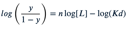
Hill coefficient (nH): slope.
nH = 1 → no cooperativity (myoglobin).
nH > 1 → positive cooperativity (hemoglobin).
nH < 1 → negative cooperativity.
👉 Think: steeper slope = more cooperative teamwork between subunits.
6. Cooperativity Models
MWC (Monod-Wyman-Changeux / concerted model)
All subunits flip together (all T or all R).
Ligand binding shifts equilibrium toward R.
👉 Like a light switch: all on or all off.
KNF (Koshland-Némethy-Filmer / sequential model)
Binding changes one subunit → induces conformational changes in neighbors.
👉 Like a domino effect.
7. R-State vs. T-State
R-state (relaxed): high O₂ affinity, smaller central cavity, stabilized by O₂ and BPG absence.
T-state (tense): low O₂ affinity, more salt bridges, stabilized by H⁺, CO₂, BPG.
👉 Memory trick: R = Ready to bind, T = Tight, hard to bind.
8. Allosteric Effects
Bohr Effect
↓pH (↑H⁺) → protonation stabilizes T-state → more O₂ released.
Physiological: active tissues release more O₂.
Carboxylation (CO₂ binding)
CO₂ reacts with N-terminal amino groups of Hb → carbamate → stabilizes T-state.
Also makes more H⁺ → reinforces Bohr effect.
2,3-BPG (bisphosphoglycerate)
Binds in central cavity of T-state → stabilizes low-affinity form.
Without BPG → Hb curve looks like myoglobin (hyperbolic).
Condition | Effect on curve | Oxygen affinity | Why |
|---|---|---|---|
High pH (basic) | Shifts left | ↑ Higher affinity | Less H⁺, enzyme stays in relaxed R-state |
Low pH (acidic) | Shifts right | ↓ Lower affinity | Bohr effect: more H⁺ binds hemoglobin and pushes O₂ off |
High BPG | Shifts right | ↓ Lower affinity | BPG stabilizes T-state (helps O₂ release) |
Low BPG | Shifts left | ↑ Higher affinity | R-state dominates, tighter binding |
R-state | Shifts left | ↑ Higher affinity | "Relaxed" form holds O₂ well |
T-state | Shifts right | ↓ Lower affinity | "Tense" form lets go of O₂ easily |
How to study/remember:
Mb vs Hb = storage vs transport.
Heme = Fe²⁺ in porphyrin.
Y vs [L] = hyperbola/sigmoid.
Hill plot slope = measure of teamwork.
MWC vs KNF = switch vs domino.
R vs T = high vs low affinity.
Bohr, CO₂, BPG = all stabilize T → promote O₂ release.