Innate Immunity: Phagocytosis, Pattern Recognition, and the Lymphatic System Interface
Introduction
This section continues our exploration of innate immunity, focusing on crucial effector mechanisms like phagocytosis and the sophisticated ways innate cells recognize pathogens. We will also touch upon the anatomical context provided by the lymphatic system, where innate and adaptive immune responses are often initiated. Key topics include:
The process of phagocytosis by cells like neutrophils and macrophages.
Mechanisms of intracellular killing of phagocytosed microbes (ROS, RNS, antimicrobial peptides, enzymes).
NETosis as an additional neutrophil defense mechanism.
The concept of Pattern Recognition Receptors (PRRs) and their ligands: Pathogen-Associated Molecular Patterns (PAMPs) and Damage-Associated Molecular Patterns (DAMPs).
Major families of PRRs: Toll-like Receptors (TLRs), NOD-like Receptors (NLRs), and RIG-I-like Receptors (RLRs/cytosolic PRRs for viral RNA) and cGAS (for cytosolic DNA).
Signaling pathways activated by PRRs, leading to inflammatory responses and type I interferon production.
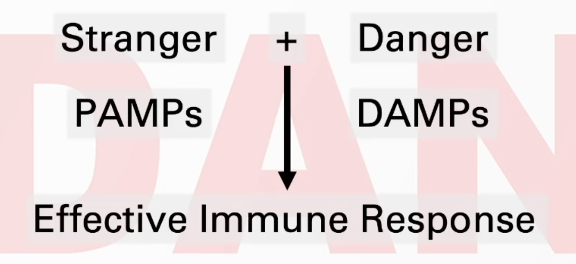
The "Danger Model" of immunity.
An overview of the anatomy of the immune system, focusing on the lymphatic system and its role in immune surveillance and response initiation.
Phagocytosis: Engulfing and Eliminating Pathogens
Phagocytosis ("cell eating") is a critical process by which specialized innate immune cells, primarily neutrophils and macrophages, engulf and destroy pathogens, cellular debris, and foreign particles.
Steps in Phagocytosis:
Recognition and Attachment:
Phagocytes recognize microbes through various mechanisms:
Opsonization: Microbes coated with opsonins (e.g., complement component C3b or antibodies) are more readily phagocytosed.
Phagocytes have receptors for opsonins, such as Complement Receptor 1 (CR1) which binds C3b.
For C3b-mediated phagocytosis via CR1, activation of the phagocyte is often required, for example, by C5a binding to its C5a receptor (C5aR).
Direct Recognition: Phagocytes can also directly recognize PAMPs on microbial surfaces via their PRRs.
Engulfment:
Upon binding, the phagocyte extends pseudopods (cytoplasmic projections) that surround and internalize the particle.
The internalized particle is enclosed within a membrane-bound vesicle called a phagosome.
Phagolysosome Formation:
The phagosome fuses with one or more lysosomes (organelles containing digestive enzymes and antimicrobial substances) to form a phagolysosome.
Killing and Digestion:
Within the phagolysosome, the microbe is killed and degraded by a combination of mechanisms:
Acidification: The pH of the phagolysosome drops, creating an unfavorable environment for most microbes and optimizing the activity of lysosomal enzymes.
Enzymatic Degradation: Lysosomal enzymes (e.g., proteases, nucleases, lipases) break down microbial components.
Antimicrobial Peptides: Such as lysozyme (degrades bacterial peptidoglycan) and defensins (disrupt microbial membranes).
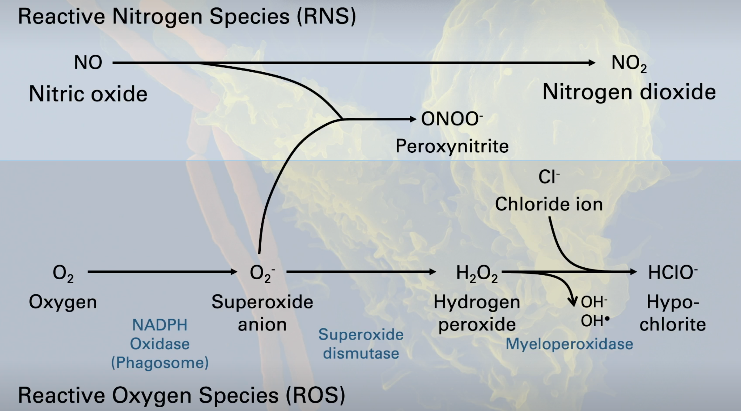
Reactive Oxygen Species (ROS) - "Respiratory Burst":
The NADPH oxidase complex (phagocyte oxidase or Phox) is assembled in the phagolysosome membrane upon activation.
It catalyzes the production of superoxide anion (O2−) from oxygen (O2).
Superoxide dismutase (SOD) converts O2− to hydrogen peroxide (H2O2).
Myeloperoxidase (MPO), abundant in neutrophil granules, uses H2O2 and chloride ions (Cl−) to produce highly toxic hypochlorite (HClO−, the active ingredient in bleach).
H2O2 can also generate hydroxyl radicals (⋅OH).
Reactive Nitrogen Species (RNS):
Inducible Nitric Oxide Synthase (iNOS) produces nitric oxide (NO).
NO can react with O2− to form peroxynitrite (ONOO−), a potent antimicrobial agent.
NO can also be oxidized to nitrogen dioxide (NO2).
Exocytosis of Waste:
Indigestible debris is eventually expelled from the phagocyte by exocytosis.
Specialized Functions:
Neutrophils: Primarily focused on rapid phagocytosis and killing of extracellular bacteria and fungi. They are short-lived and typically die after phagocytosis, contributing to pus formation.
Macrophages: Longer-lived phagocytes. Besides phagocytosis and killing, they play important roles in:
Antigen presentation: Processing and presenting microbial antigens to T cells, thus linking innate and adaptive immunity.
Cytokine release: Secreting cytokines that modulate inflammation and immune responses.
Tissue repair and cleanup.
NETosis (Neutrophil Extracellular Traps):
A unique form of cell death distinct from apoptosis or necrosis, employed by neutrophils.
Upon strong activation, neutrophils can release their decondensed chromatin (DNA) and granular contents (e.g., antimicrobial peptides, enzymes) into the extracellular space, forming web-like structures called NETs.
NETs trap and kill pathogens (bacteria, fungi, viruses), preventing their spread. This is a "suicidal" defense mechanism for the neutrophil.
Pattern Recognition in Innate Immunity
The innate immune system recognizes pathogens and danger signals by detecting conserved molecular structures.
Pathogen-Associated Molecular Patterns (PAMPs): Molecules characteristic of microbial pathogens but generally absent in host cells (e.g., bacterial lipopolysaccharide (LPS), peptidoglycan, flagellin, viral double-stranded RNA (dsRNA), microbial DNA with unmethylated CpG motifs).
Damage-Associated Molecular Patterns (DAMPs) / Alarmins / Danger Signals: Endogenous molecules released from damaged, stressed, or dying host cells (e.g., ATP, uric acid, heat shock proteins, HMGB1, self DNA/RNA in inappropriate locations).
Pattern Recognition Receptors (PRRs): Receptors expressed by innate immune cells (and other cells, like epithelial cells) that recognize PAMPs and DAMPs. Binding of a PAMP/DAMP to its PRR triggers intracellular signaling pathways, leading to activation of the cell and initiation of an immune response (e.g., inflammation, cytokine production, phagocytosis).
Major Families of Pattern Recognition Receptors:
A. Toll-like Receptors (TLRs):
A family of at least 10 functional TLRs in humans (TLR1-TLR10).
Structure: Single-pass transmembrane proteins with an extracellular leucine-rich repeat (LRR) domain (for PAMP/DAMP recognition) and an intracellular Toll/Interleukin-1 Receptor (TIR) domain (for signal transduction).
Localization:
Cell surface TLRs: Recognize PAMPs on the surface of extracellular microbes (e.g., TLR1/2, TLR2/6, TLR2, TLR4, TLR5, TLR10).
Endosomal TLRs: Located in the membranes of endosomes; recognize nucleic acids from internalized microbes (e.g., TLR3, TLR7, TLR8, TLR9).
Ligands:
Extracellular
TLR1/TLR2 heterodimer: Triacyl lipopeptides (bacterial).
TLR2/TLR6 heterodimer: Diacyl lipopeptides (bacterial), LTA (lipoteichoic acid from Gram-positive bacteria), Zymosan (fungal).
TLR2 (homodimer or with other partners): Peptidoglycan (bacterial).
TLR4 (with MD-2 and CD14): Lipopolysaccharide (LPS from Gram-negative bacteria).
TLR5: Flagellin (bacterial protein).
TLR10: Function less clear, possibly anti-inflammatory by recognizing Listeria, Influenza components.
Intracellular
TLR3: dsRNA (viral).
TLR7: ssRNA (viral, self RNA).
TLR8: ssRNA (viral).
TLR9: CpG-DNA (bacterial and viral DNA, unmethylated CpG motifs).
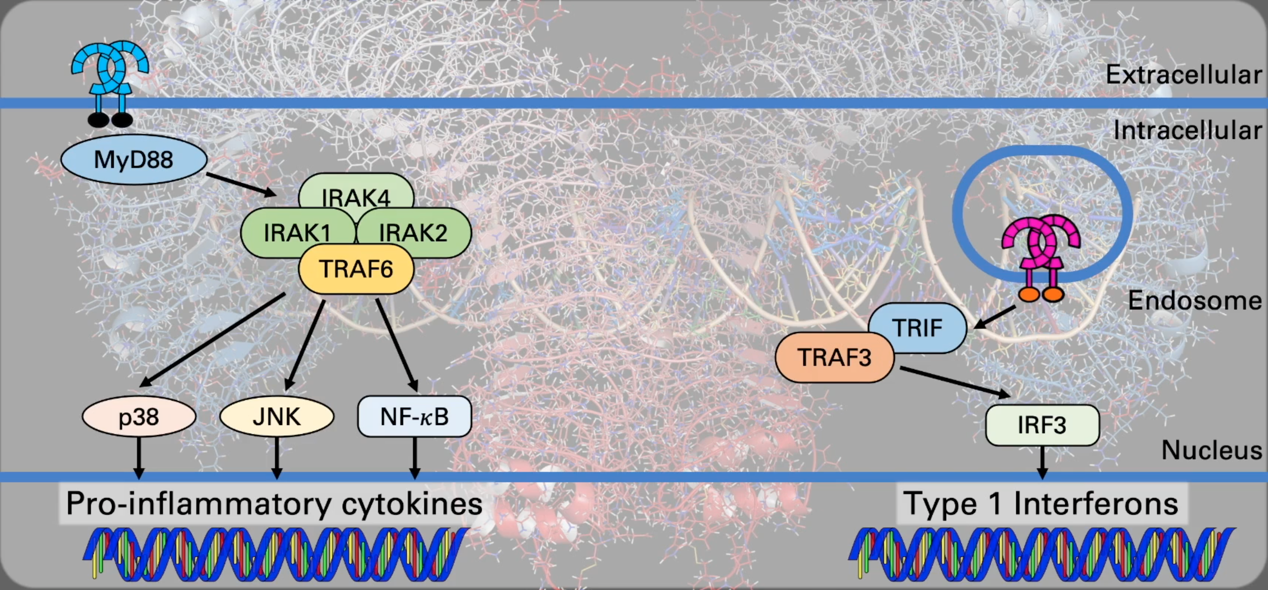
Signaling Pathways:
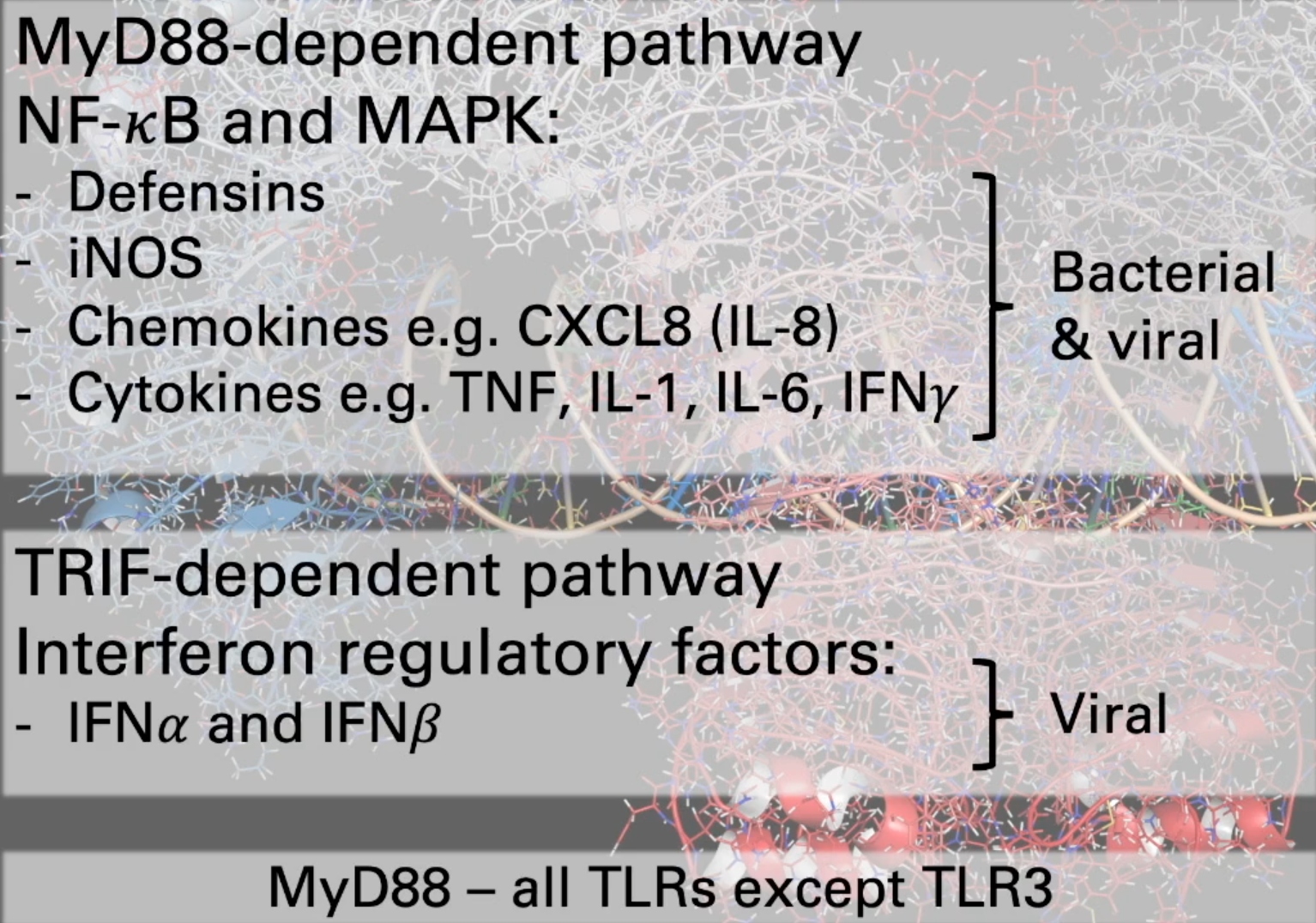
MyD88-dependent pathway: Used by all TLRs except TLR3. Leads to activation of transcription factors NF-κB (Nuclear Factor kappa B) and MAPKs (Mitogen-Activated Protein Kinases like p38, JNK).
Consequences: Production of pro-inflammatory cytokines (e.g., TNF, IL-1, IL-6), chemokines (e.g., CXCL8/IL-8), antimicrobial peptides (defensins), and inducible nitric oxide synthase (iNOS). This pathway primarily drives inflammatory responses against bacteria and viruses.
TRIF-dependent pathway: Used by TLR3 and TLR4 (from the endosome). Leads to activation of Interferon Regulatory Factors (IRFs), particularly IRF3.
Consequences: Production of Type I Interferons (IFN-α and IFN-β), which are crucial for antiviral defense. This pathway is primarily involved in viral responses.
B. Cytosolic Pattern Recognition Receptors: These PRRs detect PAMPs/DAMPs that have entered the cytoplasm.
RIG-I-like Receptors (RLRs):
Include RIG-I (Retinoic acid-Inducible Gene I) and MDA5 (Melanoma Differentiation-Associated protein 5).
Structure: Contain RNA helicase domains and CARD (Caspase Activation and Recruitment Domain) domains.
Ligands: Recognize viral RNA in the cytoplasm (e.g., dsRNA, ssRNA with 5'-triphosphate).
Signaling: Activate pathways leading to the production of Type I IFNs (via IRF3/7) and pro-inflammatory cytokines (via NF-κB and TRAF3).
Cytosolic DNA Sensors (e.g., cGAS):
cGAS (cyclic GMP-AMP Synthase): Detects cytosolic DNA (from viruses or bacteria, or misplaced self-DNA).
Signaling: Upon DNA binding, cGAS synthesizes cGAMP (cyclic GMP-AMP), which then binds to and activates STING (Stimulator of Interferon Genes) on the ER membrane. STING signaling leads to activation of IRF3 (for Type I IFN production) and NF-κB (for pro-inflammatory cytokines like TNF, IL-6).
C. NOD-like Receptors (NLRs):
A large family of intracellular PRRs that sense PAMPs and DAMPs in the cytoplasm.
Structure: Typically contain a central Nucleotide-binding Oligomerization Domain (NOD or NACHT domain), C-terminal Leucine-Rich Repeats (LRRs for ligand sensing), and N-terminal effector domains (e.g., CARD, PYRIN domain).
Subfamilies: NLRA, NLRB, NLRC (e.g., NOD1, NOD2, NLRC4), NLRP (e.g., NLRP1-14).
NOD1 and NOD2: Recognize bacterial peptidoglycan fragments. Signal via RIPK2 to activate NF-κB and MAPKs, leading to pro-inflammatory cytokine production.
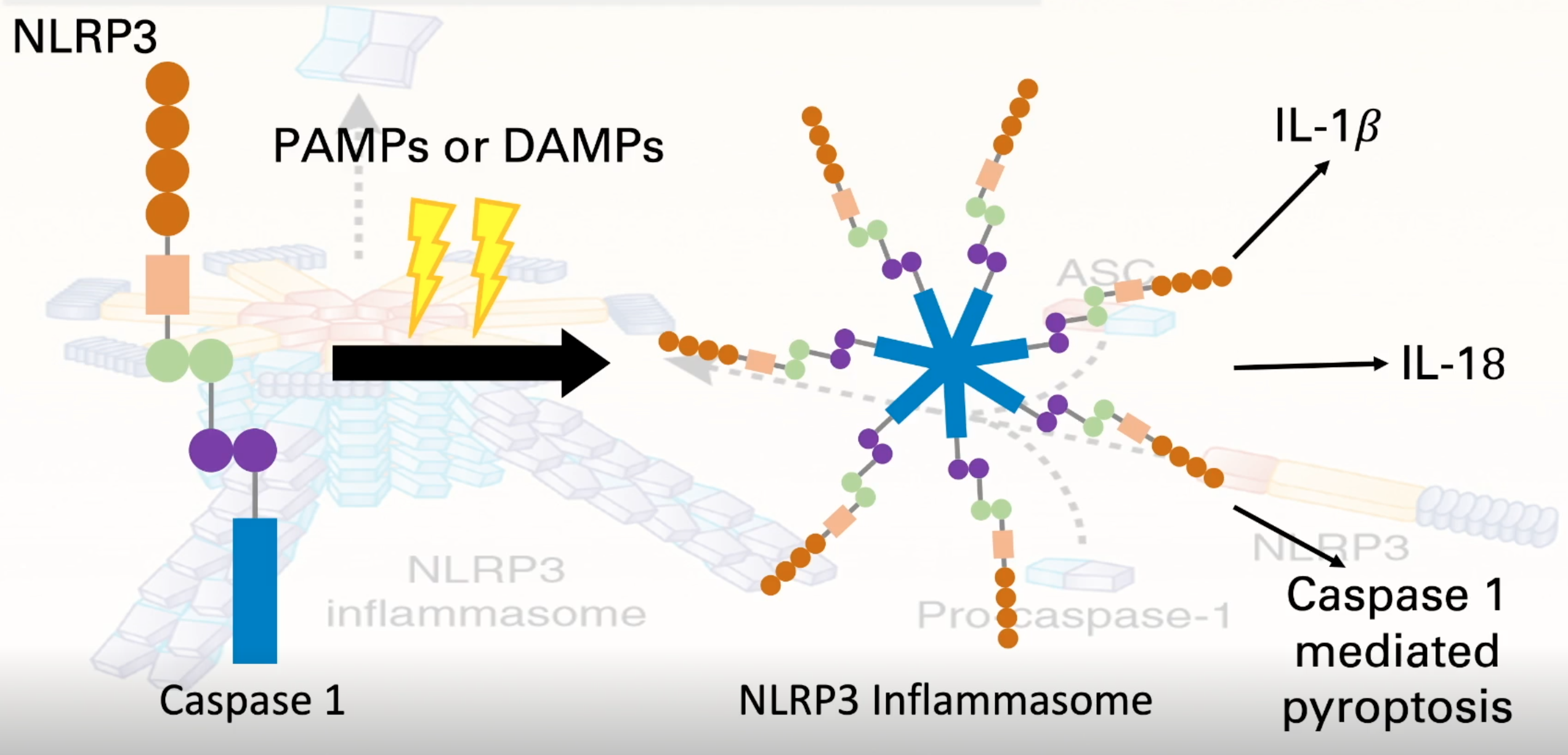
NLRPs and Inflammasomes:
Some NLRPs (e.g., NLRP3) can assemble into large multiprotein complexes called inflammasomes upon detection of PAMPs or DAMPs.
NLRP3 Inflammasome Components: Typically NLRP3 (sensor), ASC (adaptor protein), and Pro-caspase-1.
Activation: A wide range of stimuli can activate the NLRP3 inflammasome (e.g., bacterial toxins, viral RNA, ATP, uric acid crystals, particulate matter).
Function: Assembly of the inflammasome leads to the activation of Caspase-1.
Caspase-1 Activity:
Cleaves pro-IL-1β and pro-IL-18 into their active, mature forms, which are potent pro-inflammatory cytokines.
Can induce a pro-inflammatory form of programmed cell death called pyroptosis, characterized by cell swelling, membrane rupture, and release of DAMPs and pro-inflammatory mediators.
D. DAMPs / Danger Signals / Alarmins:
These are endogenous host molecules released from stressed, damaged, or dying cells that can activate PRRs (often the same ones that recognize PAMPs) and trigger an inflammatory response, even in the absence of infection (sterile inflammation).
Examples: ATP (extracellular), uric acid (crystals), High Mobility Group Box 1 (HMGB1), heat shock proteins (HSPs), components of the extracellular matrix, self DNA/RNA released into the cytoplasm or extracellular space, granule contents from immune cells.
Danger Model (Polly Matzinger): Proposes that the immune system primarily responds to "danger" signals (DAMPs) released by damaged tissues, rather than solely distinguishing "self" from "non-self" (stranger model/PAMPs). An effective immune response often requires detection of both PAMPs (stranger) and DAMPs (danger), leading to robust dendritic cell maturation and activation of adaptive immunity.
Anatomy of the Immune System: Focus on Lymphatics
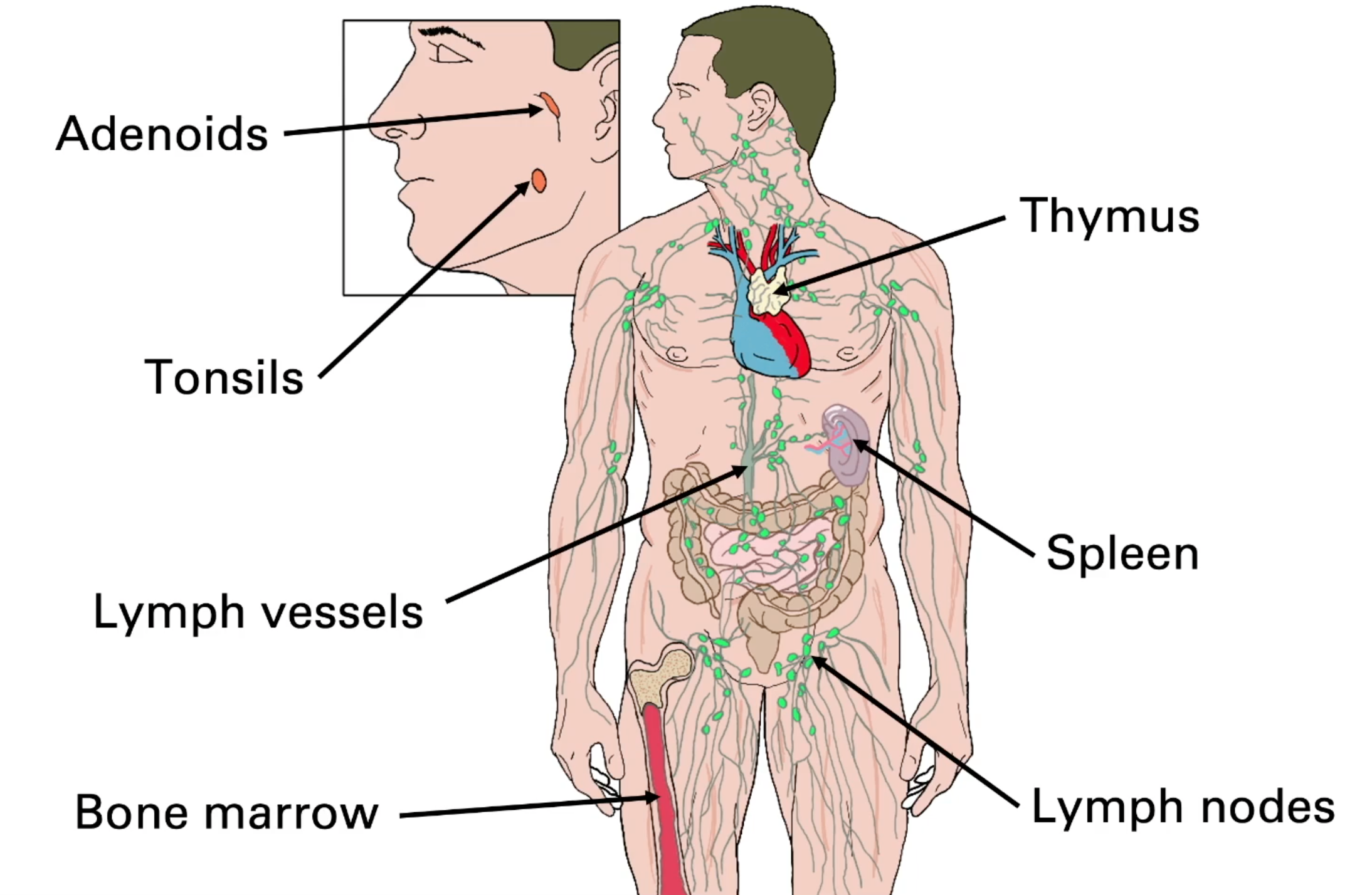
The lymphatic system is a crucial network of vessels and organs that plays vital roles in fluid balance, fat absorption, and, critically, immunity.
Major Components:
Lymph: A watery fluid originating from interstitial fluid (which itself is derived from blood plasma that has leaked out of capillaries). It circulates within lymphatic vessels.
Lymphatic Vessels: A network of vessels that collect lymph from tissues and return it to the bloodstream.
Lymphatic Capillaries: Blind-ended vessels with overlapping endothelial cells that act as one-way valves, allowing interstitial fluid, proteins, and even cells (like dendritic cells and pathogens) to enter.
Larger Lymphatic Vessels: Have smooth muscle in their walls and one-way valves to ensure unidirectional flow of lymph, propelled by muscle contractions and breathing movements.
Lymph Nodes: Small, bean-shaped organs (600-700 in the body) located along lymphatic vessels. They act as filters for lymph and are critical sites for immune surveillance and the initiation of adaptive immune responses.
Architecture:
Capsule: Outer connective tissue covering.
Afferent lymphatic vessels: Bring lymph into the node.
Efferent lymphatic vessel: Carries lymph out of the node.
Cortex: Outer region, primarily containing B-cell rich areas (primary lymphoid follicles and germinal centers if an immune response is active).
Paracortex: Area beneath the cortex, rich in T-cells and dendritic cells.
Medulla: Inner region, containing medullary cords (with plasma cells and macrophages) and medullary sinuses.
Lymphoid Organs:
Primary Lymphoid Organs: Bone marrow and Thymus (where lymphocytes develop and mature).
Thymus Structure: Bi-lobed organ with a capsule, cortex (dense with developing thymocytes and cortical epithelial cells), and medulla (fewer thymocytes, medullary epithelial cells, dendritic cells, macrophages, and Hassall's/thymic corpuscles). Site of T-cell maturation and selection.
Secondary Lymphoid Organs: Spleen, tonsils, Peyer's patches, appendix, and other Mucosa-Associated Lymphoid Tissues (MALTs) – sites where mature lymphocytes encounter antigens and initiate adaptive immune responses.
Spleen Structure:
Capsule.
Red Pulp: Filters blood, removes old/damaged red blood cells, contains macrophages.
White Pulp: Lymphoid tissue surrounding arteries. Consists of:
Periarteriolar Lymphoid Sheath (PALS): T-cell rich area.
Primary Follicles and Germinal Centers: B-cell rich areas.
Marginal Zone: Surrounds white pulp, contains specialized macrophages and B cells.
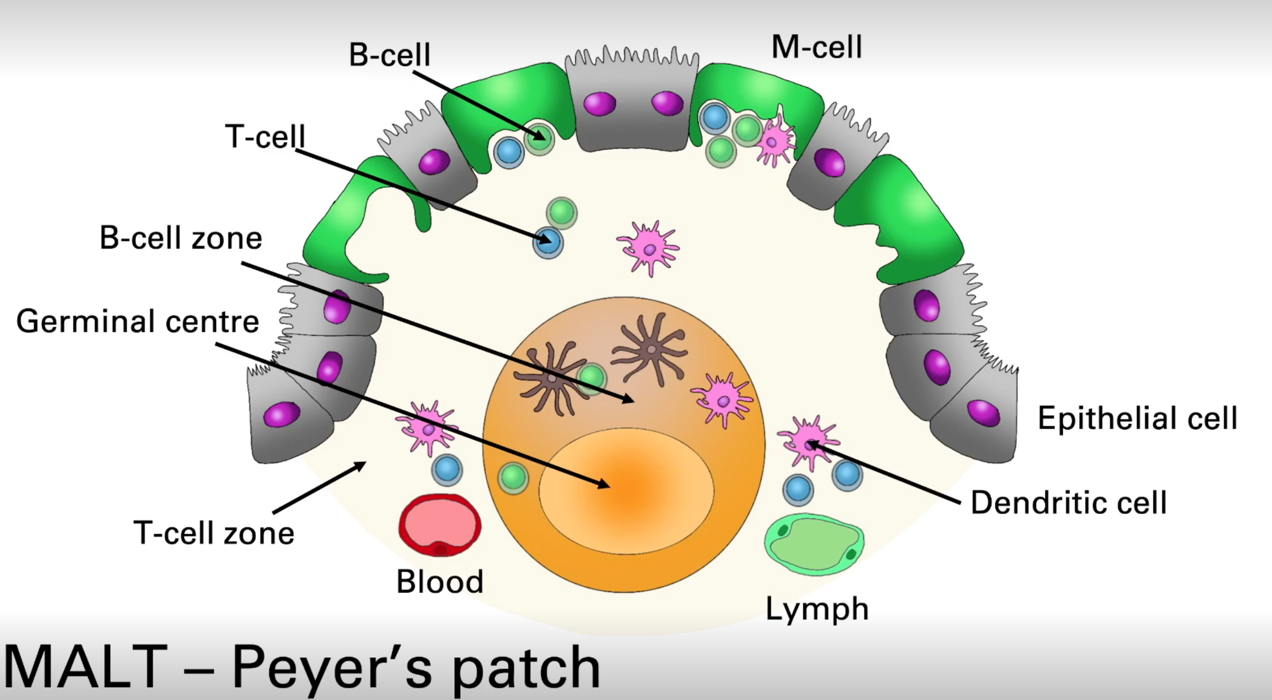
MALT (e.g., Peyer's Patch in gut, NALT in nasal cavity, BALT in bronchus): Organized lymphoid tissues strategically located at mucosal surfaces.
M-cells (Microfold cells): Specialized epithelial cells overlying Peyer's patches that actively transport antigens and microbes from the gut lumen to the underlying lymphoid tissue, where dendritic cells and lymphocytes are located. Peyer's patches contain B-cell follicles (some with germinal centers) and T-cell zones.
Function of the Lymphatic System in Immunity:
Fluid Balance: Returns excess interstitial fluid to the blood.
Immune Surveillance: Lymph nodes and spleen filter lymph and blood, respectively, trapping pathogens and antigens.
Antigen Presentation and Initiation of Adaptive Immunity: Dendritic cells pick up antigens in peripheral tissues, migrate via lymphatic vessels to draining lymph nodes, and present these antigens to T-cells, initiating adaptive immune responses. B-cells are also activated in lymph nodes and spleen.
Recirculation of Lymphocytes: Lymphocytes continuously circulate between blood, tissues, and lymphoid organs, maximizing the chances of encountering their specific antigen.
Summary
Phagocytosis is a cornerstone of innate cellular defense, involving engulfment and destruction of pathogens by cells like neutrophils and macrophages, utilizing a potent arsenal of antimicrobial mechanisms (acidification, enzymes, ROS, RNS, AMPs). NETosis provides an additional defense strategy for neutrophils.
Pattern Recognition Receptors (PRRs) like TLRs, NLRs, RLRs, and cGAS enable innate immune cells to detect conserved microbial PAMPs and host-derived DAMPs, triggering appropriate responses such as inflammation, cytokine production, and Type I interferon release. The "Danger Model" emphasizes the importance of DAMPs in initiating immunity.
The Lymphatic System, with its network of vessels, lymph nodes, and lymphoid organs (including MALTs like Peyer's patches), provides the anatomical framework for immune surveillance, fluid drainage, and the crucial interface where innate immune cells (like antigen-presenting dendritic cells) transport information to adaptive immune cells (T and B lymphocytes) to initiate specific and long-lasting immunity.
Further Reading
Innate immunity provides rapid defense against pathogens through pattern recognition receptors (PRRs) that detect conserved microbial components (Akira et al., 2006). PRRs include Toll-like receptors, C-type lectin receptors, and cytoplasmic receptors (Brubaker et al., 2015). Phagocytosis plays a crucial role in innate immunity, forming a platform for PRR activation and inflammasome assembly (Moretti & Blander, 2014). Mannose-binding lectin (MBL) is a key soluble pattern recognition molecule that links innate and adaptive immunity (Fraser et al., 1998; Ip et al., 2009). The innate immune system involves various cell types and humoral factors, orchestrating anti-infectious and antitumor responses (Zänker, 2008). While innate immunity is essential for host protection, an imbalanced response can lead to immunopathology (Suresh & Mosser, 2013). Understanding innate immune mechanisms can improve vaccine development and prevent autoimmunity (Suresh & Mosser, 2013). The innate immune system's molecular machinery is highly conserved across species, offering insights into its function in health and disease (Zänker, 2008).