knowt logo

Lecture 11 Actin World

Page 2: Learning Objectives

  • Characterize actin filament structure and composition.

  • Formation and dynamics of thin filaments: Understanding how they are formed in cells and the roles of associated motor proteins.

  • Actin-associated proteins: List and distinguish their functions.

  • Role of thin filaments: Distribution and diverse functions in cellular contexts.

  • Muscle tissue structure and function: Analyzing actin's contribution to muscle activity.

  • Cellular movement mechanisms: Outlining how actin facilitates cellular motility.

Actin Filaments

  • Major Protein Component: Actin is integral to all cells, located at cell junctions and throughout cellular structures.

Cytoskeletal Filaments

  • Types of cytoskeletal filaments:

    • Actin: Also known as the basic unit of thin filaments/microfilaments, vital for a multitude of functions. (side roads)

    • Intermediate filaments: Provide tensile strength against stress.

    • Microtubules (thick filaments): Serve various functions, acting as pathways for intracellular transport. (major highways)

Actin Dynamics

  • Dynamic structure: Actin constitutes 10% of muscle cell protein and 1-5% in non-muscle cells.

  • Filament formation and disassembly: Actin filaments are continuously formed and disassembled, tailored to cellular needs and environmental stimuli.

  • Actin genes: Humans have six actin genes that contribute to the diversity of actin functions.
Functions of Actin

  • Diverse roles: Actin is involved in cellular structure, movement, phagocytosis (“eating”), and cell division.

  • Three types of actins:

    • α-actin: Found in muscle fibers.

    • β-actin: Present in the leading edge of cells.

    • γ-actin: Associated with stress fibers.

Actin-Binding Proteins

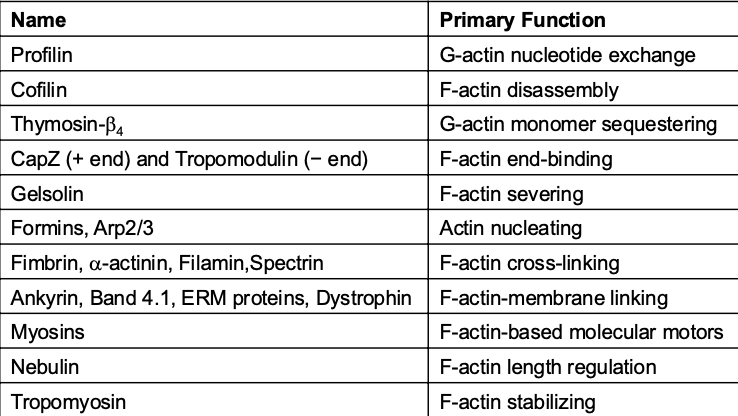
  • Table of Proteins: Overview of actin-binding proteins and their functions, e.g., Profilin (nucleotide exchange), Cofilin (disassembly), and various capping and cross-linking proteins.

  • Functionality: Proteins that can promote actin polymerization, stabilize filaments, or sever actin structures. Proteins associated with actin can modify it by nucleating, stabilizing, serving, capping, etc.
Actin Monomers

  • Actin exists as G-actin: Globular actin binds ATP and Mg2+.

  • Polymerization: Forms filamentous actin (F-actin) with 2 helical structures wrapped around one another. .

Polarization of Actin Filaments

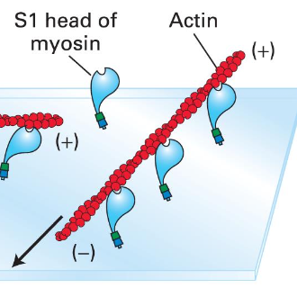
  • Polar characteristics: Myosin S1 regions coat F-actin, creating a polarized structure with distinct ends for filament dynamics.

    • form arrow like structures:

      • (-) pointed end: end has cleft exposed to the solution

      • (+) barbed end: has the cleft contacting other subunits

Filament Dynamics

  • End addition preference: G-actin adds more readily to the (+) end of the filament compared to the (-) end, influencing overall growth.

    • to form filaments G-actin-ATP units are added to both ends.

    • (+0) end addition is almost 10x greater than the (-) end

Actin Filaments need a Critical Concentration

  • Requirement for filament growth: Actin filaments need a critical concentration of G-actin-ATP. Each different Cc which much lower [Cc] required in the (+) end.

  • ATP is hydrolyzed quickly when actin monomers are added creating 3 distinct regions.

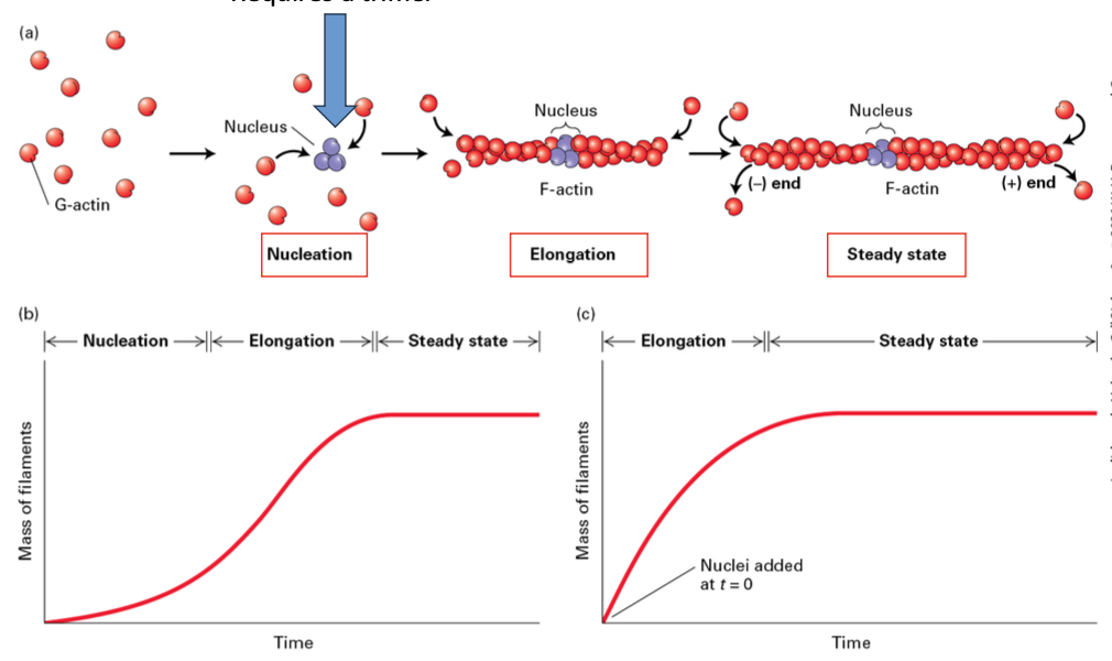
  • Treadmilling is a dynamic equilibrium process in filament assembly and disassembly. Seen at steady state the (-) end loses monomers while new ones are added to the (+) end

Steps of Actin Filament Formation

    1. Nucleation

    2. Elongation

    3. Steady State

Actin Recycling

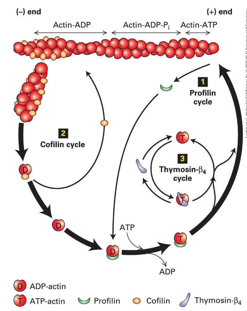
  • Profilin's role: Enhances actin recycling and promotes the binding of ATP-actin at the (+) end, contributing to dynamic filament reassembly.

  • Profilin binds G-actin lost from filaments and opens the cleft enhancing loss of ADP

    • Profilin-ATP-actin complex binds the (+) end

  • Cofilin binds 2 ADP-actin towards the (-) end, causes a twist and a break

    • Generates more (-) ends for disassembly

  • Thysomin - B4 binds free ATP-actin to keep it all from assembling needless into filaments

    • Forms dynamic equilibrium with the free subunits
Capping Proteins

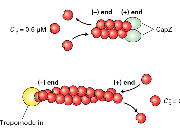
  • To regulate the formation and length of filaments/dynamics

  • CapZ binds the (+) end

    • Formed from two related proteins

  • Cells regulate CapZ ability to stop growth

    • Phosphatidylinositol 4,5- bisphosphate (PIP2)

    • Regulator proteins

  • Tropomodulin binds the (-) end and can module filament assembly and disassembly

    • Mostly found where actin needs to be stabilized
Actin-Nucleating Proteins

  • 2 major classes of actin-nucleating proteins help in the assembly of F-actin cells.

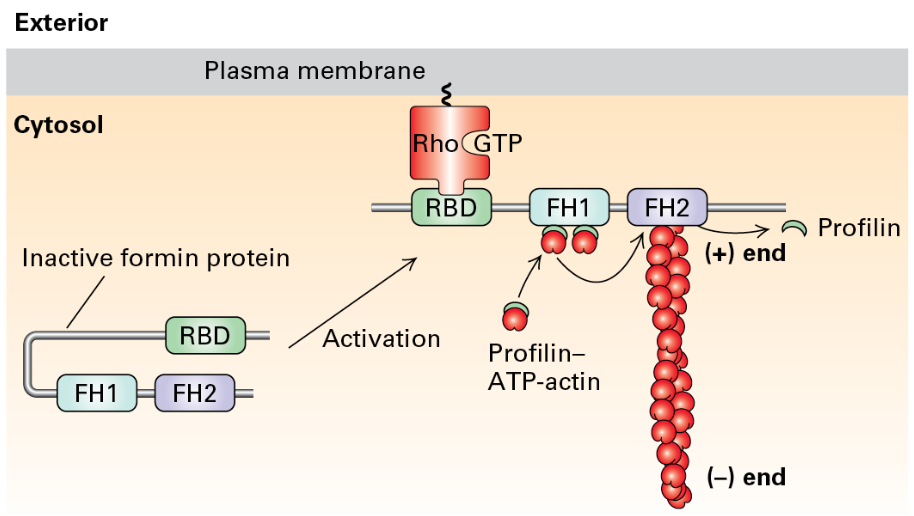
    • Formin proteins: Facilitate long filament assembly.

    • Arp2/3 complex: Promotes branched filament networks.

Formins Homology 1, 2 Domains in Formins

  • FH1 domains bind profilin to increase ATP-actin

  • FH2 domains form a ring that rocks back and forth promoting addition of ATP actin to the (+) end

Regulatory Signaling

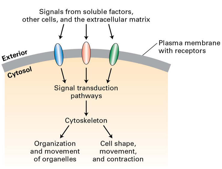
  • Signal pathways: Actin nucleation and filament formation can be controlled through receptor-mediated signaling pathways, leading to the activation of formin proteins.
ARP2/3 and NPFs

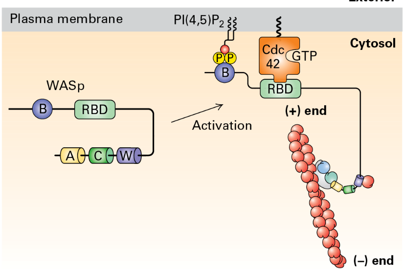
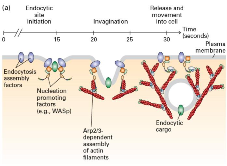
  • Complex function: The ARP2/3 complex promotes nucleation and creation of branched F-actin structures, activated by nucleation-promoting factors. This binds to existing F-actin and promotes branch formation.

WASp Activation

  • WASp's (Wiskott-Aldrich syndrome) role: Serves as a nucleation-promoting factor by activating signaling cascades that stimulate Arp2/3 near cellular membranes.
Listeria Monocytogenes

  • Overview of Gram-positive bacteria: Causes listeriosis with significant health risks, particularly for pregnant women and immunocompromised individuals.

  • ActA protein mimcs an NPF and activated and recruits Arp2/3. Actin polymerization propels the bacteria through the cell.

Actin Role in Membrane Dynamics

  • Membrane mechanisms: Actin polymerization generates force and movement and aids in endocytosis, with contributions from signaling factors that assist in membrane dynamics.

  • Bursts of Arp 2/3 activation recruits other factors that can help power this energetically unfavorable membrane dynamic
Phagocytosis

  • Immune function: Describes how actin aids immune cells in engulfing pathogens through receptor-mediated signaling and network formation.

    • Opsonized pathogen are immobilized by the cell and receptor mediate signaling activates local Arp2/3 activation and network formation. creates a contractile network that leads to isolation of the pathogen and subsequent destruction.

Cross-Linking Proteins

  • Structural diversity: Actin cross-linking proteins mold filaments into varied structures, essential for functions such as sound perception in stereocilia.

Erythrocyte Structure

  • Stress handling: Actin networks are crucial for erythrocyte integrity, maintaining its shape and structure under stress. Actin membrane is anchored by actin associated proteins such as a/B spectrin. Actin gives cell shape and structure.

Actin Filaments and Muscle

  • Myosin II was the first discovered motor protein, integral in muscle cell function by converting chemical energy to mechanical work. All family motor proteins can bind to actin.

  • Immobilized ATPase containing mysoin S1 subunits will move actin filaments under experimental conditions. 40 myosin genes in the human genome.
  • Myosin Classes

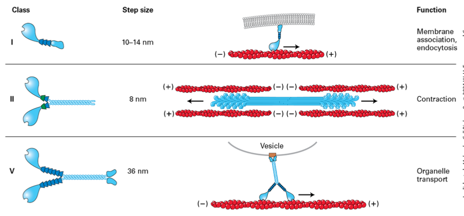
    • Myosin I – only one with asingle head domain and it is associated with the membrane

    • Myosin II – mostly in muscle (thick fibers) but also non-muscle in less organized fashion, used for contraction

    • Myosin V – motor protein to hall things towards the membrane

    • Myosin VI – only member that moves towards the negative end, moving things away from the membrane
Page 29: Myosin-Actin Interaction

  • Power stroke dynamics: Myosin-ADP binds to F-actin. ATP binds releasing myosin from F-actin Hydrolysis cocks the head domain. Head domain binds actin. Release of Pi moves it back into its original position. In muscle 10% of the myosin is contacting f-actin when activated.

  • Myosin Steps

    • Step size of myosin head is relative to the length of the neck domain. (longer legs = bigger step),

      • Myosin II moves about 8nm

      • Myosin V takes longer 72 nm steps, hauling cargo 36nm ata. step

  • Muscle Fiber Composition

    • muscles —> muscle fibers (multinucleated muscle cells) —> myofibril structures composed of sacromere

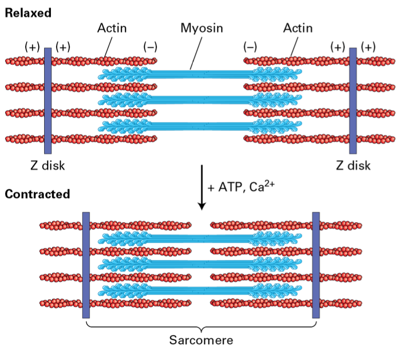
    • Z disk structures flank: Myosin II thick filaments alternating with F-actin thin filaments attached to the Z disk

    • Thick filaments are bipolar with heads out toward Z disks. F-actin is attached to Z-disk. During contraction the mysoin II moves towards the (+) shortening the distance between the (-) end of F-actin.

      • Controlled by ATP availability and Ca2+ influx
Accessory Proteins in Muscle

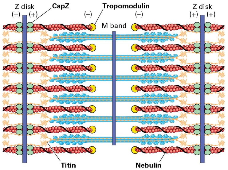
  • Structural support: CapZ, Tropomodulin, Titin, and Nebulin play crucial roles in maintaining muscle structure and function.

    • CapZ helps bind F-actin (+) and Tropomodulin (-)

    • a giant elastic titin protein binds Z disk and extends toward the M band on either side (holds the thick filaments in place)

    • Giant nebulin protein wraps around the F-actin
Cytokinesis

  • Role of actin: Highlighting the involvement of actin-myosin in cytokinesis, forming contractile rings that assist in cell division.

  • Sarcoplasmic reticulum (SR) holds a high store of Ca2+ for skeletal muscle regulation

  • Neural impulse travels down the transverse tubules and opens voltage- gated Ca2+ channels in the SR

  • The release of Ca2+ allows muscle contraction by binding Troponin

    • Troponin shifts Tropomyosin

  • Actin-Mysoin II is involved in Cytokinesis. A contractile ring forms after chromosomal partition and eventually pinches off.

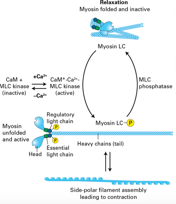
Smooth Muscle Contraction

  • Regulatory mechanisms: Smooth muscle contractions are controlled by signaling pathways rather than calcium levels alone.

    • Myosin is inactive during relaxation

  • Phosphorylation activates it forming filaments that can induce contraction in smooth muscle.

    • Dephosphorylation relaxes it again.

RM

Lecture 11 Actin World

Page 2: Learning Objectives

  • Characterize actin filament structure and composition.

  • Formation and dynamics of thin filaments: Understanding how they are formed in cells and the roles of associated motor proteins.

  • Actin-associated proteins: List and distinguish their functions.

  • Role of thin filaments: Distribution and diverse functions in cellular contexts.

  • Muscle tissue structure and function: Analyzing actin's contribution to muscle activity.

  • Cellular movement mechanisms: Outlining how actin facilitates cellular motility.

Actin Filaments

  • Major Protein Component: Actin is integral to all cells, located at cell junctions and throughout cellular structures.

Cytoskeletal Filaments

  • Types of cytoskeletal filaments:

    • Actin: Also known as the basic unit of thin filaments/microfilaments, vital for a multitude of functions. (side roads)

    • Intermediate filaments: Provide tensile strength against stress.

    • Microtubules (thick filaments): Serve various functions, acting as pathways for intracellular transport. (major highways)

Actin Dynamics

  • Dynamic structure: Actin constitutes 10% of muscle cell protein and 1-5% in non-muscle cells.

  • Filament formation and disassembly: Actin filaments are continuously formed and disassembled, tailored to cellular needs and environmental stimuli.

  • Actin genes: Humans have six actin genes that contribute to the diversity of actin functions.

Functions of Actin

  • Diverse roles: Actin is involved in cellular structure, movement, phagocytosis (“eating”), and cell division.

  • Three types of actins:

    • α-actin: Found in muscle fibers.

    • β-actin: Present in the leading edge of cells.

    • γ-actin: Associated with stress fibers.

Actin-Binding Proteins

  • Table of Proteins: Overview of actin-binding proteins and their functions, e.g., Profilin (nucleotide exchange), Cofilin (disassembly), and various capping and cross-linking proteins.

  • Functionality: Proteins that can promote actin polymerization, stabilize filaments, or sever actin structures. Proteins associated with actin can modify it by nucleating, stabilizing, serving, capping, etc.

Actin Monomers

  • Actin exists as G-actin: Globular actin binds ATP and Mg2+.

  • Polymerization: Forms filamentous actin (F-actin) with 2 helical structures wrapped around one another. .

Polarization of Actin Filaments

  • Polar characteristics: Myosin S1 regions coat F-actin, creating a polarized structure with distinct ends for filament dynamics.

    • form arrow like structures:

      • (-) pointed end: end has cleft exposed to the solution

      • (+) barbed end: has the cleft contacting other subunits

Filament Dynamics

  • End addition preference: G-actin adds more readily to the (+) end of the filament compared to the (-) end, influencing overall growth.

    • to form filaments G-actin-ATP units are added to both ends.

    • (+0) end addition is almost 10x greater than the (-) end

Actin Filaments need a Critical Concentration

  • Requirement for filament growth: Actin filaments need a critical concentration of G-actin-ATP. Each different Cc which much lower [Cc] required in the (+) end.

  • ATP is hydrolyzed quickly when actin monomers are added creating 3 distinct regions.

  • Treadmilling is a dynamic equilibrium process in filament assembly and disassembly. Seen at steady state the (-) end loses monomers while new ones are added to the (+) end

Steps of Actin Filament Formation

    1. Nucleation

    2. Elongation

    3. Steady State

Actin Recycling

  • Profilin's role: Enhances actin recycling and promotes the binding of ATP-actin at the (+) end, contributing to dynamic filament reassembly.

  • Profilin binds G-actin lost from filaments and opens the cleft enhancing loss of ADP

    • Profilin-ATP-actin complex binds the (+) end

  • Cofilin binds 2 ADP-actin towards the (-) end, causes a twist and a break

    • Generates more (-) ends for disassembly

  • Thysomin - B4 binds free ATP-actin to keep it all from assembling needless into filaments

    • Forms dynamic equilibrium with the free subunits

Capping Proteins

  • To regulate the formation and length of filaments/dynamics

  • CapZ binds the (+) end

    • Formed from two related proteins

  • Cells regulate CapZ ability to stop growth

    • Phosphatidylinositol 4,5- bisphosphate (PIP2)

    • Regulator proteins

  • Tropomodulin binds the (-) end and can module filament assembly and disassembly

    • Mostly found where actin needs to be stabilized

Actin-Nucleating Proteins

  • 2 major classes of actin-nucleating proteins help in the assembly of F-actin cells.

    • Formin proteins: Facilitate long filament assembly.

    • Arp2/3 complex: Promotes branched filament networks.

Formins Homology 1, 2 Domains in Formins

  • FH1 domains bind profilin to increase ATP-actin

  • FH2 domains form a ring that rocks back and forth promoting addition of ATP actin to the (+) end

Regulatory Signaling

  • Signal pathways: Actin nucleation and filament formation can be controlled through receptor-mediated signaling pathways, leading to the activation of formin proteins.

ARP2/3 and NPFs

  • Complex function: The ARP2/3 complex promotes nucleation and creation of branched F-actin structures, activated by nucleation-promoting factors. This binds to existing F-actin and promotes branch formation.

WASp Activation

  • WASp's (Wiskott-Aldrich syndrome) role: Serves as a nucleation-promoting factor by activating signaling cascades that stimulate Arp2/3 near cellular membranes.

Listeria Monocytogenes

  • Overview of Gram-positive bacteria: Causes listeriosis with significant health risks, particularly for pregnant women and immunocompromised individuals.

  • ActA protein mimcs an NPF and activated and recruits Arp2/3. Actin polymerization propels the bacteria through the cell.

Actin Role in Membrane Dynamics

  • Membrane mechanisms: Actin polymerization generates force and movement and aids in endocytosis, with contributions from signaling factors that assist in membrane dynamics.

  • Bursts of Arp 2/3 activation recruits other factors that can help power this energetically unfavorable membrane dynamic

Phagocytosis

  • Immune function: Describes how actin aids immune cells in engulfing pathogens through receptor-mediated signaling and network formation.

    • Opsonized pathogen are immobilized by the cell and receptor mediate signaling activates local Arp2/3 activation and network formation. creates a contractile network that leads to isolation of the pathogen and subsequent destruction.

Cross-Linking Proteins

  • Structural diversity: Actin cross-linking proteins mold filaments into varied structures, essential for functions such as sound perception in stereocilia.

Erythrocyte Structure

  • Stress handling: Actin networks are crucial for erythrocyte integrity, maintaining its shape and structure under stress. Actin membrane is anchored by actin associated proteins such as a/B spectrin. Actin gives cell shape and structure.

Actin Filaments and Muscle

  • Myosin II was the first discovered motor protein, integral in muscle cell function by converting chemical energy to mechanical work. All family motor proteins can bind to actin.

  • Immobilized ATPase containing mysoin S1 subunits will move actin filaments under experimental conditions. 40 myosin genes in the human genome.

  • Myosin Classes

    • Myosin I – only one with asingle head domain and it is associated with the membrane

    • Myosin II – mostly in muscle (thick fibers) but also non-muscle in less organized fashion, used for contraction

    • Myosin V – motor protein to hall things towards the membrane

    • Myosin VI – only member that moves towards the negative end, moving things away from the membrane

Page 29: Myosin-Actin Interaction

  • Power stroke dynamics: Myosin-ADP binds to F-actin. ATP binds releasing myosin from F-actin Hydrolysis cocks the head domain. Head domain binds actin. Release of Pi moves it back into its original position. In muscle 10% of the myosin is contacting f-actin when activated.

  • Myosin Steps

    • Step size of myosin head is relative to the length of the neck domain. (longer legs = bigger step),

      • Myosin II moves about 8nm

      • Myosin V takes longer 72 nm steps, hauling cargo 36nm ata. step

  • Muscle Fiber Composition

    • muscles —> muscle fibers (multinucleated muscle cells) —> myofibril structures composed of sacromere

    • Z disk structures flank: Myosin II thick filaments alternating with F-actin thin filaments attached to the Z disk

    • Thick filaments are bipolar with heads out toward Z disks. F-actin is attached to Z-disk. During contraction the mysoin II moves towards the (+) shortening the distance between the (-) end of F-actin.

      • Controlled by ATP availability and Ca2+ influx

Accessory Proteins in Muscle

  • Structural support: CapZ, Tropomodulin, Titin, and Nebulin play crucial roles in maintaining muscle structure and function.

    • CapZ helps bind F-actin (+) and Tropomodulin (-)

    • a giant elastic titin protein binds Z disk and extends toward the M band on either side (holds the thick filaments in place)

    • Giant nebulin protein wraps around the F-actin

Cytokinesis

  • Role of actin: Highlighting the involvement of actin-myosin in cytokinesis, forming contractile rings that assist in cell division.

  • Sarcoplasmic reticulum (SR) holds a high store of Ca2+ for skeletal muscle regulation

  • Neural impulse travels down the transverse tubules and opens voltage- gated Ca2+ channels in the SR

  • The release of Ca2+ allows muscle contraction by binding Troponin

    • Troponin shifts Tropomyosin

  • Actin-Mysoin II is involved in Cytokinesis. A contractile ring forms after chromosomal partition and eventually pinches off.

Smooth Muscle Contraction

  • Regulatory mechanisms: Smooth muscle contractions are controlled by signaling pathways rather than calcium levels alone.

    • Myosin is inactive during relaxation

  • Phosphorylation activates it forming filaments that can induce contraction in smooth muscle.

    • Dephosphorylation relaxes it again.

robot