June 08 Muscle Tissue Histology and Physiology
Muscle Tissue: Overview and General Characteristics
Specialization: Muscle cells are specialized for contractility.
Origin: Muscle tissue has a mesodermal origin.
Differentiation: The process of differentiation involves:
Cell lengthening.
Synthesis of myofibrillar proteins, specifically actin and myosin.
General Nomenclature of Muscle Tissue
Muscle fiber: Referred to as the muscle cell.
Sarcolemma: The cytoplasmic membrane (plasma membrane) of the muscle cell.
Sarcoplasm: The cytoplasm of the muscle cell.
Sarcoplasmic reticulum (SR): The specialized smooth endoplasmic reticulum of the muscle cell.
Calcium 2+ ions: Essential for muscle contraction, they are released from the sarcoplasmic reticulum in response to an action potential, allowing sliding filament theory to take place.
Classification and Comparison of Muscle Tissues
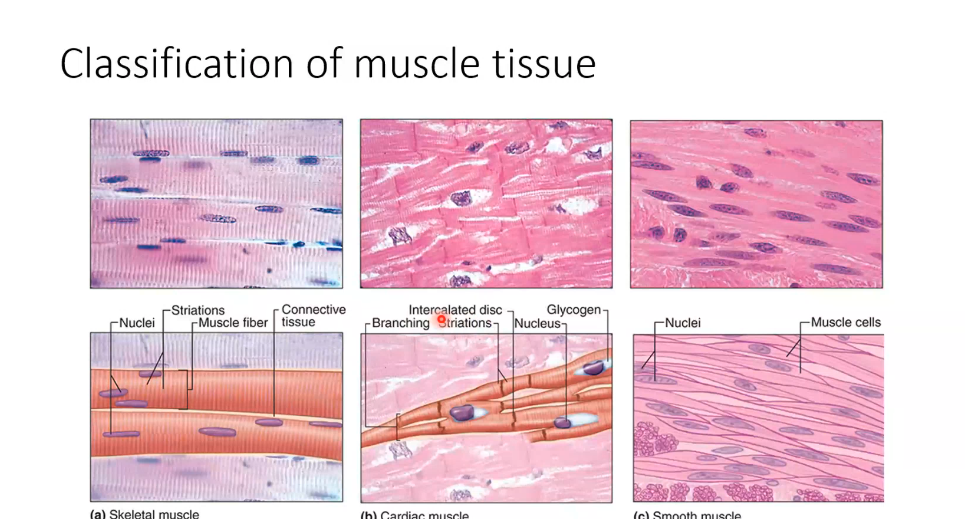
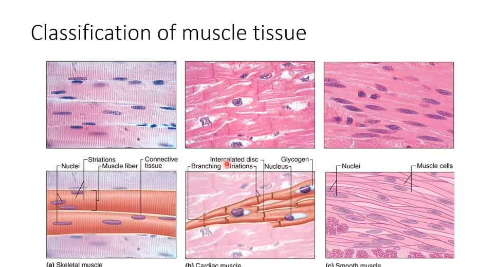
Skeletal Muscle
Structure: Composed of bundles of very long, cylindrical fibers (cells).
Nuclei: Multinucleated cells with nuclei located peripherally, adjacent to the sarcolemma.
Appearance: Displays prominent cross-striations.
Contraction: Fast and strong contractions; usually under voluntary control.
Location: Associated with the skeletal system, tongue, diaphragm, eyes, and upper esophagus.
Cardiac Muscle
Structure: Elongated, often branched cells.
Nuclei: Typically contains one central nucleus per cell.
Inclusion of glycogen granules which serve as an energy source for the muscle tissue during contraction. The glycogen does not stain with hematoxylin, eosin, stain.
Cardiomyocytes are more sensitive to the absence of oxygen than to the absence of glucose. However, because they contain glycogen, which serves as a rapidly accessible energy source, they can maintain contraction for a short period when oxygen levels are limited. Glycogen provides an emergency fuel reserve, but prolonged oxygen deprivation will still lead to impaired function and cell damage.
Appearance: Displays cross-striations.
Cell Junctions: Joined by intercalated discs that include desmosomes, adherens junctions (transverse regions), and gap junctions (longitudinal regions) for electrical synapses.
Contraction: Vigorous, rhythmic, and involuntary (automatic) pumping of blood.
Location: Found exclusively in the heart what part mycardium.

Smooth Muscle
Structure: Collection of fusiform (spindle-shaped) cells, typically small and closely packed.
Nuclei: One central, elongated nucleus located at the widest part of the cell.
Appearance: No striations (non-striated).
Contraction: Slow, steady, and involuntary contractions; can be spontaneous, wavelike, and rhythmic.
Location: Found in the walls of hollow internal organs (e.g., blood vessels, digestive and respiratory tracts, uterus, bladder).
Skeletal Muscle: Development and Organization
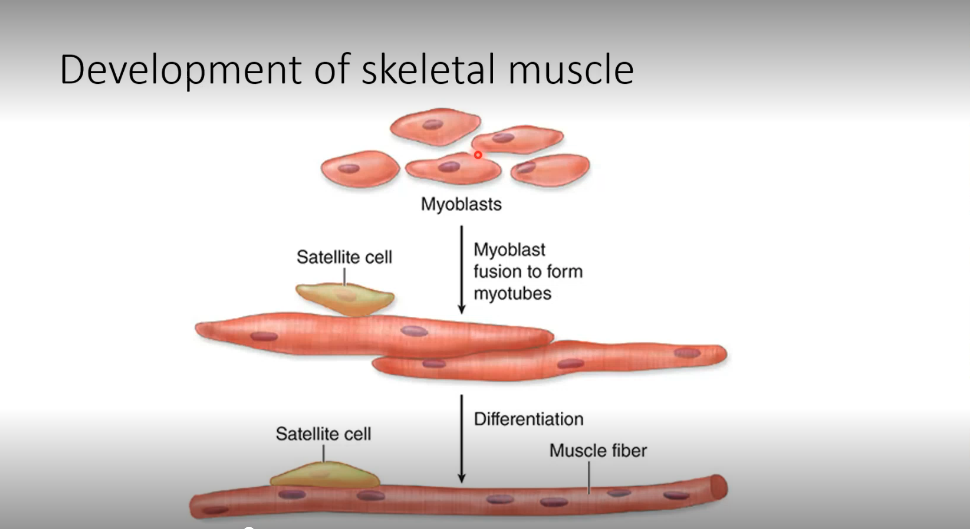
Development
Mechanism: Myoblasts undergo fusion to form myotubes.
Differentiation: Myotubes differentiate into mature muscle fibers.
Satellite Cells: These are reserve cells that remain associated with the muscle fiber for regeneration.
Connective Tissue Layers
Function: Collagen fibers in these layers transmit mechanical forces resulting from cell contraction.
Whole Muscle→Fascicles→Muscle Fibers (Muscle Cells)→Myofibrils
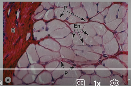
Epimysium: Dense irregular connective tissue that surrounds the entire muscle.
Perimysium: Thin connective tissue that surrounds fascicles (bundles of muscle fibers).
Endomysium: Composed of reticular fibers and fibroblasts; it surrounds individual muscle fibers and carries a rich vascular network.
Surrounds each individual muscle fiber (muscle cell)
Contains small blood vessels and nerves that supply the muscle fiber
Question:
What surrounds each muscle fiber? Answer: Endomysium
What surrounds a fascicle?Answer: Perimysium
What surrounds the entire muscle? Answer: Epimysium
Diagram
Epimysium
┌───────────────────┐
│ │
│ Perimysium │
│ ┌─────────────┐ │
│ │ Fascicle │ │
│ │ │ │
│ │ Endomysium │ │
│ │ ○ ○ ○ │ │
│ │ Muscle │ │
│ │ Fibers │ │
│ └─────────────┘ │
│ │
└───────────────────┘

The Sarcomere and Contractile Apparatus
Myofibrils and Striations
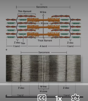
Myofibrils: Highly organized sarcoplasmic structures running parallel to the long axis of the cell. Made of myofilaments, one thin filaments, which are the active filaments, and then the thick filaments, which are called the myosin filaments
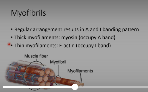
Banding Pattern: Created by the lateral registration of sarcomeres in adjacent myofibrils.
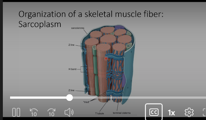
A band (anisotropic): Dark bands containing thick filaments (myosin)
Both thick and thin
overlapping zone within the A band but outside the H band
I band (isotropic): Light bands containing thin filaments (actin) but no thick filaments.
Z disc (or Z line): A dark line bisecting the I band; seen clearly with Transmission Electron Microscopy (TEM).
H zone: Lighter central zone in the A band where no thin filaments are present.
Exclusively thick filaments
M line: Bisects the H zone; contains myomesin (myosin-binding protein) and creatine kinase.
Middle of the H band
Sarcomere: The functional subunit of the contractile apparatus, defined as the segment between two Z discs.
Sarcolema and Transverse tubule system
Sarcoplasmic Reticulum (SR)
Specialized smooth endoplasmic reticulum (SER) of muscle cells.
Surrounds each myofibril.
Organized as a tubular network (like smooth ER).
Stores and regulates Ca²⁺ (calcium) needed for muscle contraction.
Contains calsequestrin, a calcium-binding protein that sequesters and stores Ca²⁺ within the SR.
T-Tubules (Transverse Tubules)
Deep invaginations (infoldings) of the sarcolemma.
Penetrate deep into the sarcoplasm.
Encircle each myofibril.
Located at the A band–I band junction of the sarcomere.
Rapidly conduct action potentials from the cell surface to the interior of the muscle fiber.
T-Tubules & Sarcoplasmic Reticulum
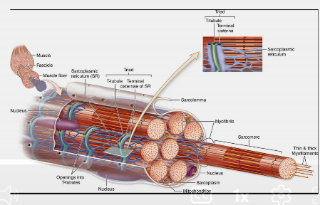
T-tubules (transverse tubules) are deep invaginations of the sarcolemma that penetrate into the muscle fiber.
T-tubules are closely associated with the sarcoplasmic reticulum (specialized smooth ER).
A triad consists of:
1 T-tubule
2 terminal cisternae of the sarcoplasmic reticulum
Function: Carry the action potential deep into the muscle fiber, triggering Ca²⁺ release from the sarcoplasmic reticulum.
Mitochondria in Muscle
Muscle contraction requires large amounts of ATP.
Mitochondria produce ATP through aerobic respiration.
Skeletal muscle fibers contain numerous mitochondria to meet the high energy demands of contraction.
Mitochondria are often located between myofibrils near sites of ATP use
Terminal Cisternae
Enlarged sacs of the sarcoplasmic reticulum.
Located on either side of a T-tubule.
Major storage sites for Ca²⁺.
Triad
Consists of:
1 T-tubule
2 terminal cisternae of the sarcoplasmic reticulum
Located at the A-I band boundary.
Surround each myofibril.
Function of the Triad
Depolarization travels along the sarcolemma and down the T-tubules.
Depolarization of the T-tubule affects the adjacent terminal cisternae.
Causes release of Ca²⁺ from the sarcoplasmic reticulum.
Initiates muscle contraction.
I due to the action potential
Thick Filaments: Myosin
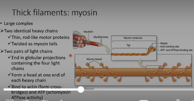
Structure: A large complex consisting of two identical heavy chains and two pairs of light chains.
Heavy Chains: Thin, rod-like motor proteins twisted as myosin tails.
Heads: Globular projections at one end of each heavy chain that contain the four light chains.
Functions: Bind to actin (forming cross-bridges) and bind ATP (possessing actomyosin ATPase activity).
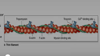
Thin Filaments: F-actin
Composition: Formed by monomers of G-actin, each having a binding site for myosin.
Regulatory Proteins:
Tropomyosin: A coil of two polypeptides located in the groove between twisted actin strands.
Troponin: A complex of three subunits:
TnT: Attaches the complex to tropomyosin.
TnC: Binds to .
TnI: Regulates the actin-myosin interaction.
Accessory Proteins
Titin: The largest protein in the body; an accessory protein of the I band that anchors myosin to the Z disc.(green)
-actinin: Anchors actin filaments to the Z discs.
More information
I Band (Light Band)
Contains thin (actin) filaments only.
Bisected by the Z disk (Z line).
Actin filaments are attached to the Z disk.
Contains titin (largest protein in the body).
Titin extends from the Z disk toward the thick filament and helps anchor myosin to the Z disk.
Accessory proteins help maintain filament alignment.
Z Disk (Z Line)
Boundary of each sarcomere.
Anchors actin (thin) filaments.
Anchors titin.
Connects adjacent sarcomeres.
Titin
Largest protein in the body.
Extends from Z disk to thick filament (myosin).
Anchors myosin to the Z disk.
Provides elasticity and prevents overstretching.
A Band (Dark Band)
Contains the entire length of thick (myosin) filaments.
Includes regions where thick and thin filaments overlap.
H Zone
Light central region within the A band.
Contains thick (myosin) filaments only.
No thin (actin) filaments present.
M Line
Located in the center of the H zone.
Bisects the H zone.
Contains:
Myomesin (myosin-binding protein)
Creatine kinase
Holds thick filaments together and maintains alignment.
Muscle Contraction: Physiology and the Triad
The T-Tubule System and Triad
T-tubules (Transverse tubules): Infoldings of the sarcolemma that penetrate deep into the sarcoplasm, encircling myofibrils at the A-I band boundaries.
Triad Components: Consists of one T-tubule and two terminal cisternae of the sarcoplasmic reticulum.
Step-by-Step Contraction Mechanism
Impulse: A nerve impulse triggers the release of Acetylcholine (ACh) into the synaptic cleft. ACh binds to receptors on the motor end plate, initiating a muscle impulse.
Release: The impulse spreads along T-tubules, triggering the release of from the terminal cisternae of the SR into the sarcoplasm.
Binding: binds to troponin, causing it to change shape and move tropomyosin, exposing active sites on actin.
Cross-bridge: Myosin heads attach to exposed active sites. They pivot and slide the thin filaments toward the center of the sarcomere (M line).
Cycle: ATP binds to myosin, causing detachment. ATP hydrolysis (to ADP + P) provides energy to return the head to its prepivot position. The cycle repeats as long as remains bound to troponin.
Relaxation: When the impulse stops, is actively transported back into the SR. Tropomyosin re-covers the active sites, and filaments slide back to a relaxed state.

More information
Calcium & Muscle Contraction
Calcium (Ca²⁺) is stored at high concentrations inside the sarcoplasmic reticulum (SR).
The concentration of Ca²⁺ is much higher in the SR than in the sarcoplasm.
When a muscle fiber is stimulated, the sarcolemma depolarizes.
The depolarization travels down the T-tubules.
Depolarization of the T-tubules affects the adjacent sarcoplasmic reticulum.
Ca²⁺ is released from the SR into the sarcoplasm through calcium-release channels (facilitated diffusion).
This increases the concentration of Ca²⁺ in the sarcoplasm.
Neuromuscular Junction (Motor End Plate)
Structure: Nerves branch in the perimysium, and unmyelinated terminal fibers pass through the endomysium to contact individual muscle fibers.
Axon Terminal: Dilation within the muscle cell trough containing mitochondria and synaptic vesicles filled with ACh.
Schwann Cells: Cover the small axon branches at the contact points.
Junctional Folds: Folds in the sarcolemma adjacent to the synaptic cleft that contain ACh receptors.
Muscle Proprioceptors
Muscle Spindle:
Encapsulated by modified perimysium.
Contains intrafusal fibers (thin muscle cells) surrounded by sensory axons.
Acts as a stretch receptor within the skeletal muscle.
Muscle Spindle: Intrafusal vs Extrafusal Fibers
Intrafusal Fibers
Muscle fibers located inside a muscle spindle
Specialized for detecting muscle stretch (sensory function)
Smaller and thinner than regular muscle fibers
Contained within the connective tissue capsule of the muscle spindle
Help provide information about:
Muscle length
Rate of muscle stretch
Innervated by sensory neurons and gamma motor neurons
Extrafusal Fibers
Regular skeletal muscle fibers
Located outside the muscle spindle
Make up the bulk of the muscle
Responsible for muscle contraction and force production
Innervated by alpha motor neurons
The muscle spindle detects stretching of a muscle.
When the muscle stretches:
The intrafusal fibers inside the muscle spindle also stretch.
The stretch is detected by sensory nerve endings (sensory axons) wrapped around the intrafusal fibers.
The sensory neurons then send signals to the CNS.
Sensory Axons
Sensory axons surround individual intrafusal fibers.
These nerve endings detect:
Muscle length
Changes in muscle length (stretch)
Muscle Spindle Structure
Contains:
Intrafusal fibers
Sensory nerve endings
Gamma motor neuron endings
Enclosed by a connective tissue capsule
Capsule
Made of dense connective tissue.
Professor's note:
The capsule is a modified perimysium.
It surrounds and protects the contents of the muscle spindle.
Muscle stretches → Intrafusal fibers stretch → Sensory axons detect stretch → Signal sent to CNS
Golgi Tendon Organ:
Encapsulated axons found among collagen fibers at the myotendinous junction.
Smaller than the muscle spindle; also acts as a stretch receptor.
Skeletal Muscle Fiber Types
Feature | Slow Oxidative (Type I) | Fast Oxidative-Glycolytic (Type IIa) | Fast Glycolytic (Type IIb) |
|---|---|---|---|
Contraction Speed | Slow | Fast | Fast |
Fatigue Rate | Slow | Intermediate | Fast |
ATP Source | Oxidative phosphorylation | Oxidative phosphorylation | Anaerobic glycolysis |
Mitochondria | Numerous | Numerous | Sparse |
Capillaries | Numerous | Numerous | Sparse |
Myoglobin | High (Red fibers) | High (Red fibers) | Low (White fibers) |
Glycogen | Low | Intermediate | High |
Fiber Diameter | Small | Intermediate | Large |
Major Location | Postural muscles (Back) | Major muscles of legs | Extraocular muscles |
Ultrastructure of Cardiac Muscle cells (Cardiocytes)
Mitochondria: Extremely abundant, making up approximately of the sarcoplasm.
SR and T-tubules: Sarcoplasmic reticulum is well-organized. T-tubules are longer in ventricular cells.
Dyads: Composed of one T-tubule and one terminal cisterna of the SR, located at the Z discs.
Storage: Contains small lipid droplets (triglycerides) and perinuclear glycogen granules.
Secretory Granules: Atrial cardiocytes contain granules of Atrial Natriuretic Peptide (ANP).
Smooth Muscle: Structure and Function
General Features
Functions: Slow steady contraction and synthesis of Extracellular Matrix (ECM) components (Type I and Type III collagen).
Connective Tissue: Thick branching perimysium and endomysium composed of collagen types I and III.
Gap Junctions: Numerous junctions link adjacent cells for stimulus propagation.
Ultrastructure and Contractile Apparatus
Organelles: Perinuclear mitochondria, RER, Golgi apparatus, and polyribosomes.
Caveolae: Plasmalemma invaginations with channels that control release (replaces T-tubule system).
Dense Bodies: Found in the sarcoplasm and sarcolemma; they anchor actin filaments through -actinin and intermediate filaments (desmin). They function like Z discs to transmit contractile force.
Mechanism: Myosin filaments are less regular with fewer cross-bridges. Contraction is regulated by Calmodulin and Myosin Light-Chain Kinase (MLCK) instead of troponin.
Regeneration and Response to Load
Skeletal Muscle: Slow regeneration via satellite cells. Responds to load through Hypertrophy (increase in fiber size).
Cardiac Muscle: Very poor regeneration; lacks satellite cells. Responds to load through Hypertrophy.
Smooth Muscle: Rapid regeneration; cells are relatively undifferentiated and can undergo mitosis. Responds to load through Hypertrophy and Hyperplasia (increase in cell number).