Equids and Ruminants Parasitology
Equine Gastrointestinal Nematodes
Habronema species

Common Name: Stomach worm.
Definitive Host: Horses and all members of the equid family (including mules and donkeys).
Location: Stomach.
Life Cycle: Indirect life cycle.
Intermediate Host: Flies.
Transmission & Cycle:
Eggs are passed in the feces.
Flies lay maggots in the feces; as the maggots mature into adult flies, they ingest the parasite eggs or larvae.
Adult flies feed around the mouthparts, face, lips, and eyes of the host.
The larvae are deposited by the fly on the face/lips. When the horse licks its lips, it ingests the larvae.
Larvae mature into adult parasites inside the stomach.
Cutaneous Infection ("Summer Sores"):
Occurs if larvae are deposited on the skin by the fly but are not ingested by the horse (e.g., horse does not lick its lips).
The larvae reside in the skin and cause circular lesions known as subcutaneous infections or "summer sores."
Diagnosis:
Detection of eggs or larvae in feces.
Eggs and larvae are rarely found via routine fecal analysis because most eggs hatch prior to fecal passage, and larvae are difficult to detect.
Best Diagnostic Technique: Fülleborn (Ferremon) technique.
Infective Stage: larval stage contained within the fly, which is deposited while feeding around the host's face.
Pre-Patent Period: .
Treatment: Ivermectin or moxidectin, combined with environmental fly control.
Clinical Signs: Gastric upset and stomach inflammation caused by mucosal irritation (unless presenting as cutaneous summer sores).
Parascaris equorum
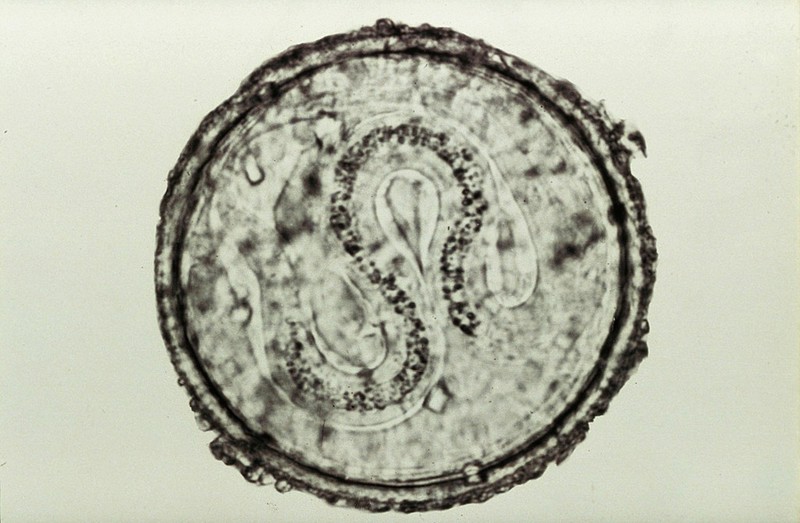
Common Name: Roundworm of horses.
Location: Small intestine.
Prevalence: Significantly more prevalent in foals than in adult horses.
Life Cycle: Direct life cycle.
Eggs are passed in feces and larvate in the environment.
The horse ingests the infective eggs while grazing on pasture.
Diagnosis: Detection of eggs or adult worms in feces.
Adult worms do not migrate up into the stomach or mouth because horses are physically incapable of vomiting.
Egg Morphology: Typical roundworm egg appearance featuring a dark, thick, pitted shell.
Infective Stage: larval stage that hatches into the pasture.
Pre-Patent Period: .
Treatment: Pyrantel, Panacur (fenbendazole), oxibendazole, mebendazole, ivermectin, or moxidectin.
Pathology & Clinical Signs:
Competes directly with the host for nutrients, leading to an unthrifty appearance.
Heavy worm burdens cause severe abdominal pain (colic) and intestinal impactions.
Larval migration through the respiratory tract causes coughing, where larvae are coughed up and re-swallowed.
Size: The largest parasite affecting horses (larger and substantially longer than a large nightcrawler).
Strongylus species (Strongyles)

Common Name: Strongyles.
Classification: Divided into Large Strongyles and Small Strongyles.
Location: Large intestine and cecum.
Life Cycle: Direct life cycle.
Eggs are passed in feces and hatch in the environment.
Larvae progress from to to .
The host ingests while grazing grass.
Infective Stage: larva on pasture grass.
Diagnosis: Egg detection in feces.
Individual species eggs cannot be differentiated microscopically; fecal results are reported generically as Strongylus species.
Morphology: Oval shape with a dark centered morula. Resembles canine hookworm eggs but is slightly larger. Strongyles must never be called hookworms in equine medicine.
Pre-Patent Period: .
Treatment: Pyrantel, fenbendazole, oxibendazole, moxidectin, or ivermectin.
Clinical Signs: Anemia, weight loss, colic, and intestinal pathology (signs occur predominantly during larval migration).
Pathogenic Large Strongyle Species:
Strongylus edentatus
Strongylus equinus
Strongylus vulgaris
Pathogenicity of Strongylus vulgaris:

Highly pathogenic species. Larvae penetrate the intestinal wall and migrate directly to the cranial mesenteric artery (the primary artery supplying blood to the intestines contained within the abdominal mesentery).
Migration induces severe arterial inflammation, blood clots, and vessel blockages.
Disruption of intestinal blood supply leads to intestinal necrosis, acute colic, and death if untreated.
Strongyloides westeri

Common Name: Threadworm.
Location: Small intestine.
Environmental Requirements: Requires warm, moist soil to survive in the environment.
Life Cycle: Direct life cycle.
Diagnosis: Fülleborn (Ferremon) technique (used to detect larvae in feces, as larvae are far more common than eggs).
Pre-Patent Period: .
Treatment: Ivermectin or oxibendazole.
Transmission Routes:
Transmammary: Primary route of infection in foals through the mare's milk.
Percutaneous: Skin penetration.
Oral: Direct ingestion.
Note: Transplacental transmission does not occur in horses.
Parasitic Stage: Adult females only (parthenogenetic; capable of producing viable ova without a male present).
Clinical Signs: Diarrhea, weight loss, anemia, and anorexia. Most common and severe in foals.
Differential Diagnosis: Frequently misdiagnosed as "foal heat diarrhea" (diarrhea attributed to hormonal changes in mare's milk during her post-foaling heat cycle) when it is actually transmammarily transmitted Strongyloides westeri. Adults usually maintain light, asymptomatic infections.
Oxyuris equi

Common Name: Pinworm.
Location: Cecum, colon, and rectum.
Life Cycle: Direct life cycle.
Gravid adult females travel out of the rectum and lay eggs around the perianal skin.
Eggs fall off into the environment via rubbing or defecation.
Host ingests infective eggs from the environment, which mature into adults in the cecum, colon, or rectum.
Diagnosis:
Scotch Tape Method: Clear scotch tape is wrapped backward around a tongue depressor (sticky side out) and pressed around the perianal region. The tape is placed sticky side down on a microscope slide to view concentrated eggs.
Eggs are infrequently found in routine fecal samples.
**
Ruminant Oral Anatomy
Upper Jaw Structures: Ruminants do not possess upper incisor teeth; instead, they have a thick, fibrous tissue layer known as the dental pad.
Gastrointestinal Nematodes: Trichostrongyles
Taxonomic Grouping and Identification:
Ruminant trichostrongyles are analogous to equine strongyles, comprising numerous distinct genera and species whose eggs cannot be morphologically differentiated during routine fecal flotation.
Eggs are collectively classified and reported as Trichostrongylus species or trichostrongyles.
Genera and Common Names:
Haemonchus species: Commonly known as the barber pole worm.
Ostertagia species: Commonly known as the brown stomach small intestinal worm.
Bunostomum species: Referred to as a hookworm.
Chabertia species (Schibertia): Commonly known as the large mouth bowel worm.
Oesophagostomum species (Esophagastinum): Commonly known as the nodular worm.
Trichostrongylus species: Commonly known simply as trichostrongyle.
Anatomical Location: Primary sites of infection are the abomasum and the small intestine.
The abomasum is the fourth chamber of the ruminant stomach and functions as the true stomach.
Definitive Hosts: All ruminants (cattle, sheep, goats).
Life Cycle: Direct.
Infective Stage: Free-living larva in the environment.
Diagnostic Features:
Identified by detecting eggs in feces.
Egg morphology: Oval to slightly oblong shape with a dark, segmented center (morula) and a small space between the cell mass and shell.
Because species individualization is impossible on fecal examination, findings must be reported as Trichostrongylus species.
Prepatent Period (PPP): .
Treatment Options: Levamisole, ivermectin, and benzimidazoles (e.g., fenbendazole).
Pathology and Clinical Signs:
Most species feed on host blood.
Clinical signs include anemia, lethargy, severe weakness, and an overall unthrifty appearance.
Represent the most common gastrointestinal nematodes found in ruminants.
Gastrointestinal Nematodes: Nematodirus Species
Common Names: Thread neck intestinal worm or thread neck trichostrongyle.
Morphological Differentiation:
Easily distinguished from typical trichostrongyle eggs due to size and internal structure.
At least to larger than typical trichostrongyle eggs.
Shape is characteristically football-like (elongated with tapered ends).
Contains large, distinctly visible, individual blastomeres/oospheres rather than an undifferentiated cellular mass.
Anatomical Location: Abomasum, small intestine, and large intestine.
Definitive Hosts: All ruminants.
Life Cycle: Direct.
Infective Stage: larva located on pasture vegetation.
Diagnosis: Identification of characteristic large eggs in feces.
Prepatent Period (PPP): .
Treatment Options: Fenbendazole, ivermectin, and levamisole.
Clinical Signs: Unthriftiness, dehydration, severe diarrhea, and anorexia.
Gastrointestinal Nematodes: Trichuris Ovis
Common Name: Whipworm.
Anatomical Location: Cecum and colon.
Definitive Hosts: All ruminants (cows, sheep, goats).
Life Cycle: Direct.
Infective Stage: larva within the environmental egg.
Diagnostic Features:
Demonstration of eggs in feces.
Egg morphology is identical to Trichuris vulpis (lemon-shaped/football-shaped with prominent bi-polar plugs).
Treatment Options: Ivermectin and fenbendazole.
Clinical Signs: Ranges from asymptomatic subclinical infection to dark melenic feces, anorexia, anemia, and unthriftiness.
Respiratory Nematodes: Dictyocaulus Species
Species and Host Specificity:
Dictyocaulus filaria: Primary lungworm of sheep and goats.
Dictyocaulus viviparus: Lungworm of cattle and cattle-family species (e.g., buffalo).
Common Name: Lungworm.
Anatomical Location: Bronchi and bronchioles of the lungs.
Dictyocaulus viviparus is the only parasite that reaches sexual maturity as an adult within the bovine lungs.
Life Cycle: Direct.
Infective Stage: larva in the environment.
Path of Migration: Ingested larvae penetrate the intestinal mucosa, enter the lymphatics and bloodstream, and migrate directly to the lungs to mature.
Diagnostic Features:
Female worms produce larvated eggs containing fully developed larvae.
Eggs hatch almost immediately inside the host respiratory or digestive tract.
Diagnosis relies on detecting larvae in nasal discharge or in fresh feces using the Baermann technique.
Prepatent Period (PPP): .
Treatment Options: Fenbendazole and ivermectin.
Clinical Pathology and Field Misdiagnosis:
D. filaria causes chronic coughing and severe unthriftiness in sheep and goats.
D. viviparus causes severe bronchitis and parasitic pneumonia in cattle.
Infected cattle present with severe dyspnea, extending their neck downwards with their mouth open, unable to close their mouth to chew or swallow.
Diagnostic Pitfall: Newly purchased auction/sale barn cattle presenting with dyspnea are frequently misdiagnosed with bacterial respiratory disease and treated unsuccessfully with antibiotics, allowing parasitic pulmonary destruction to progress to fatal levels.
Cestodes (Tapeworms) of Ruminants
Moniezia Species:
Moniezia benedeni: Definitive host is cattle.
Moniezia expansa: Definitive hosts are sheep, goats, and cattle.
Common Name: Moniezia tapeworm.
Life Cycle: Indirect.
Intermediate Host: Environmental oribatid grain mite (orbated grain mite).
Infective Stage: Cysticercoid larva residing within the grain mite.
Transmission Cycle: Host sheds gravid proglottids or eggs in feces -> proglottids rupture -> grain mites ingest eggs -> cysticercoids develop inside mites -> ruminant ingests infected grain mites while grazing.
Diagnostic Features: Detection of square or triangular eggs (Type A morphology) with a prominent inner circular pyriform apparatus, or observation of proglottid segments in feces.
Prepatent Period (PPP): .
Treatment Options: Benzimidazole derivatives including fenbendazole, albendazole, and oxfendazole (praziquantel is not standard in ruminants).
Clinical Signs: Often asymptomatic; massive worm burdens can cause intestinal impaction, severe colic, and fatal intestinal rupture.
Thysanosoma Actinioides:
Common Name: Fringed tapeworm (named for the fringe-like projections along the posterior border of each proglottid segment).
Anatomical Location: Bile ducts, pancreatic ducts, and small intestine.
Definitive Hosts: All ruminants.
Life Cycle: Indirect.
Intermediate Hosts: Oribatid grain mites or environmental bark lice.
Infective Stage: Cysticercoid larva within the intermediate host.
Diagnostic Features: Identification of fringed proglottid segments or characteristic egg packets in feces.
Treatment Options: Fenbendazole, oxfendazole, and albendazole.
Economic Implications: Infection is generally asymptomatic in live animals; however, presence in bile and pancreatic ducts leads to complete liver condemnation at slaughter, causing significant financial loss to livestock producers.
Trematodes (Flukes) of Ruminants
Fasciola Hepatica:
Common Name: Liver fluke.
Anatomical Location: Bile ducts of the liver.
Definitive Hosts: All ruminants.
Life Cycle: Indirect.
Intermediate Host: Aquatic snails.
Infective Stage: Encysted metacercariae attached to aquatic vegetation.
Diagnostic Features:
Detected by fecal sedimentation testing (fluke eggs are heavy and sink, failing to float in standard flotation solutions).
Egg morphology: Large, oval, yellow-brown, operculated egg completely filled with internal contents.
Treatment Option: Albendazole.
Clinical Signs and Pathology: Causes severe hepatic damage, chronic anemia, weight loss,