kin
1. What are the 3 types of muscle?
Skeletal muscle – voluntary, attached to bones, produces movement
Cardiac muscle – heart muscle, involuntary
Smooth muscle – organs & blood vessels, involuntary
2. Fascia coverings
Epimysium – covers entire muscle
Perimysium – surrounds fascicles (bundles of fibers)
Endomysium – surrounds individual muscle fibers
3. Why does the plasmalemma have shallow folds?
The folds increase surface area and help distribute force across the membrane during contraction. They reduce stress and prevent tearing.
4. What does the plasmalemma fuse with at the end of the muscle fiber?
It fuses with the tendon, helping transmit force from muscle to bone.
5. Satellite cells
Located between the sarcolemma (plasmalemma) and basal lamina
They are muscle stem cells
Help with muscle repair, growth, and regeneration
6. Extensions of the plasmalemma
Called T-tubules (transverse tubules)
They:
Carry the action potential deep into the muscle fiber
Trigger calcium release from the sarcoplasmic reticulum
7. What are myofibrils made up of?
Myofibrils are made of repeating units called sarcomeres, which contain:
Actin (thin filaments)
Myosin (thick filaments)
8. Sarcomere Structures


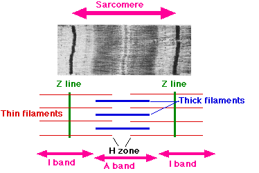

Structure | Location |
|---|---|
Z-disks | Boundary of each sarcomere; anchor actin filaments |
H-zone | Middle of sarcomere; only myosin present |
M-line | Center of sarcomere; holds myosin filaments together |
Titin | Runs from Z-disk to myosin; stabilizes & provides elasticity |
9. Functions of skeletal muscle
Produce movement
Maintain posture
Stabilize joints
Generate heat (thermogenesis)
Store nutrients (glycogen, amino acids)
10. Role of calcium in contraction
Calcium is released from the sarcoplasmic reticulum
Binds to troponin
Moves tropomyosin off actin binding sites
Allows myosin to bind actin → cross-bridge formation → contraction
No calcium = no contraction.
11. What does nebulin do?
Stabilizes actin filaments
Helps regulate thin filament length
12. Excitation–Contraction Coupling (quick summary)
Motor neuron releases acetylcholine
Action potential travels along sarcolemma
Down T-tubules
Sarcoplasmic reticulum releases calcium
Cross-bridge cycling begins
13. Role of ATP in contraction
ATP:
Detaches myosin from actin
Provides energy for the power stroke
Powers calcium pumps to return Ca²⁺ to SR
No ATP = rigor (like rigor mortis).
14. Type 1 vs Type 2 Fibers
Type 1 | Type 2 | |
|---|---|---|
Myosin ATPase | Slow | Fast |
Contractile speed | Slow | Fast |
Fatigue resistance | High | Low |
Motor unit strength | Small | Large |
Size of motor unit | Small | Large |
Sarcoplasmic reticulum | Less developed | Highly developed |
Force (same diameter) | Same | Same |
Power (same diameter) | Lower | Higher |
(Remember: force per cross-sectional area is the same; power differs because speed differs.)
15. Principle explaining orderly recruitment
Size Principle
Small motor units (Type 1) are recruited first → larger units (Type 2) added as force demand increases.
16. How is muscle force graded?
Two ways:
Motor unit recruitment
Rate coding (frequency of stimulation)
17. Two mechanisms explaining muscle memory
Myonuclear addition – nuclei gained during hypertrophy remain even after detraining
Epigenetic changes – DNA modifications that make retraining faster