Vision
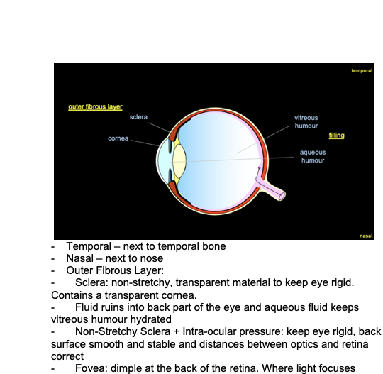
- Temporal – next to temporal bone
- Nasal – next to nose
- Outer Fibrous Layer:
- Sclera: non-stretchy, transparent material to keep eye rigid. Contains a transparent cornea.
- Fluid ruins into back part of the eye and aqueous fluid keeps vitreous humour hydrated
- Non-Stretchy Sclera + Intra-ocular pressure: keep eye rigid, back surface smooth and stable and distances between optics and retina correct
- Fovea: dimple at the back of the retina. Where light focuses
- The retina is generated from the neural tube and links the photoreceptor that detect the light to the retinal ganglion cells that take the signal to the brain
- Too much pressure = glaucoma
__
- As the eye is a camera its needs a stable shape to bring the image of the world into focus on a layer of light sensitive cells at the back of the eye. If the eyeball was wiggly, that layer would go in and out of focus.
- To have this stable shape, the eyeball has a tough, fibrous outer layer. Most of this layer is the white sclera but at the front the sclera becomes transparent and becomes the cornea. As the outer layer is tough, it isn’t stretchy therefore if you can generate a certain amount of pressure inside the eyeball it will maintain a rigid state.
- This rigid state is maintained through the production and drainage of the aqueous humour. The back of the eye is filled with a smooth transparent jelly called the vitreous humour which is hydrated by the aqueous humour.
- However if that pressure does build up it can damage neural structures at the back of the eye
- Optical Apparatus:
- Lens – rounded, transparent structure which is suspended in the light path and bends the light rays to bring them into focus. Can change shape due to it being suspended all the way around from the ciliary body. The ciliary body contains a ring of smooth muscle, so if the smooth muscle contracts, the ring is smaller which allows the lends to adopt that shape. If the ring of muscles relaxes, the ring gets larger, and the suspension of ligaments pull the lens into a flatter shape.
Cornea – curved shape and a air-water interface carries out most of the focussing.
- Adjustable aperture: Pupil – size is adaptable due to contraction and relaxation of the iris (coloured part of the eye).
__
What is the optical apparatus of the eye and how does it achieve a good focus of the image?
- Light rays are focused onto the cornea which is curved and transparent
- Lens bends the light further and is suspended by a ring of ligaments (ciliary bodies)
- Muscle contracts and relaxes to fatten the lens, so the light refracts more
- Fovea: brings light into focus
- Cornea: refracts most of the light
- Lens: produce a fine focus
- Iris: controls how much light enters the pupil and cuts out the light rays that go through edge of the lens therefore it stays focused an image
- Darker Place: Opens up the pupil but the image gets blurrier as there is less light to focus on the image
__
- When the light strikes our eye in all directions, some will strike the cornea and pass through it. When it passes through, it will be refracted and bent, so that the light that came from a single point has diverged and will begin to converge again. Some light rays will be blocked by the iris but that that passes through the pupil will be refracted a bit more than the lens, just enough to ensure that the light that is from a single point in the visual field is brought back into a single point on the retina into focus.
- Cornea – acts as a major refracting surface
- Iris – prevents light from passing through the edges of the lens
- Lens – gives additional “fine” focusing.
__
What is the organisation of a primary visual pathway?
Fields...
Cards...
Preview
Front
What is the organisation of a primary visual pathway?
Back
__ - To capture the image and pattern of brightness and wavelengths we need light sensitive cells such as rods and cons which are photoreceptors. They sit on the outside edge of the neural retina which is adjacent to the retinal pigment epithelium. This is passed on to retinal ganglion cells, which are the afferents of the eye. The retinal ganglion cells have axons which run alongside the inside of the retina and exit as a bundle to form the optic nerve. - The bundles of optic nerve from each retina run medially and converge at the optic chiasm and continue through a pathway known as the optic tract. The optic tract carries on into the brain into specific nucleus of the thalamus (lateral geniculate nucleus). In the LGN, the axons from the eyes, makes fast excitatory synapses with the lamex (??) cells which have axons that continue up through the cortical white matter (optic radiation) to the primary visual cortex (area 17). - Axons from the ganglion cells of the nasal side cross over to the over side as those from the temporal side stay on the same side. - Most of these axons have branches that end up on the brainstem where they innervate nuclei that are involved in eye reflexes movement, - Dysfunction of the RPE can lead to many retinal degenerative and RPE dysfunction kills photoreceptors. __
- Rods: very sensitive photoreceptors to night vision - Cones: less sensitive day vision. Less saturated and non-functional levels of them are activated twilight onwards. |
3 Tags
7-Vision
Neuro
Phsyio2
What is the structure of a cone photoreceptor and how does it respond to increase and decrease in illumination?
- Has lots of cells which falls into three layers. The purple and black dots show the cell bodies and the outer layer is where the photoreceptors would be. The inner layer is where you find the ganglion cells with the axons running through this area and the middle layer contains interneurons.
- Rods – used for scotopic vision (night vision). These saturate and become useless at anything above twilight levels. Their pathway are designed for maximum sensitivity at the expense of any fine detail vision
- Cones:
- Outer segment: packed with multiple layers of a phospholipid membrane which is attached by a thin cilium to the inner segment
- Inner segment: cell keeps all of its apparatus for making proteins
- Nucleus: produces proteins
- “Axon” – not exactly an axon but has the anatomy of an axon but doesn’t fire action potentials so not really classed as an axon
- Synaptic terminal – releases glutamate
- Infolding Membrane at right angles to the light path and hold light capturing molecules in a certain way to gather light – present in outer segment
- Has a resting membrane potential, which is brought by the leakage of potassium ions. Has a low RMP of around -45mV. This is because the outer segments contain sodium channels that are open at default and allow positive charge back into the cell.
- An increase in illumination: there is an increase in light hitting the outer segment. It closes some of the sodium channels which causes the cell to hyperpolarise and reduce its release of glutamate.
- A decrease in illumination: has the opposite effect where more channels are opened and the cell is depolarised, increasing the release of glutamate.
What is the outline of the phototransduction?
- This takes place in one of the layers of the phospholipid membranes in the outer segment. The oval represents the membrane disc in the outer segment and the blue represents the out membrane and has sodium channels between this and the cell membrane.
- These sodium channels are held together by cyclic GMP which is an intracellular molecule.
- The membrane disc is embedded within its phospholipid membrane a molecule called opsonin which binds with retinal to make a photo pigment, which response to light.
- Retinal is also known an 11-cis retinaldehyde, where the 11th bond is unstable and when a photon of light hits the bond, it activates the photo pigment which reforms in the trans configuration as all-trans retinaldehyde.
- This activated photopigment, activated a G protein which activates an enzyme which reduces the concentration of cyclic GMP within the cell.
- As these levels falls, some of the cyclic GMP that had been attached to the outer membrane sodium channels, diffuse away from them causing the channels to close.
- Some of the cyclic GMP that has been attached to the outer membrane sodium channel diffuse away from them and the channels close.
- When the GTP, G protein and associated enzyme runs out and stops working, a second enzyme helps to rebuild these cyclic GMP levels and reopen those channels.
- Trans retinal is removed and a new molecule of 11 cis retinal is attached.
- The photoreceptors gives a very rapid response when activated by light. Each activated photo pigment molecules will activate many G proteins, and the enzymes involved are very powerful. At first, the cell responds with hyperpolarisation, by over a short period of time will be restored back to resting membrane.
What is an overview of the retinal structure and circuitry?
- Fovea: the only part of our retina that can see fine detail and takes place in the centre.
- From the fovea, the circuitry gets messier, and your vision gets more and more blurred.
- To deal with this blur, our eyes automatically scan around the image very rapidly, gathering information so the brain reconstructs the rest of the image of what it thinks by adding extra details.
- Loss of peripheral vision: Glaucoma or advanced retinitis pigmentosa may have a loss of peripheral vision but still able to retain the ability to see in fine detail.
Age-related macular degeneration: macular is the centre of the visual mid and in AMD it can destroy the centre of the retina, therefore there is no way of gathering the fine details in that image.
What are differences between retinal structure and circuitry in the central vs peripheral region?
Peripheral Retina:
- The big gaps between the cones are filled with rods (no function in daylight) therefore this sampling array is filled with holes.
- The photoreceptors (cones) make synaptic contact with an interneuron cell called bipolar which passes the information on to the ganglion cells.
- The image created is known as the ganglion cells receptive field.
- As you go from the field to the far periphery, the convergence gets greater and receptive field size gets bigger, so our ability to resolve fine details gets smaller and smaller. This doesn’t matter as light passes from inside the eye towards the outside and therefore by the time it hits the outer segment of the photoreceptor, it is blurred and there is no point in sampling with fine detail a blurred image
Central Retina:
- 1st image: all the blood vessels converge on the centre point (fovea). This is surrounded by the macula lucida which is where AMD occurs.
- 2nd image: The central vision histology shows a foveal pit, where the interneurons and ganglion cells are pushed over to one side of the retina. The light hitting the fit doesn’t have the pass through and blood vessels or dense nuclei. This is one place the image has no blur, and has an excellent sampling array. There are only red and green cones and these are extra thin so they can be packed together at high density. The ganglion cells that serve this part of the retina do not have any convergence points, they receive input from a single cone (extra thin spindly cone) so have tiny receptive fields.
How do differences in the structure affect central and peripheral visual resolution?
How does the retina maps onto primary visual pathway, resulting retinotopic map and why does it have a distorted scale?
- PICTURE: the two eyes focus on the panel of coloured lights. Due to the inversion of optics, the left side of the image is on the right side of BOTH retinae. The centre of the panel is focussed on the fovea of the eye. Everything on the right-hand side of the fovea will end up on the right hand cortex and the same for the left hand cortex due to crossing of the axons from the nasal retina.
- The map is orderly so that the two points in the retina are looking at the same part of the visual space will project to the same location in the cortex. There will be binocular cells present here which serve out stereoscopic vision.
- As adjacent points in the retina project to adjacent points in the cortex we now have a “retinotopic map”, as there is so much information coming up it requires a larger part of their cortex.
What are the centre-surround receptive fields of retinal ganglion and lateral geniculate nucleus cells?
- Retinal ganglion cells can also report changes in brightness but with an extra constraint
- They are also designed to respond only when they detect a difference in brightness between their receptive field.
- These cells only respond if there is contrast in the visual image. This works due to the cones being linked through an interneuron called a horizontal cell which is inhibitory.
- EXAMPLE: When it gets darker over all of these cells, it will activate the horizontal cell which will inhibit and hyperpolarise the central cone. The effect of the first light response from the surrounding will cancel out and the ganglion cells won’t respond.
What are the main types of retinal ganglion cell (RGC) in human, how their receptive field properties are generated by the retinal circuitry and their role in vision
- We also have differentiation between cones through short, medium and long wavelength cones.
- The colours that we see are perceived from our cerebral cortex.
- In order to determine what wavelength of light is reflecting off an object within our visual image, the visual system needs to compare the output of two different types of cone which is done by the ganglion cells.
How can the increased stimulus selectivity be seen in visual cortex?
- In the primary visual fields, the cells elongate as they respond to both an increase and decrease in illumination, they often have separate zones which respond to one or the other.
- As they are elongated, they are extremely sensitive to the orientation of a contour and it’s not unusual to find a cell that responds really well to a contour at one orientation. It won’t respond to all the same contour when its tilted by as little as 15 degrees.
- Cells in the cerebral cortex also prefer moving images and respond in a directional selective manner as well and some of them are very fussy about them and some will only respond well if their receptive field is on a corner.
1. Increased Stimulus Selectivity in the Visual Cortex
The visual cortex is like a layered team of experts. As signals travel through the visual system, neurons become more specialized and “fussy” about what they respond to. Let’s break this down based on the details you’ve provided:
Cells Elongate in the Primary Visual Cortex:
• What This Means: Neurons in the primary visual cortex (V1) have receptive fields that are shaped like elongated bars or rectangles. These cells respond best to edges or contours of specific orientations.
• Example Analogy: Imagine holding a flashlight and shining it on a rod. The rod casts a sharp shadow when the light is angled just right. Similarly, these neurons “light up” only for specific edge orientations.
Sensitivity to Orientation:
• Highly Selective Response: Neurons in V1 are like musicians tuned to a single note. A neuron might respond strongly to a vertical line but become silent if the line tilts even slightly, like 15 degrees off-axis.
• Why This Matters: This sensitivity allows the brain to construct a precise map of edges and shapes in a scene.
Preference for Moving Images:
• Directional Selectivity: Some neurons in V1 and higher areas (e.g., MT/V5) respond only if a line or edge moves in a specific direction.
• Analogy: Imagine someone tossing a ball. If you’re tracking it with your eyes, some neurons fire based on the ball’s direction and speed, helping you predict where it’s going.
Specialization for Corners:
• Receptive Fields: Certain cells in V1 and beyond are particularly tuned to intersections or corners of objects, as these are rich sources of visual information about object structure.
• Analogy: Corners are like landmarks in a city. If you’re trying to figure out what a building looks like, the corners give you essential clues about its shape.
What are the higher cortical areas that are specialised for different aspects of vision?
-
- In the cerebral cortex, in the primary visual cortex, the cells are extracting more complex information about the features of the visual image and that information is passed on to higher visual areas.
- There are around 20 different higher cortical areas in vision and each one has a slightly different and, in some cases, quite specific role in visual perception.
- They receive different patterns of input from the retinal ganglion cells from the visceral cells.
- There is a string of higher cortical areas running along the base of the temporal lobe. These intra temporal areas establish the exact appearance of an object, including the wavelength from the retina to ascribe colour and then object identity.
- They get a lot of input from these cells especially the parvocellular cells
2. Higher Cortical Areas Specialized for Different Aspects of Vision
The brain doesn’t stop at detecting edges or motion. It passes this information to higher cortical areas, which are specialists for different aspects of visual processing. Here’s a deeper look:
Primary Visual Cortex (V1):
• Role: Extracts basic features like edges, contours, and motion.
• Output: Sends this information to higher areas for more advanced processing.
Temporal Lobe Pathway (Ventral Stream – “What” Pathway):
• Purpose: Focuses on object recognition, color, and detailed shape analysis.
• Intra-Temporal Areas:
• Object Identity: Neurons in the inferotemporal cortex (IT) respond to specific objects, such as faces or tools.
• Example: A neuron might respon
d only to a dog’s face but not to a cat’s face.
• Color Processing: Areas like V4 integrate wavelength information from retinal input (via parvocellular cells) to assign color perception.
• Analogy: This is like mixing paint colors to identify an exact hue.
Parietal Lobe Pathway (Dorsal Stream – “Where” Pathway):
• Purpose: Analyzes spatial location, motion, and interaction with objects.
• MT/V5 (Motion): Specialized for detecting motion and its direction.
• Spatial Awareness: Areas in the parietal lobe (e.g., LIP) guide attention and help you interact with objects in space.
• Example: Judging the distance to grab a coffee mug or dodging a ball.
Distinct Roles of Higher Areas:
• There are about 20 specialized areas in total, and they act like a network of departments in a company. Each department has a specific role:
• V4: Processes color and complex shapes.
• FFA (Fusiform Face Area): Detects faces.
• PPA (Parahippocampal Place Area): Analyzes visual scenes, such as landscapes or rooms.
Input Sources:
• These higher areas receive inputs from retinal ganglion cells (via LGN) and specialized cells like parvocellular cells:
• Parvocellular Cells: Detect fine details and color, crucial for object recognition.
• Magnocellular Cells: Focus on motion and spatial information, critical for tracking movement.