Digestion 7 INS and ENS
Intrinsic nervous system (Enteric nervous system (ENS)
brain of the gut
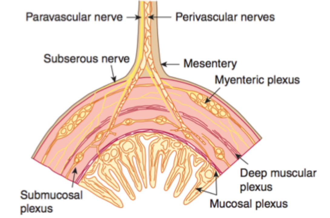
the enteric nervous system can regulate GI motility and secretion independent of the extrinsic sympathetic and parasympathetic input. its within the walls of the GI tract
consists of two neural plexuses that extend from the esophagus to the anus
these are:
1 the myenteric plexus (Auerbach's plexus) which lies between the longitudinal and circular smooth muscle layers of the muscularis externa
2 submucosal (Meissner's) plexus, found in the submucosa
Myenteric
myenteric ganglia exert control primarily over digestive tract motility
the myenteric plexus forms a continuous network that extends from the upper esophagus to the internal anal sphincter
Submucisal
the submucosal plexus helps to control local intestinal secretion, absorption, and contraction of the submucosal muscle
submucosal ganglia and connecting fiber bundles form plexuses in the small and large intestines, but not in the stomach and esophagus
neurons in the two plexuses are linked by interganglionic strands
the enteric nervous system includes:
1 intrinsic primary afferent neurons (IPANs) or sensory neurons which receive information from the sensory receptors in the mucosa and muscle,
2 motor neurons which control gastrointestinal motility, secretion and absorption, and come in three main types:
muscle motor neurons
secretomotor neurons
vasodilator neurons
3 interneurons which primarily convey signals from the IPANs to the motor neurons and also synapse with other interneurons
afferent fibers from sensory nerve endings in the epithelium are received by both plexuses, which cause local reflexes within the gut
both plexuses receive CNS output
pathways from the CNS reach the ENS and gastrointestinal effector tissues through vagal, sympathetic and pelvic pathways
Extrinsic nervous system
Both plexuses receive CNS output from the parasympathetic and sympathetic system
parasympathetic innervation to the gut is via the vagus and pelvic nerves
the vagus nerve innervates the esophagus, stomach, gallbladder, pancreas, small intestine and first part of the large intestine, cecum, and the proximal part of the colon
the pelvic nerves innervate the distal part of the colon and the anorectal region, in addition to the other pelvic organs that are not part of the GI tract
parasympathetic stimulation increase motor and secretory functions of GIT and relaxation of GIT sphincters
the preganglionic sympathetic fibers to the ENS originate in the spinal segments between T5 and L2
they synapse with postganglionic neurons at the prevertebral (celiac, superior, and inferior mesenteric) ganglia
fibers spread from these ganglia and terminate on the neurons in all portions of the ENS
some sympathetic fibers directly innervate blood vessels of the GI tract, and glandular structures in the wall of the gut
sympathetic stimulation causes inhibition of gastrointestinal secretion, motor activity, and sphincter and blood vessel contraction
afferent fibers from the ENS run through afferent vagal and sympathetic (spinal) nerves transmitting to CNS
these transmit information such as the nature of the luminal contents, such as acidity, nutrient content, and osmolality of the luminal contents, as well as the degree of stretch or contraction in smooth muscle
afferent innervation is also responsible for transmitting painful stimuli to the CNS
neurons also project from the ENS to prevertebral ganglia, the gallbladder, pancreas and trachea