Epithelial Cell Specializations Notes
Epithelial Cell Surface Domains
Different cell surfaces are in contact with differing extracellular environments.
Apical surface: abuts the lumen.
Lateral surfaces: abut other epithelial cells.
Basal surface: abuts the basement membrane and underlying connective tissue. Functions are linked to structural features.
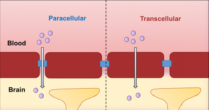
Pathways Across Epithelia
The pathway taken is influenced by the membrane domain (proteins and channels present) and the presence/absence of surface specializations.
Paracellular pathway: between cells.
Transcellular pathway: through the cell cytoplasm.
Apical Surface Specializations
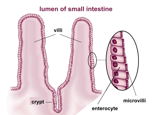
Microvilli
Increase surface area for absorption.
Approximately long.
Finger-like protrusions on the apical surface.
Contain a central dense core of actin filaments.
Increase the number of enzymes and carrier proteins to maximize absorption.
Cilia
Motile processes on the cell surface, covered with a cell membrane; approximately in length.
Beat in unison to create a unidirectional current along the cell surface.
Contain a core of microtubular structures (axoneme) anchored to a basal body (similar to a centriole).
Examples are found in the trachea, bronchi, and oviducts.
Work with mucus-producing goblet cells to move contaminants away from the lungs in the respiratory tract.

Stereocilia
Similar in structure to microvilli (dense actin core) but longer and branched.
Involved in bulk absorption and sensory functions. Sensory functions come from having mechanosensitive properties that allow them to detect and respond to changes in the environment, such as fluid movement and sound vibrations.

Specializations of Lateral Surfaces
Lateral surfaces are the surfaces between adjacent epithelial cells.
Specializations span the space between cells, forming intercellular junctions.
Important for barrier integrity and coordinated cellular activity.
Lateral Surface Specializations = Intercellular Junctions
Tight Junctions
Define cell polarity and control the passage of substances between adjacent cells.
Have a belt-like distribution, like a ribbon internally bracing the cells, and are associated with actin filaments.
Seal the intercellular space.
Adjacent cell membranes fuse together.
Block intercellular passage.
Structure
Transmembrane proteins:
Junctional adhesion molecule (JAM)
Occludin
Claudin
ZO-associated proteins (ZO-1, ZO-2) in the cell cytoplasm interact with actin filaments of the cytoskeleton.
ZO = zonula occludens (continuous band of tight junctions around a cell).

Role
Create a barrier between two environments.
Prevent the destruction of the gut lining by digestive secretions in the lumen.
Substances outside cells cannot diffuse between cells past tight junctions.
Apical membrane proteins cannot migrate past tight junctions to the lateral cell surface.
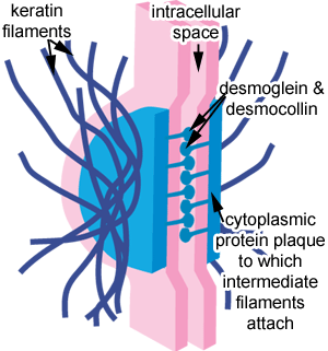
Desmosome
Protein fibers hold adjacent cells together, but the intercellular space is not sealed.
Intermediate filaments reinforce the cytoplasmic surface of the membrane.
Structure
Localized spot-like junction.
Attachment plaque on the cytoplasmic side of the cell membrane:
Desmoplakins
Plakoglobins
Anchorage site for intermediate filaments (keratin).
Transmembrane proteins are cadherins (desmoglein and desmocollin.
Wide intercellular space between adjacent cell membranes.
Functions
Provide strong attachment that holds adjacent cells together.
Numerous in epithelia subjected to abrasion and tearing stresses, e.g., epidermis, oral cavity, esophagus.
Aka macula adherens, spot desmosomes
Adherens junctions
Provide additional structural support and maintain tissue integrity by linking the actin cytoskeleton of adjacent cells.
Structure similar to desmosome, except adherens junctions involve cadherin proteins that connect to the actin filaments, whereas desmosomes connect via intermediate filaments, thus supporting tissue architecture more dynamically.
Also instead of attachment plaque, it has linking proteins such as catenins that aid in the regulation of cellular signaling and can influence cellular behavior.
Aka zonula adherens, belt desmosome

Gap Junction
Aligned channel protein pores allow the passage of small molecules from the cytoplasm of cell A to cell B.
Allows a sheet of epithelial cells to function in unison.
Allows cell-to-cell communication.
Ionic coupling.
Composition
Composed of many transmembrane channels (connexons) in close array. One connexon is made of six proteins called connexins, which form a channel that connects the cytoplasm of adjacent cells, allowing for the passage of ions and small molecules.
Connexons in adjacent cells pair up.

Appearance
Connexons of 2 cell membranes align & bridge extracellular space
Hydrophilic channels (each wide, creating a gap)
Connexons open based on voltage (electrical potential)
Junctional Complexes
Definite order of junction types from the apical towards the basal aspect of cells.
Extensive strands of tight junctions (zonula occludens) and modified desmosomes/adherent junctions (zonula adherens) form encircling bands around the apical ends of cells.
Proteins of junctions (plaques) are anchored to the cell’s cytoskeleton.
Deeper are desmosomes and gap junctions. (i.e. adherens more apical than desmosomes)
Functional Implications
Bands of tight junctions ensure:
All passage across the epithelium must occur through the cytoplasm of cells (selectivity of passage).
No intercellular seepage (seals are also protective with respect to the external environment).
Ions can be transported against a concentration gradient.
Membrane proteins remain localized to the correct domain.
Zonula adherens:
Enable adhesion of cellular sheets to maintain barrier integrity.
Attach to actin filaments that can change cell shape and bring about the contour of the epithelial surface.
Other Functions
Desmosomes:
Attach to intermediate filaments of the cytoskeleton to provide cytoskeletal support and tissue integrity.
Gap junctions (communicating junctions):
Allow direct passage of small molecules from one cell to the next = cell-to-cell transport.
Chemical and electrical coupling of cellular activities leads to syncytium.
Note: junctions are not confined to epithelial cells.
Specializations of Basal Surfaces
Hemidesmosomes
Responsible for cell-matrix adhesion at the basal surfaces of epithelia.
Very important in providing immobility to gingival epithelium and enabling its strong attachment to underlying connective tissue.
Structure
Similar to half a desmosome.
Plaques (plectins and bp230) attach to intermediate filaments (keratins) in the cytoplasm.
Link to transmembrane proteins that are integrins and heterdimers.
Integrins bind to extracellular laminin molecules of the basal lamina.

1. Epithelial Cell Surface Domains
Description: Specialized regions (apical, lateral, basal) enabling specific functions.
Apical Surface:
Specializations: microvilli, cilia, stereocilia.
Functions: absorption, creating currents, bulk absorption, sensory functions.
Lateral Surfaces:
Intercellular Junctions: tight junctions, desmosomes, gap junctions.
Functions: barrier integrity, coordinated cellular activity.
Basal Surface:
Hemidesmosomes: mediate cell-matrix adhesion.
Importance: strong attachment, e.g., gingival epithelium.
2. Structure-Function Correlation
Structural Features: Directly correlate with tissue function.
Microvilli in the small intestine: increase absorptive surface.
Cilia in the trachea: clear contaminants with goblet cells.
3. Dynamic Tissue Response
Epithelial Cells: Dynamic, can renew and respond to changes.
Metaplasia: cell type transforms to withstand changing conditions.
Adaptation: Maintains tissue integrity and function in response to environmental demands.