L6: Extracellular Appendages & Biofilms Notes
Extracellular Appendages & Biofilms
Overview
Definition: Group of organisms that adhere to surfaces and each other, forming a matrix-enclosed structure.
Components: Proteins, DNA, extracellular polymeric substances (EPS), cell wall material, organelles.
Types of Biofilm Organisms
Bacillus subtilis
Lineages are mathematically tractable (Haseloff group, Cambridge).
Growth can manifest in a 'rope' outreach mode.
Vibrio fischeri
Notable for its wrinkled surface morphology (Visick group, Loyola, Chicago).
Halorubrum lacusprofundi
Antarctic archaea.
Cellulophaga lytica
Notable for size comparisons in microscopy (e.g., 2 μm, 0.5 μm).
Biofilm Formation
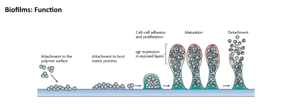
The stages of Biofilm formation are as follows:
Initial Attachment: Bacteria adhere to a surface through weak van der Waals forces and other interactions.
Irreversible Attachment: After a period, the attachment becomes more permanent through the production of polysaccharides and other biofilm matrix components.
Maturation: The biofilm develops distinctive structures, forming microcolonies and channels which aid in nutrient flow.
Dispersion: Cells detach from the biofilm to colonize new surfaces, ensuring the continuation of the biofilm lifecycle.
Characterized by:
Adhesion/Attachment : ability to stick to surfaces through molecular interactions.
Protection: enhanced resistance to environmental stresses, including antibiotics.
Dormancy: can enter a dormant state, contributing to persistence.
Mixed biofilms foster growth and horizontal gene transfer (HGT).
Biofilm Formation and Disease
Biofilm-related diseases include:
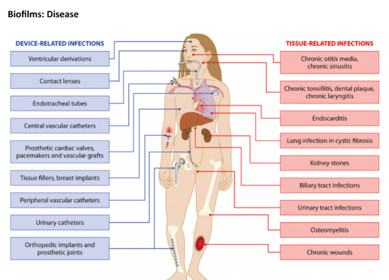
Device-related infections: artificial devices such a implants.
Tissue-related infections: e.g., chronic sinusitis, endocarditis, chronic wounds.
Pathogens like Staphylococcus epidermidis are known for their biofilm-forming ability, causing issues in prosthetic devices (e.g., endocarditis, kidney stones).
Extracellular Appendages
Pili and Fimbriae
Pili/Fimbriae: hair like appendages on surface of cells for attachment and motility.
Fimbriae = means adhesive with tips that almost always have another protein
Pili are antigenic. They are also fragile and constantly replaced, sometimes with pili of different composition, resulting in altered antigenicity. Specific host responses to old pili structures are not effective on the new structure.
Types:
Type I: Adhesive pili, common in many Gram-negative bacteria.
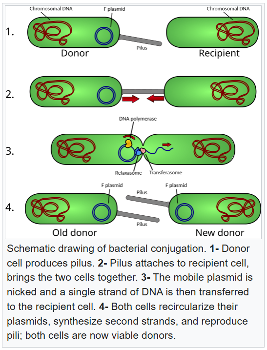
Type II: Involved in DNA transfer aka Conjugative Pili: These pili facilitate the horizontal gene transfer between bacteria, promoting genetic diversity and adaptability. They are tunnel like.
Conjugative pili allow for the transfer of DNA between bacteria, in the process of bacterial conjugation. They are sometimes called "sex pili", in analogy to sexual reproduction, because they allow for the exchange of genes via the formation of "mating pairs". Perhaps the most well-studied is the F-pilus of Escherichia coli, encoded by the F sex factor.
Type IV: Involved in twitching motility (extension, tethering, retraction)
Structure: Composed of pilin monomers, can vary greatly in morphology and function.
They are always a sight of recognition for the host immune system. When they feel like the immune system recognise them they can change the shape of the pili.
Difference between pili and flagella =

Molecular Pathways
Chaperone-Usher Pathway:
This pathway is responsible for the assembly and secretion of pili, utilizing chaperone proteins to stabilize the structure during transit and usher proteins to facilitate their incorporation into the growing pilus.
The Chaperone-Usher Pathway is utilized by Gram-negative bacteria for protein export, relying on sec translocase to cross the inner membrane (IM) and featuring multiple terminal branches for outer membrane (OM) crossing.
The usher is an outer membrane protein
Biofilms in Pathogenicity
Major players in diseases due to their ability to resist antibiotics and evade the immune system.
Notable articles discuss the role of Geobacter pili in extracellular electron transfer.
Functional Amyloids
Curli (from Enterobacteriaceae): insoluble, fibrous protein aggregates that contribute to adhesion/aggregation in biofilms.
Gram-positive organisms like Bacillus subtilis form amyloid fibers that aid in structural integrity of biofilms.
Anti-biofilm Strategies
Dispersal promoting agents: Enzymes, chelators, D-amino acids.
Killing persister cells: Combination therapies (e.g., sugar and antibiotics).
Surface modification: Non-antibiotic coatings to inhibit adhesion.
Quorum-sensing inhibitors (e.g., honey, which contains natural antibacterial properties).
C-di-GMP Signaling in Biofilm Regulation
Cyclic-di-GMP serves as a secondary messenger, influencing bacterial adhesion, motility, and biofilm formation.
High levels of C-di-GMP often lead to a sessile lifestyle (biofilm) instead of a planktonic lifestyle.
Conclusion
Understanding biofilm structures and their functions is critical for developing strategies against biofilm-associated diseases. This change in perspective towards biofilms highlights their ecological and clinical importance, as well as their role in microbial pathogenicity and resilience.