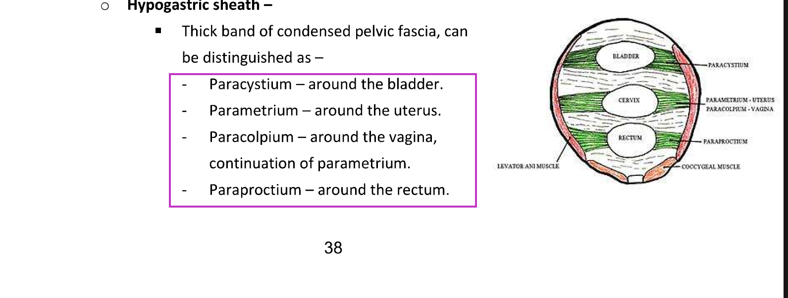
HYPOGASTRIC SHEATH
1. Hypogastric Sheath
The hypogastric sheath is a thick band of condensed pelvic fascia that runs on the lateral pelvic wall.
Its main functions:
Acts as a support framework for pelvic organs
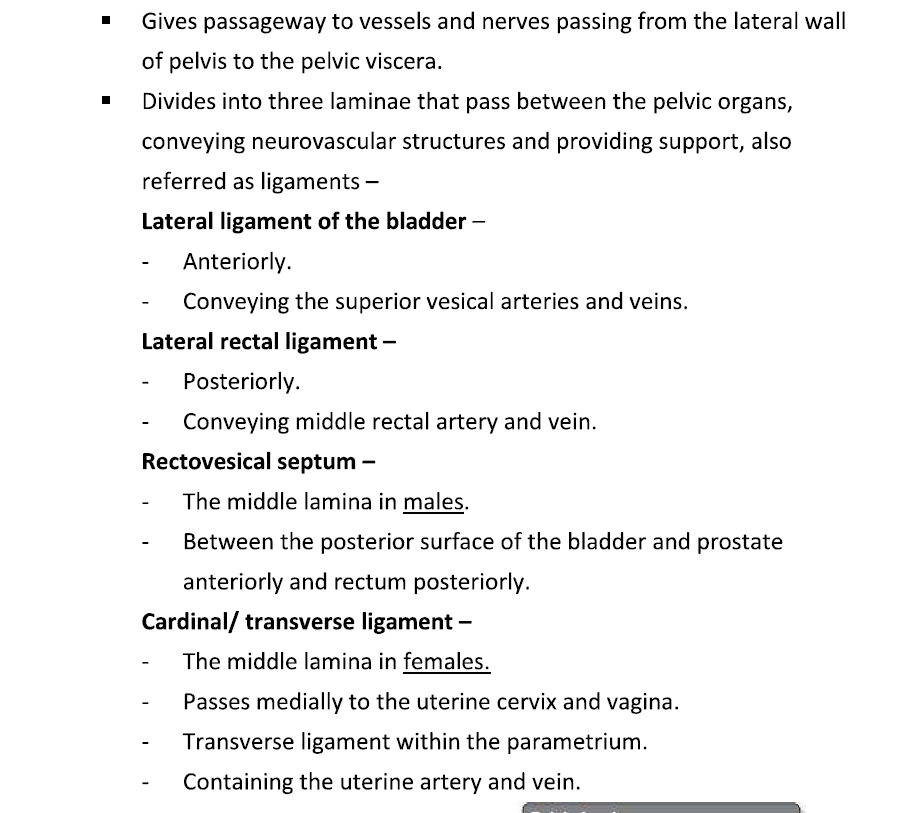
Provides pathways for vessels and nerves from the lateral wall of the pelvis to the pelvic viscera
2. Laminae of the Hypogastric Sheath
The hypogastric sheath splits into laminae, which surround different pelvic organs and are named accordingly:
Lamina | Surrounds | Notes / Continuation |
|---|---|---|
Paracystium | Bladder | Supports bladder; carries superior vesical vessels |
Parametrium | Uterus | Lateral connective tissue around cervix & uterus; carries uterine vessels, nerves, lymphatics; forms cardinal ligament |
Paracolpium | Vagina | Continuation of parametrium; supports vagina; contains vessels & nerves |
Paraproctium | Rectum | Supports rectum; carries middle rectal vessels |
Visual cue: Think of a “laminar sandwich” — bladder anteriorly, uterus in the middle, rectum posteriorly, with the fascia forming a sheath connecting all of them laterally to the pelvic wall.
3. Ligaments Derived from These Laminae
These laminae condense to form the named ligaments:
Ligament | Lamina / Organ | Contents |
|---|---|---|
Lateral ligament of bladder | Paracystium | Superior vesical arteries & veins |
Cardinal / transverse cervical ligament | Parametrium (middle lamina in females) | Uterine artery & vein; nerves; supports cervix and upper vagina |
Rectovesical septum | Middle lamina in males | Between posterior bladder/prostate & rectum |
Lateral rectal ligament | Paraproctium | Middle rectal artery & vein; supports rectum |
Key idea: The dense connective tissue of the parametrium, paracolpium, and paraproctium forms the main ligaments supporting pelvic organs and acts as conduits for vessels and nerves.
4. How to visualize
Imagine looking at a cross-section of the pelvis:
Anterior: Bladder → paracystium → lateral ligament of bladder
Middle: Uterus → parametrium → cardinal ligament → paracolpium (vagina)
Posterior: Rectum → paraproctium → lateral rectal ligament
All these structures connect to the lateral pelvic wall, forming a hypogastric sheath that acts like a suspension system for pelvic organs.
Summary in simple terms
The hypogastric sheath is condensed pelvic fascia that splits into laminae surrounding the bladder, uterus, vagina, and rectum. These laminae (paracystium, parametrium, paracolpium, paraproctium) condense into ligaments like the lateral ligament of bladder, cardinal ligament, and lateral rectal ligament, carrying vessels, nerves, and lymphatics and providing support to pelvic organs.