Muscle Tissue
Muscle Tissue Overview
Definition: Muscle tissue in animals permits active movement of the body or transport of materials within the body.
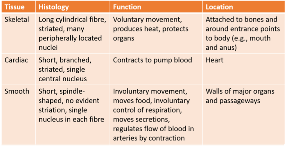
Types of Muscle Tissue: There are three main types:
Skeletal Muscle: Responsible for voluntary movements, attached to bones, and constitutes about 40% of body mass.
Cardiac Muscle: Found in the heart, involuntary control responsible for pumping blood.
Smooth Muscle: Involuntary control, involved in movements like digestion and responses to external stimuli.
Common Properties of Muscle Tissue
Excitability: All muscle types can change electrical states (polarized to depolarized) and propagate action potentials along membranes.
Contractility: Ability to shorten and exert force.
Extensibility: Can be stretched.
Elasticity: Ability to return to original length after contraction.
Muscle Contraction Mechanism
Initiation: Requires an increase in calcium ions inside muscle cells.
Process: Contraction begins with actin being pulled by myosin:
In skeletal and cardiac muscle, calcium enables binding sites on actin by interacting with proteins covering these sites.
In smooth muscle, calcium activates enzymes to trigger contractions.
Energy Requirement: All muscles require ATP for sustained contraction, and relaxation occurs when calcium levels drop.
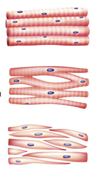
Structural Differences Among Muscle Types
Skeletal Muscle
Cells (Myocytes): Multinucleated cells formed by fusion of myoblasts.
Striations: Regular striped pattern visible under a microscope.
Cardiac Muscle
Cells (Cardiomyocytes): Single nucleus, striated appearance, and contract autonomously.
Intercalated Discs: Connect cells with anchoring and gap junctions for synchronized contraction.
Smooth Muscle
Cells: Spindle-shaped, single nucleus, and non-striated (no visible stripes).
Functionality: Responsible for slow, sustained contractions in various organs (e.g., blood vessels and digestive tract).
Key Functions of Skeletal Muscle
Movement: Major role in moving body and resisting gravity for posture.
Joint Stability: Prevents injuries by stabilizing joints.
Internal Functions: Assists with controlling openings (e.g., swallowing, urination).
Protection: Shields internal organs from damage.
Homeostasis: Generates heat during contraction.
Components of Skeletal Muscle
Organ Structure: Composed of muscle fibers, blood vessels, nerve fibers, and connective tissue.
Connective Tissue Sheaths:
Epimysium: Outer layer, provides structural integrity and separates muscle from other tissues.
Perimysium: Surrounds bundles of muscle fibers (fascicles).
Endomysium: Encases individual muscle fibers, containing nutrients supplied by blood.
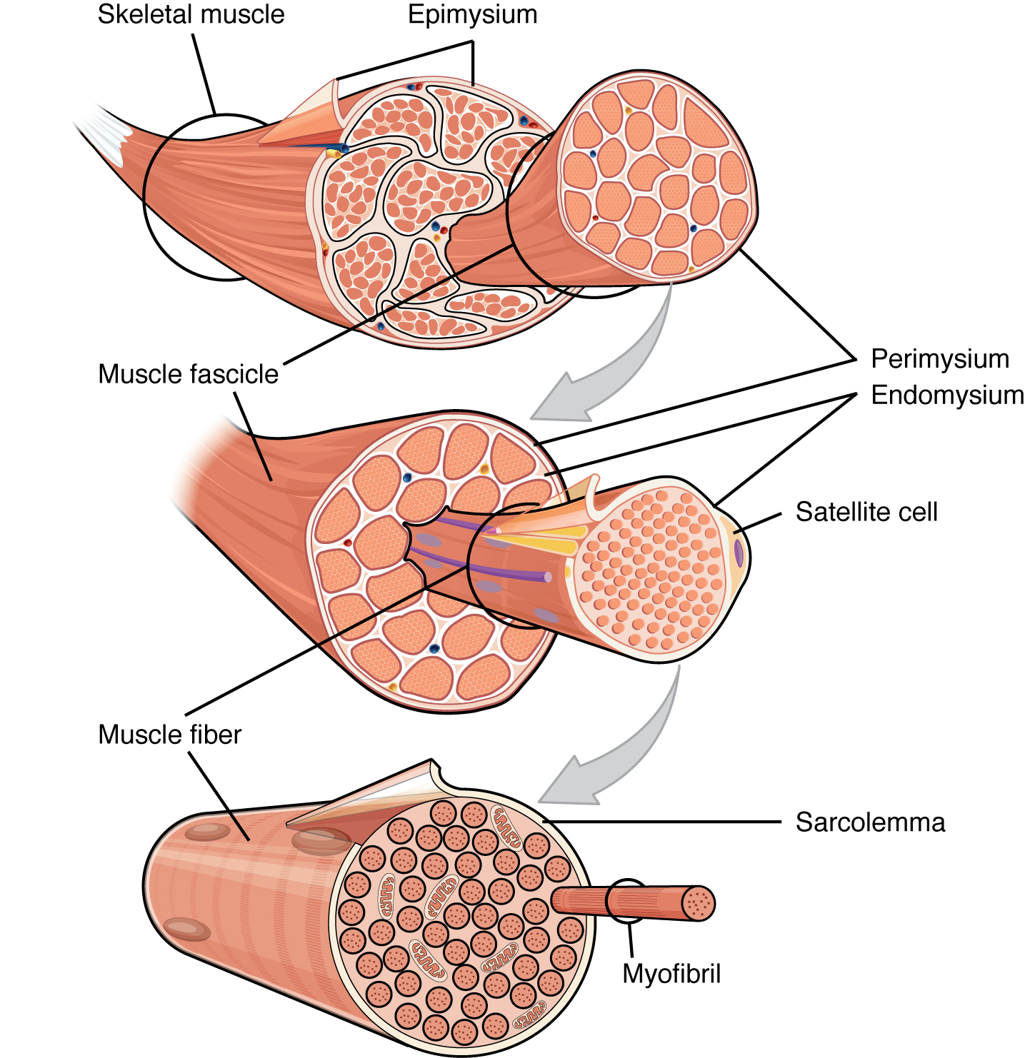
Largest to smallest
Epimysium → in epimysium are muscle fasicles → in the fascicle, muscle fibers are grouped together, each encased by endomysium for optimal function and support → then myofibrils encased by plasma (sarcolemma) - Sar
Connective Tissue Attachments
Skeletal muscles work with tendons to pull on bones.
Collagen in the three tissue layers (mysia) intertwines with tendon collagen.
Tendon fuses with the periosteum, the tissue coating the bones.
Muscle fiber contraction creates tension, which is transferred through mysia to tendon, then to periosteum, producing movement.
Mysia may also fuse with aponeurosis or fascia, which are broad tendon-like sheets.
Example: Latissimus dorsi muscles fuse into a broad sheet of connective tissue in the lower back.
Neuromuscular Junction
Function: Site where motor neuron meets muscle fiber, leading to action potential generation.
Acetylcholine (ACh) is released from the motor neuron, causing sodium ions to enter the muscle fiber, depolarizing the membrane.
Sets off a sequence of events for muscle contraction through excitation-contraction coupling.
Excitation-Contraction Coupling
Action Potential: Initiates contraction; spreads via T-tubules, triggering calcium release from the sarcoplasmic reticulum.
Muscle Fiber Shortening: Calcium binds to troponin, moving tropomyosin and allowing myosin to pull actin filaments, contracting the muscle.
Sliding Filament Model
Process Description: Thick (myosin) and thin (actin) filaments interact, leading to contraction toward the sarcomere center.
Cross-Bridge Cycle: Myosin heads attach and pull actin filaments; ATP is required for detachment and resetting.
ATP's Role in Contraction
Energy Source: Provides the energy necessary for muscle contraction and relaxation.
Myosin Head Resetting: ATP binding allows detachment of myosin from actin, and hydrolysis re-cocks the myosin head for further contraction.
Muscle contraction steps:
Acetylcholine is released from the axon terminal and binds to receptors in the motor end plate.
Action potential travels along the sarcolemma and into transverse tubules.
Sarcoplasmic reticulum releases calcium.
Calcium ions combine with troponin, uncovering myosin-binding sites.
Energized myosin head (cross-bridges) attach to actin.
Thin filaments slide toward the center of the sarcomere.