Transport across the cell membrane
Properties of the membrane transport
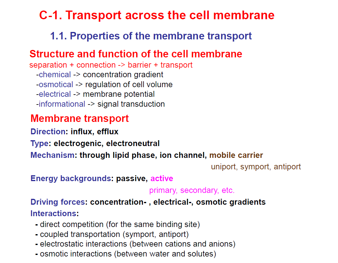
🔹 Structure and function of the cell membrane
The cell membrane isn’t just a barrier – it also enables selective transport of molecules and ions.
It has four main functions based on gradients:
Chemical → maintains concentration gradients (e.g., higher Na⁺ outside, higher K⁺ inside).
Osmotical → regulates cell volume by controlling water and solute balance.
Electrical → maintains the membrane potential (charge difference across the membrane).
Informational → involved in signal transduction (communication between cells).
So the cell membrane is both a barrier and a connection.
🔹 Membrane transport
Direction:
Influx = movement into the cell
Efflux = movement out of the cell
Type:
Electrogenic → changes membrane potential (charge moves unevenly)
Electroneutral → no net change in charge
Mechanism (how substances cross):
Through lipid phase (simple diffusion – small, nonpolar molecules like O₂, CO₂)
Ion channel (specific pore proteins for ions)
Mobile carrier (proteins that bind and shuttle molecules across)
➝ Can be uniport (one substance), symport (two substances same direction), or antiport (two substances in opposite directions).
🔹 Energy backgrounds
Passive transport → no energy needed (down a gradient).
Active transport → requires energy (ATP or other).
Primary active transport → directly uses ATP (e.g., Na⁺/K⁺ pump).
Secondary active transport → uses energy stored in gradients created by primary transport.
🔹 Driving forces
Transport is driven by different gradients:
Concentration gradient (chemical difference)
Electrical gradient (charge difference)
Osmotic gradient (water movement driven by solute concentration differences)
🔹 Interactions in transport
Direct competition → molecules compete for the same transporter.
Coupled transport → one molecule’s movement drives another’s (symport, antiport).
Electrostatic interactions → attraction/repulsion between charged particles (cations/anions).
Osmotic interactions → water follows solute movement.
Diffusion through the lipid phase
Simple Diffusion: This is the passive transport of small, neutral, hydrophobic, lipid-soluble substances across the membrane (lipid phase) down an electrochemical gradient.
This does not require any metabolic energy; thus, it is a passive transport.
Example of substances that pass through the membrane by simple diffusion includes: O2, CO2, N2.
Saturation: cannot be saturated
Calculated by: Fick’s law. SEE BELOW:
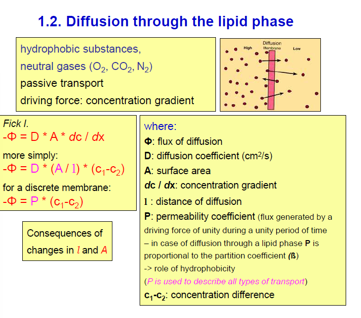
Take note:
C1-C2 = is the concentration difference allowing the diffusion.
P = the permeability coefficient - this describes the permeability of a solute through the membrane, which depends on the characteristics of the solute and the membrane.
Transport mediated by carrier proteins
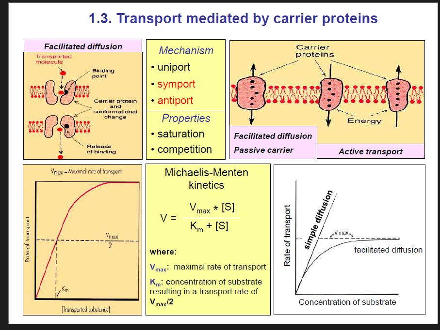
Transport that is mediated by carriers includes:
Facilitated diffusion,
primary active transport and
secondary active transport.
CARRIER FACILITATED DIFFUSION:
Passive: does not require metabolic energy and occurs down an electrochemical gradient.
Example: Glucose uses a carrier called GLUT to enter the cell.
Characteristics of carrier-mediated transport:
Saturation: Carriers can be saturated, so the transport rate will increase until they become saturated, then it will stop at this rate, which is called the transport maximum.
Michaelis-Menten Kinetics: This can be used to describe the transport via carriers, representing the capacity of the carriers and their sensitivity.
Vmax- maximal rate of transport
Km- Concentration of the substrate at half the Vmax. SEE DIAGRAM ABOVE FOR EQUATION.
Take note: A molecule with a high Km has a lower affinity and vice versa.
Competition
Difference between carrier-mediated transport and simple diffusion:
1. At low solute concentrations
Carrier-mediated transport is much faster than simple diffusion.
Because the transporter has specific binding sites and can “grab” solutes efficiently, even when there aren’t many around.
This gives it an initial steep rate of transport.
Simple diffusion through the lipid bilayer is relatively slow at low solute concentrations, since it depends only on how much solute is present and its lipid solubility.
2. As solute concentration increases
Carrier-mediated transport becomes saturated:
All binding sites get occupied.
Transport rate reaches a maximum (Vmax).
This “plateau” is characteristic of carriers.
Simple diffusion does not saturate:
The more solute you add, the faster diffusion occurs (linear increase).
No plateau, because there are no binding sites.
Competition between solutes
Competition: structurally related solutes compete for the transport sites.
If two solutes can bind to the same carrier (e.g., glucose and galactose to SGLT), they will compete for the binding site.
Which solute gets transported depends on:
Affinity → how tightly the carrier binds to the solute.
Concentration of solute → more molecules increase the chance of binding. Thus, increasing the concentration of the substrate would increase its chance of binding,
A solute with a higher affinity or higher concentration will outcompete the other.
Direction of movement in carrier-mediated transport:
Uniport: one substance in one direction
Symport: 2 substances in the same direction.
Antiport: 2 substances in opposite directions.
ACTIVE TRANSPORT:
Active transport is when the cell membrane moves ions against a concentration gradient.
Active transport is divided into two types:
Primary active transport.
Secondary active transport.
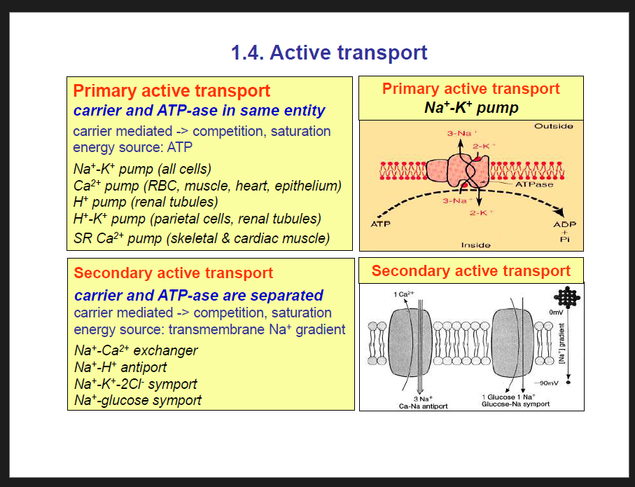
PRIMARY ACTIVE TRANSPORT:
In primary transport, the energy is derived directly from the breakdown of Adenosine triphosphate (ATP) or of some other high-energy phosphate compound.
Primary active transport is carrier-mediated.
Active: requires the use of metabolic energy and occurs against the electrochemical gradient.
ATPase: can break ATP molecules and use their energy store.
Examples:
Na+/K+ pump- maintains low intracellular Na+ and high K+ since it pumps 3 Na+ out and 2 K+ in. It is found in all cells.
H+/K+ pump- helps to maintain the acidity of the stomach. This pump is found in parietal cells; renal tubules.
Ca2+ pump- Found in RBCs, muscle, heart, and epithelium.
Sarcoplasmic reticulum Ca2+ pump- found in skeletal and cardiac muscle.
IMPORTANT NOTE: The carrier and the ATPase are in the same place.
SECONDARY ACTIVE TRANSPORT
In secondary active transport, the energy is derived secondarily from energy that has been stored in the form of ionic concentration differences of secondary molecular or ionic substances between the two sides of a cell membrane, created originally by primary active transport.
IMPORTANT NOTE: The carrier and the ATPase are separated.
Active indirectly: the energy required for this transport is indirectly provided by the Na+ gradient that is maintained by the primary active transport. Without the primary active transport, the secondary active transport will be inhibited.
Coupled- one solute moves down the concentration gradient (Na mostly) while the other is moved against the concentration gradient.
Energy source: Transmembrane Na+ gradient.
Examples:
Na+/Ca2+ exchanger
Na+/H+ antiport
Na+-K+-2Cl- symport
Na+- glucose symport
Na+/amino acid symport
Na+/K+ Pump
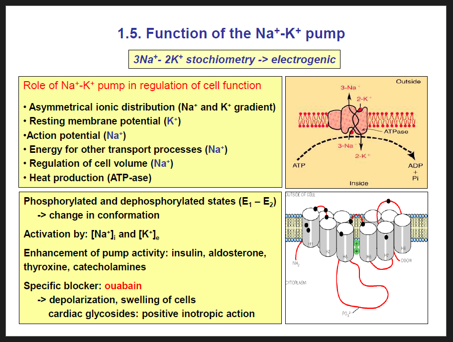
Characteristics of the Na+/K+pump:
Activation: This pump is activated due to high Na+ inside the cell and high K+ outside the cell.
So 3 Na+ ions are pumped out of the cell while 2 K+ ions are pumped into the cell, thus it is electrogenic.
Enhanced by: insulin, aldosterone, thyroxine and catecholamine.
Blocked by: Ouabain. Ouabain is excreted by the kidney.
Also, digitoxin and digoxin are other toxins that block the Na+/K+ pump.
What happens when the Na+/K+ pump is blocked?
When the Na+/K+ pump is blocked, Na+ accumulates inside the cell because Na+ cannot be pumped out of the cell.
Then, chloride ions follow as well as water, thus causing cell swelling
Another consequence of the blockage of the Na+/K+ pump is depolarisation
Function of Na+/K+ pump:
Assymetrical ionic distrubution (Na+ and K+ gradient)- Uneven ionic distribution of Na+ and K+ allows for the following:
Resting membrane potential
Secondary active transport
regulation of cell volume
Helps in maintaining the Resting membrane potential
Action potential
Energy for other transport processes
Helps in the regulation of cell volume- when Na+ ions move into the cell or outside the cell, water also enters by osmosis, thus affecting the volume of the cell, i.e. the cell volume either increases or decreases. For instance:
If too much Na⁺ stays inside the cell, water will flow in → the cell swells and could burst.
If Na⁺ is pumped out, water follows → preventing swelling and keeping cell volume stable.
Heat production (ATPase) - The breakdown of ATP causes heat generation. Examples of hormones that increase heat production by enhancing Na+/K+ pump include: Thyroxine and catecholamines.
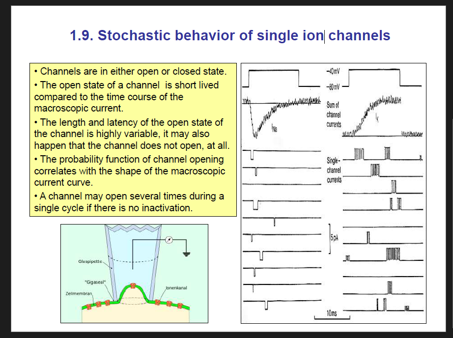
ION CHANNELS
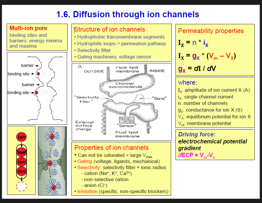
Ion channels
Structure of ion channels:
Ion channels are made up of 4 transmembrane domains.
Each of the transmembrane domains have 6 segments.
Each segment has its various functions such as acting as a selective filter, gating, etc.
Properties of Ion channels:
They cannot be saturated.
Gating:
Voltage gated ion channel: change in membrane potential causes this ion channel to open or close
Ligand gated ion channel: Open or close when a specific chemical (ligand) binds to the channel.
Stretch gated ion channel: Open in response to mechanical deformation of the cell membrane (stretch, pressure, vibration).
Examples:
Mechanoreceptors in skin (touch, pressure).
Hair cells in the inner ear (sound and balance).
Baroreceptors (blood pressure sensing).
Selectivity:
Inhibition: specific and non-specific inhibitors
Permeability Properties
I = i x n
i.e Total current = current from one channel x total number of channels.
I = g x (Vm-Ve); where g is conductance. If there is high conductance, i.e. the channel is open, then there would be high current (passage of ions).
Why can’t ion channels get saturated?
They are like pores/holes in the membrane that open and allow ions to flow down their electrochemical gradient.
Once open, ions simply diffuse through — no binding, conformational change, or transport cycle is needed.
The rate of transport depends only on:
How many channels are open?
The electrochemical gradient.
Since ions just flow freely, there is no "maximum transport rate" due to binding sites.
So, ion channels behave like an open doorway — the more ions push, the more flow you get.
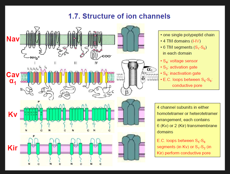
Structutre Of voltage-gated Na+ ion channel and voltage gated calcium ion channel
Each ion channel has 4 transmembrane domains, and each domain has 6 segments
And each segment has different functions:
S4 = voltage sensor
S3 = activation gate
S6 = inactivation gate
E.C.- Loops between S5-S6: conductive pore
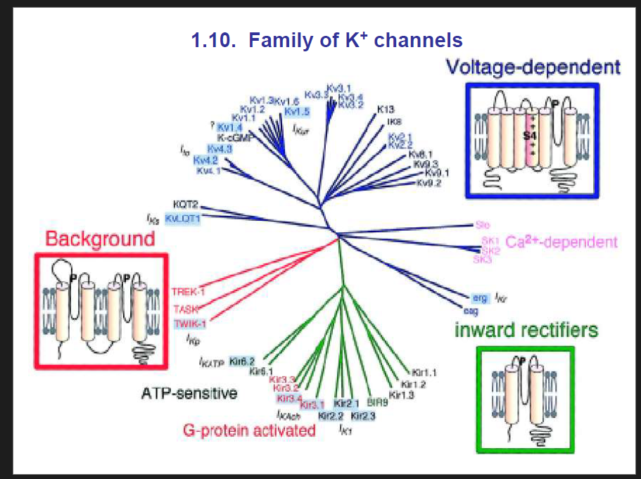
Potassium Channel (K+ channel) Structure
Subunit arrangement
K⁺ channels are formed by 4 subunits arranged as a tetramer.
Can be:
Homotetramer → all 4 subunits identical.
Heterotetramer → subunits are different.
Transmembrane domains
Each subunit has multiple transmembrane segments:
Voltage-gated K⁺ channels (Kv): 6 transmembrane segments (S1–S6).
Inward rectifier K⁺ channels (Kir): only 2 transmembrane segments (M1 & M2).
Pore-forming region
The extracellular (E.C.) loop between:
S5–S6 in Kv channels, or
M1–M2 in Kir channels
forms the selectivity filter / conductive pore.
This region is highly conserved and is responsible for K⁺ selectivity.
✅ Summary (easy recall):
4 subunits → tetramer (homo/hetero).
Kv: 6 TMs per subunit, pore between S5–S6.
Kir: 2 TMs per subunit, pore between M1–M2.
Extracellular loop = selectivity filter for K⁺.
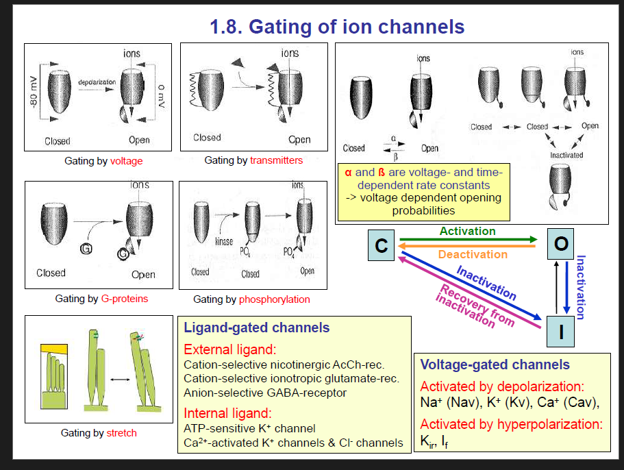
Gating by ion channels:
Gating by voltage
Gating by tranmitters
Gating by G-proteins
Gating by phosphorylation
Gating by stretch
Ligand gated channels:
External ligand
Internal ligand.
There are three states of ion channel:
Closed state: This state can be stimulated to open state
Open state:
Inactivated state: This state CANNOT be stimulated to an open state.
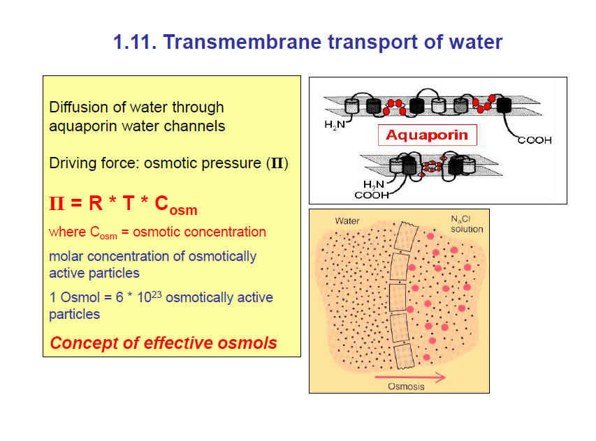
Transmembrane transport of water
This is simply osmosis.
This is the movement/ diffusion of water from the more dilute to more concentrated compartment through water channels called aquaporins.
The driving force of osmosis is osmostic pressure
π=RTCosm
where:
π = osmotic pressure
R = gas constant (0.082 L·atm·K⁻¹·mol⁻¹ or 8.314 J·K⁻¹·mol⁻¹)
T = absolute temperature in Kelvin (K)
C = osmotic concentration of the solute (mol/L)
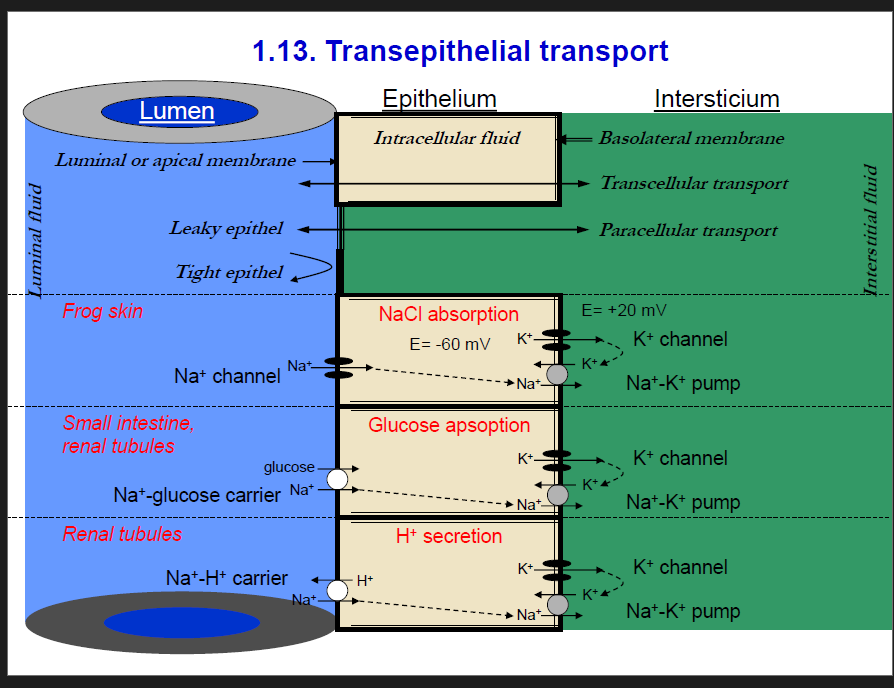
Transepithelial transport:
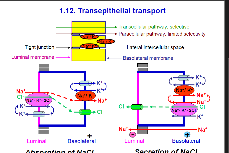
Transepithelial transport is the movement from the lumen into the interstitium or vice versa. This could be absorption or secretion.
2 types of transepithelial transport:
Transcellular transport: This is a selective transport that involves the movement through the cell
Ions move through the epithelial cell, crossing both the luminal (apical) and basolateral membranes.
This pathway is selective because transport proteins (channels, pumps, and cotransporters) control what passes.
Paracellular transport:
Ions move between cells through the tight junctions.
This is less selective, allowing small ions and water to pass depending on junction properties.
Absorption and secretion:
Cells in different tissues in our body have different functions, and that is why some of them need to absorb a certain substance while others secrete it.
Furthermore, the kidney cells can choose if they want to absorb or secrete substances depending on the state of the body.
Absorption of NaCl (left diagram)
This shows how NaCl is absorbed from the lumen → blood (basolateral side).
Na⁺-K⁺-2Cl⁻ cotransporter (NKCC, green/purple):
Located on the luminal side.
Brings Na⁺, K⁺, and 2 Cl⁻ into the cell.
Na⁺/K⁺ pump (red):
On the basolateral membrane.
Pumps Na⁺ out of the cell into the blood, while bringing K⁺ into the cell.
This keeps intracellular Na⁺ low so absorption continues.
Cl⁻ channels (green):
Move Cl⁻ into the basolateral side (blood).
K⁺ channels:
Let K⁺ leak back out to keep the cycle running.
Net effect: NaCl is absorbed into the blood, water usually follows by osmosis.
Secretion of NaCl (right diagram)
This shows how NaCl is secreted into the lumen (opposite direction).
Na⁺/K⁺ pump (red):
Maintains low intracellular Na⁺, bringing K⁺ in.
Na⁺-K⁺-2Cl⁻ cotransporter (NKCC):
Located on the basolateral side now.
Brings Na⁺, K⁺, and 2 Cl⁻ into the cell from the blood.
Cl⁻ channel (green):
On the luminal side, moves Cl⁻ into the lumen.
Na⁺ movement paracellularly:
Positive Na⁺ follows Cl⁻ through the paracellular pathway, maintaining electroneutrality.
Net effect: NaCl is secreted into the lumen, pulling water with it. This is important in places like the airways, sweat glands, and intestines.
HINT: When the Na⁺-K⁺-2Cl⁻ cotransporter is on the luminal side, water absorption occurs by when the Na⁺-K⁺-2Cl⁻ cotransporter is on the basolateral side, then secretion occurs.
Take note:
Interstitial fluid (interstitium) = the fluid that surrounds the cells in tissues, outside blood vessels but not inside cells.
It is formed when plasma leaks out of capillaries.
It bathes the cells and allows exchange of nutrients, oxygen, and waste between blood and cells.
1. General layout of the slide
Left side (blue): Lumen (e.g., gut lumen, kidney tubule, etc.)
Middle (yellow): Epithelial cell (intracellular fluid).
Right side (green): Interstitium (interstitial fluid → eventually blood).
Top labels:
Luminal/apical membrane = faces the lumen.
Basolateral membrane = faces the interstitium (blood side).
Transcellular transport = through the cell.
Paracellular transport = between cells.
Tight vs leaky epithelia:
Tight = less paracellular movement (e.g., urinary bladder).
Leaky = more paracellular movement (e.g., proximal tubule).
2. Specific examples in the diagram
(a) NaCl absorption – Frog skin
On the luminal side:
Na⁺ channel lets Na⁺ enter the cell (driven by the negative membrane potential, E = –60 mV).
On the basolateral side:
Na⁺/K⁺ pump pumps Na⁺ out into the interstitium and brings K⁺ in.
K⁺ channel allows K⁺ to leak out, maintaining balance.
Result: NaCl is absorbed into the interstitium (and eventually into blood).
(b) Glucose absorption – Small intestine & renal tubules
On the luminal side:
Na⁺-glucose carrier (SGLT) brings glucose into the cell together with Na⁺ (secondary active transport).
On the basolateral side:
Na⁺/K⁺ pump removes Na⁺ into the interstitium.
Glucose then moves into the interstitium through a glucose transporter (not shown, but implied).
Result: Glucose and Na⁺ are absorbed from the lumen into the blood.
(c) H⁺ secretion – Renal tubules
On the luminal side:
Na⁺-H⁺ exchanger moves H⁺ into the lumen in exchange for Na⁺ (important for acid secretion and pH balance in the kidney).
On the basolateral side:
Na⁺/K⁺ pump again keeps Na⁺ low inside the cell.
K⁺ channel allows K⁺ to leak back into the interstitium.
Result: H⁺ is secreted into the lumen (urine side), Na⁺ reabsorbed into interstitium.