The heart
❤ BIOM2020 – HEART: The Complete, Exam‑Ready Summary
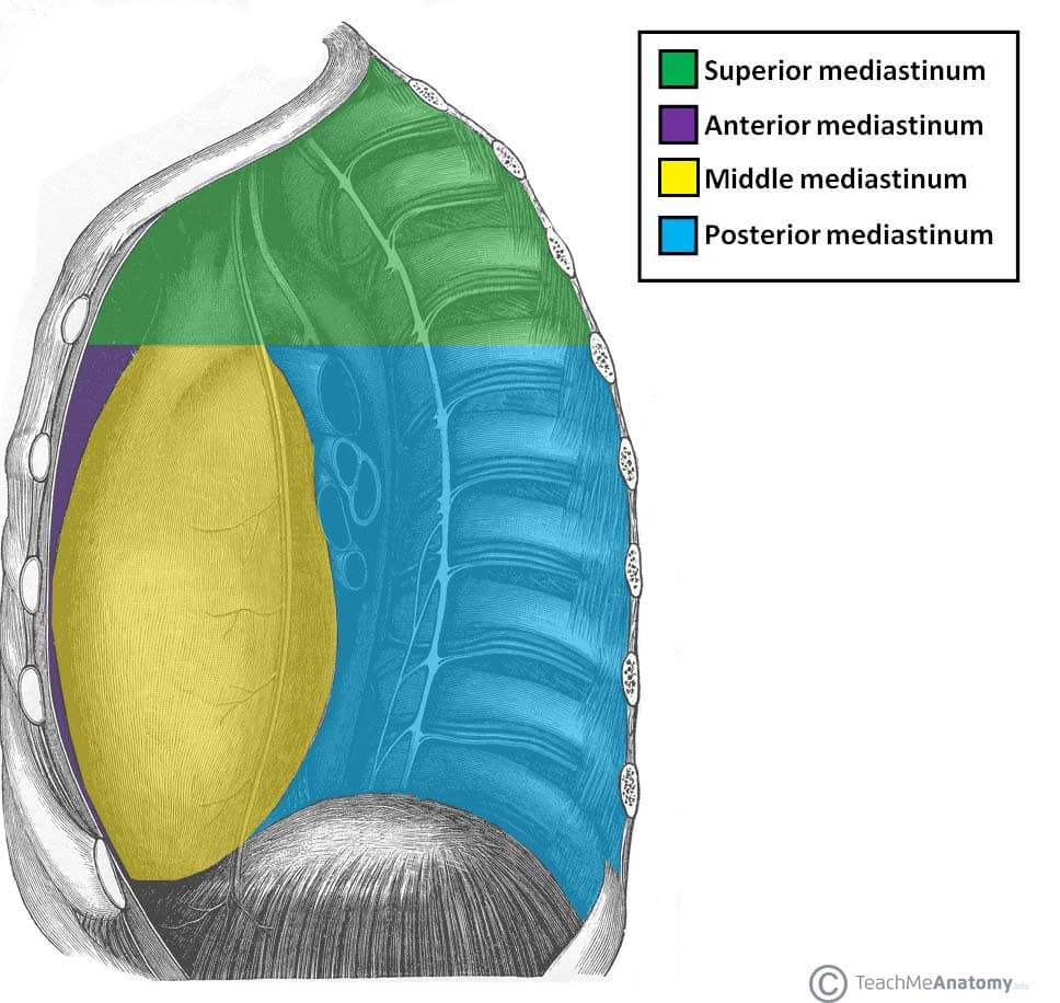
1. Location & Mediastinum
The heart sits in the middle mediastinum.
The mediastinum is divided by a plane from:
Manubriosternal joint (Angle of Louis) → T4 vertebra
Above this plane = superior mediastinum
Below = inferior mediastinum, subdivided into:
Anterior mediastinum (in front of pericardium)
Middle mediastinum (heart + pericardium)
Posterior mediastinum (behind pericardium)
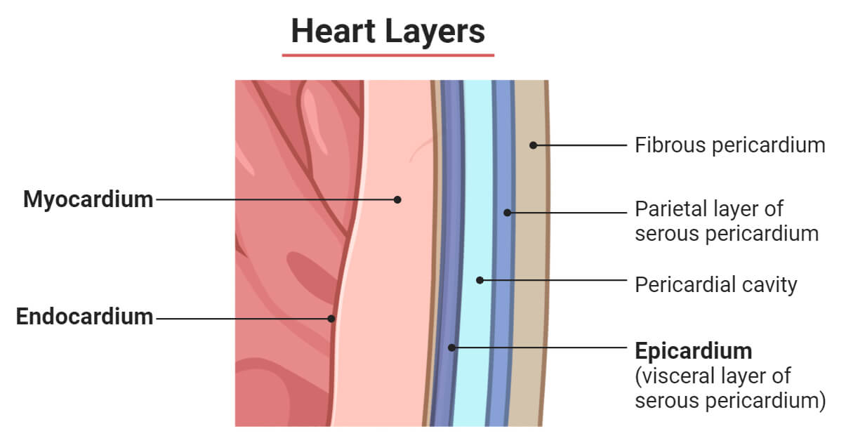
2. Pericardium
Two major layers:
A. Fibrous pericardium
Tough, collagen‑rich, non‑stretchable
Anchors heart to diaphragm & great vessels
B. Serous pericardium
Two layers:
Parietal layer – lines inside of fibrous pericardium
Visceral layer – adherent to heart surface (also called epicardium)
Between them: pericardial cavity with a thin film of fluid → reduces friction.
3. External Features of the Heart
Think of the heart as a pyramid:
Apex
Formed by left ventricle
Points inferolaterally to the left 5th intercostal space
Base
Posterior surface
Formed mainly by left atrium
Surfaces
Sternocostal surface → mostly right ventricle
Diaphragmatic surface → mostly left ventricle
4. Internal Anatomy
Atria
Right atrium
Key features:
Smooth posterior wall
Rough anterior wall with musculi pectinati
Crista terminalis – ridge separating smooth & rough regions
Openings:
SVC
IVC
Coronary sinus
Fossa ovalis – embryological remnant of foramen ovale
Left atrium
Mostly smooth
Receives 4 pulmonary veins
Small region of musculi pectinati in auricle
Ventricles
Shared features
Papillary muscles
Chordae tendineae
Trabeculae carneae
Right ventricle
Thinner wall
Pumps to low‑pressure pulmonary circulation
Outflow → pulmonary trunk via pulmonary valve
Left ventricle
Thickest wall (high systemic pressure)
Outflow → aorta via aortic valve
5. Heart Valves
Atrioventricular (AV) valves
Right AV valve = tricuspid (3 cusps)
Left AV valve = bicuspid / mitral (2 cusps)
Function:
Prevent backflow from ventricles → atria
Papillary muscles + chordae tendineae prevent cusp prolapse
Semilunar valves
Pulmonary valve
Aortic valve
Function:
Prevent backflow from arteries → ventricles
No chordae tendineae
6. Coronary Circulation
The heart receives the best oxygenated blood directly from the aorta.
Right coronary artery (RCA)
Supplies:
Right atrium
Right ventricle
SA node (in most people)
AV node (in many people)
Left coronary artery (LCA)
Branches:
Left anterior descending (LAD)
Circumflex artery
Supplies:
Left ventricle (majority)
Interventricular septum
Left atrium
Venous drainage
Great cardiac vein
Middle cardiac vein
→ drain into coronary sinus → right atrium
7. Conduction System
(Not heavily tested in BIOM2020 but know the sequence)
SA node (pacemaker)
AV node
Bundle of His
Right & left bundle branches
Purkinje fibres
8. Foetal Circulation & Adult Remnants
Foetal shunts bypass lungs & liver.
Foetal structures → Adult remnants
Foramen ovale → Fossa ovalis
Ductus arteriosus → Ligamentum arteriosum
Ductus venosus → Ligamentum venosum
Purpose:
Bypass non‑functional foetal lungs
Bypass developing liver
Closure triggered by first breath → pressure changes.
9. Clinical Notes
Angina
Caused by coronary artery narrowing
Reversible ischaemia
Myocardial infarction
Irreversible death of myocardium
Subendocardial region most vulnerable
Endocardium survives longer because it receives oxygen directly from chamber blood
Broken heart syndrome (Takotsubo cardiomyopathy)
Stress‑induced
Catecholamine surge
✔ What you should absolutely memorise for BIOM2020
Surfaces: sternocostal = RV, diaphragmatic = LV
Right atrium landmarks: crista terminalis, musculi pectinati, fossa ovalis, coronary sinus
Valve names & functions
Coronary arteries (RCA, LCA, LAD, circumflex)
Foetal → adult remnants
Differences between ventricles (wall thickness + pressure)
Pericardium layers