Thoracic Wall
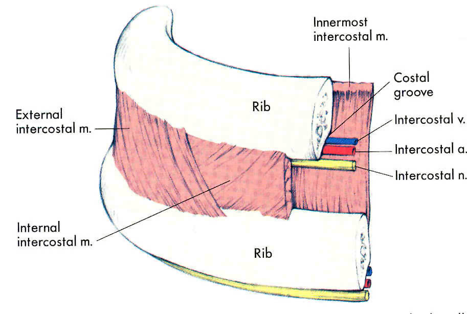
The intercostal space contains:
External intercostal muscle
Internal intercostal muscle
Innermost intercostal muscle
intercostal vessels and nerve (VAN) between 2nd and 3rd layers of muscles
External Intercostal muscle
%%Origin%%: from lower border of rib above
%%Insertion%%: upper border of rib below
%%Fibers%%: downward, forward
- Begins: posterior at the tubercle of the rib
- at costochondral junction: replaced by anterior (external) intercostal membrane
Internal Intercostal muscle
%%Origin%%: from subcostal groove of rib above
%%Insertion%%: upper border of rib below
%%Fibers%%: downward, backward
- Begins: anterior at the lateral margin of the sternum
- at angle of the ribs: replaced by posterior (internal) intercostal membrane
Innermost Intercostal muscle
%%Fibers%%: downward, backward
- ==incomplete layer==
- present only in the ^^lateral part of thoracic wall^^ (absent in anterior and posterior parts)
- ^^intercostal nerve and vessels are superficial^^ to it
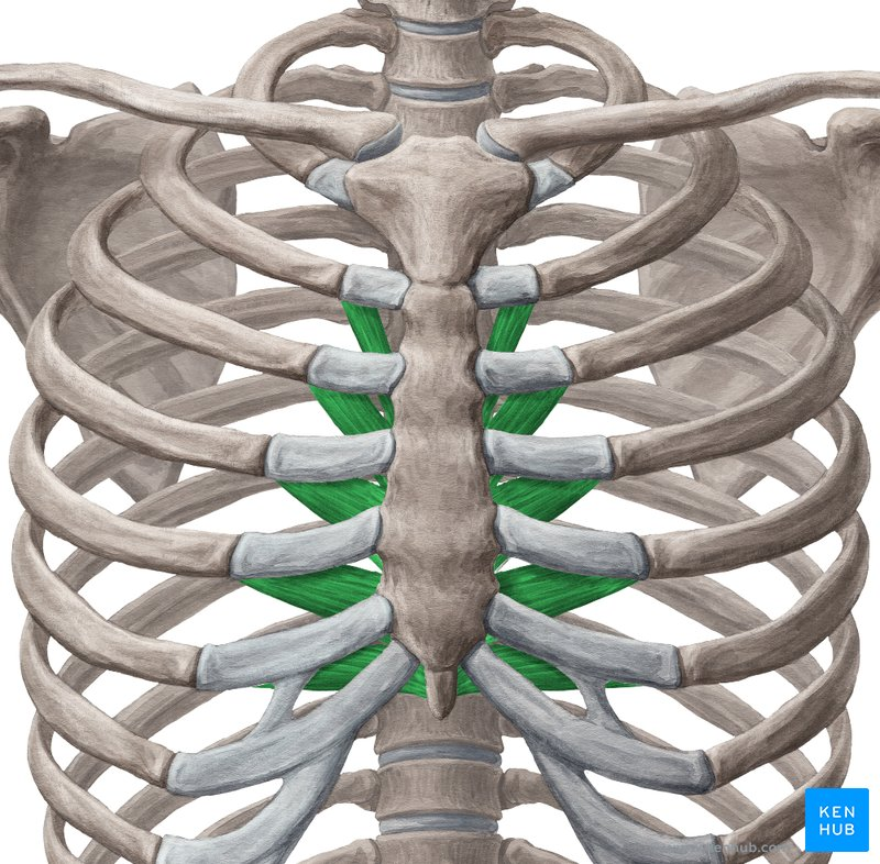
Transversus Thoracis muscle (Sternocostalis)
%%Origin%%: lower 1/2 of posterior surface of the sternum and xiphoid process
%%Insertion%%: posterior surfaces of the costal cartilages from 2nd to 6th
Nerve Supply
Intercostal nerves (T1 - T11)
Action
muscles of respiration
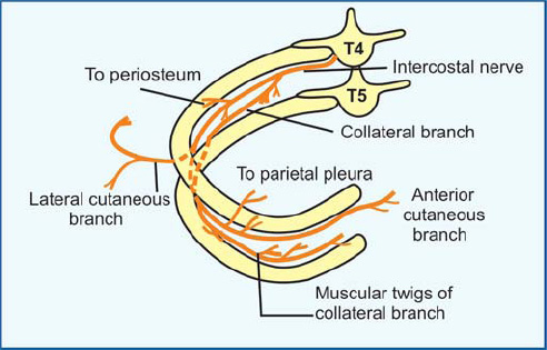
Intercostal nerve
- ventral ramus of thoracic spinal nerve (T1 - T11)
- T12: subcostal nerve
Typical intercostal nerve (3rd to 6th)
%%Origin%%: anterior (ventral) ramus of thoracic spinal nerve
%%Course%%:
- passes from intervertebral foramen
- runs anteriorly between internal and innermost muscles
- ends anterior as @@anterior cutaneous branch@@
Branches:
branches to sympathetic trunk
muscular branch to the intercostal muscles
lateral cutaneous branch (divides into anterior and posterior branches)
anterior cutaneous branch (divides into medial and lateral branches)
sensory branch to the parietal pleura
collateral branch (runs on the upper border of rib below)
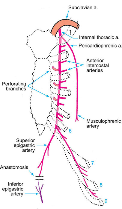
Internal Thoracic Artery
%%Origin%%: from 1st part of subclavian artery in the neck, supplies the anterior wall of the trunk from the clavicle to the umbilicus
%%Course%%: descends vertically down behind costal cartilages half an inch lateral to the sternum
%%Termination%%: in the @@6th intercostal space@@ by dividing into:
- ==superior epigastric artery==
- ==musculophrenic artery==
Branches:
anterior intercostal arteries
perforating arteries
pericardiacophrenic artery
mediastinal artery
superior epigastric artery
musculophrenic artery
Intercostal arteries (anterior and posterior)
Anterior intercostal arteries:
- in the 1st to 6th spaces are branches from: @@internal thoracic artery@@
- lower 3 spaces (7th to 11th) are branches from: @@musculophrenic artery@@
Posterior intercostal arteries:
- 1st and 2nd arteries from: @@superior intercostal artery@@
- arteries from 3rd to 11th branches from: @@descending thoracic aorta@@
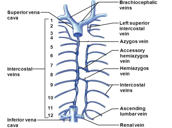
Intercostal veins
Anterior intercostal veins: drain into internal thoracic vein
internal thoracic vein:
- formed at 3rd intercostal space ( by union of vena comitants)
- ends in brachiocephalic vein
Posterior intercostal veins: drain into the azygos vein (in the right side) and to hemiazygos veins (in the left side)
Azygos vein:
begins in the abdomen : from inferior vena cava
ascends up to end in: the superior vena cava in the thorax
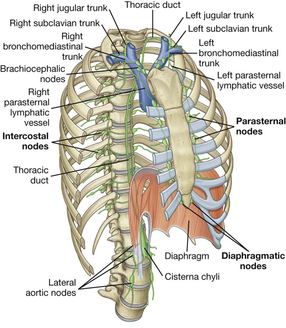
Lymphatic drainage of the thoracic wall
a. Superficial lymphatics:
- from anterior wall: into pectoral (anterior) group of axillary lymph nodes
- from posterior wall: into subscapular (posterior) group of axillary lymph nodes
b. Deep lymphatics
from anterior parts: into internal thoracic (parasternal) lymph nodes (present along internal thoracic vessels)
from posterior parts: into posterior intercostal lymph nodes (present along the posterior intercostal vessels)