Chapter 2 - Renal Function
Renal Physiology
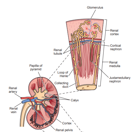
- Each kidney is made up of functional units that filter urine called nephrons.
- Cortical nephrons are situated in the outer layer where they are responsible primarily for removal of waste products and reabsorption of nutrients.
- Juxtamedullary nephrons have longer loops of Henle that extend deep into the medulla of the kidney, and their primary function is controlling the concentration of urine.
Renal Blood Flow
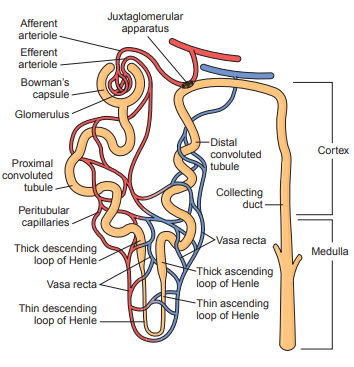
- The renal artery supplies 25% of the blood pumped from the heart at all times to the kidneys.
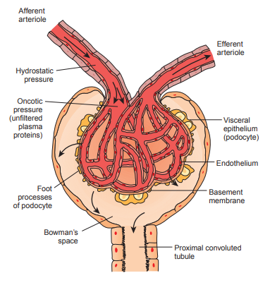
- This blood enters through the afferent arteriole into the capillaries of the nephron.
- It then flows through the glomerulus and into the smaller efferent arteriole.
- The glomerulus consists of a coil of approximately eight capillary lobes referred to collectively as the capillary tuft, and is located within the Bowman’s capsule, a structure that forms the beginning of a nephron.
- The glomerulus is non-selective and can filter plasma substances with molecular weights of less than 70,000, but it can still be influenced by some factors.
- Plasma filtrate must pass through three cellular layers: the capillary wall membrane, the basement membrane (basal lamina), and the visceral epithelium of Bowman’s capsule.
- The capillary walls contain pores but do not allow the passage of large molecules and blood cells.
- A barrier comprising of filtration slits between foot-like processes of podocytes further filter out plasma proteins.
- The renin-angiotensin-aldosterone system (RAAS) controls the regulation of the flow of blood to and within the glomerulus and responds to changes in blood pressure and plasma sodium content.
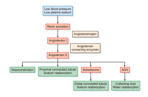 * Plasma sodium content is directly proportional to water retention and affects blood volume and blood pressure.
1. When low, renin is secreted and reacts with the blood-borne substrate angiotensinogen to produce the inert hormone angiotensin I. 2. Angiotensin converting enzyme (ACE) changes it to the active form angiotensin II which is responsible for vasodilation of the afferent arterioles and constriction of the efferent arterioles, removal of sodium in the proximal convoluted tubules, and triggering the release of aldosterone to control sodium concentration in the distal convoluted tubules and of vasopressin / antidiuretic hormone (ADH) to control water concentration in the collecting duct. * The difference in size creates hydrostatic pressure differential important for glomerular filtration and to maintain consistency of glomerular capillary pressure and renal blood flow within the glomerulus. * This pressure is necessary to overcome pressure from fluid within the Bowman’s capsule and the oncotic pressure of unfiltered plasma proteins in the glomerular capillaries. * It consists of the juxtaglomerular cells in the afferent arteriole and the macula densa of the distal convoluted tubule. 3. As systemic blood pressure and plasma sodium content increase, the secretion of renin decreases. * Every minute, approximately two to three million glomeruli filter plasma protein, any protein-bound substances and cells to produce approximately 120 mL of water-containing low-molecular weight substances. * Normal urine contains very little protein: usually, less than 10 mg/dL or 100 mg per 24 hours is excreted and is mostly albumin (see Protein in Chapter 5 - Chemical Examination of Urine). * It has a specific gravity of 1.010.
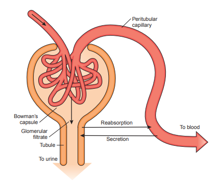
- Next, the blood enters the peritubular capillaries and the vasa recta and flows slowly through the cortex and medulla of the kidney close to the tubules.
- The peritubular capillaries undergo tubular secretion where waste products not filtered by the glomerolus are eliminated and the acid-base balance of the body is regulated through the secretion of hydrogen ions.
 * Substances bound to plasma proteins have a higher affinity to tubular cells and disassociate from their carrier proteins, allowing them to be carried into the filtrate.
* The secretion of hydrogen ions creates a concentration gradient that transfers the bicarbonate ions to the bloodstream.
* In order to maintain a normal body pH of 7.4, bicarbonate ions are needed to buffer out changes from dietary intake and metabolism, but they are filtered out by glomerolus and need to be returned to the bloodstream.
* However, no normal values are assigned to urinary pH, and it must be considered in conjunction with other patient information (see pH in Chapter 5 - Chemical Examination of Urine).
* The hydrogen ions bind with hydrogen phosphate, ammonia and weak organic acids in urine, preventing them from being removed and allowing them to be excreted in urine (see pH in Chapter 5 - Chemical Examination of Urine).
- Tubular reabsorption takes place here through active and passive transport.
- Active transport makes use of electrochemical energy from binding to a carrier protein to move across the cell membranes and back into the bloodstream.
- However, if the concentration exceeds the maximum reabsorptive capacity (Tm) of the tubules, the renal threshold is reached and it is retained in the urine.
- Passive transport makes use of concentration or electrical potential differences or gradients on opposite sides of the membrane.
- Active transport makes use of electrochemical energy from binding to a carrier protein to move across the cell membranes and back into the bloodstream.
- Each section controls the concentration of substances in urine in different ways.
| active transport | passive transport | |
|---|---|---|
| proximal convoluted tubule | glucose, amino acids, sodium, salts | water, urea |
| descending loop of Henle | water | |
| ascending loop of Henle | chloride | urea, sodium |
| distal convoluted tubule | sodium | |
| collecting duct | water |

* Plasma-bound substances eliminated by the peritubular capillaries are removed in the proximal convoluted tubule.
* The ascending loop of Henle uses a countercurrent mechanism of a water-impermeable wall to prevent water from escaping and to maintain the osmotic gradient in the medulla.
* The concentration of sodium retained in the distal convoluted tubule is controlled by aldosterone in RAAS.
* ADH controls the permeability of the distal convoluted tubule and collecting duct to water.
* A high level of ADH during dehydration increases permeability, resulting in increased reabsorption of water, and a low volume of concentrated urine. Likewise, the absence of ADH when hydrated renders the walls impermeable to water, resulting in a large volume of dilute urine.
Renal Filtration Tests
Glomerular Filtration Tests (GFR)
- The standard test used is the clearance test.
- It measures the glomerular filtration rate (GFR) or the rate at which the kidneys are able to remove a filterable substance from the blood.
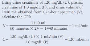
- It is reported in mL/min, and it is calculated using urine volume in mL/min (V), urine creatinine concentration in mg/dL (U), and plasma creatinine concentration in mg/dL (P).
- Plasma and urine concentrations are determined by chemical testing.
- The milliliters of plasma cleared per minute (C) times the mg/dL of plasma creatinine (P) must equal the mg/dL of urine creatinine (U) times the urine volume in mL/min (V), because all of the filtered creatinine will appear in the urine.
- CP = UV; using derivation, C = UV/P
- The average body size is 1.73 sq. m, but to account for differences, 1.73/A is multiplied to C, where log A = (0.425 x log weight)(0.725 x log height) - 2.144
- Factors to consider when choosing a substance to be analyzed is whether it is removed and secreted by the tubules, the stability of the substance in urine during a possible 24-hour collection period, the consistency of the plasma level, the substance’s availability to the body, and the availability of tests for analysis of the substance.
- Early tests made use of urea, but it was later replaced with inulin and creatinine.
- Urea is present in all urine specimens and endogenous to the body, but 40% gets reabsorbed and this needed normal values in calculations to be readjusted and patients to be hydrated to produce a urine flow of 2 mL/min to ensure that no more than 40% of the urea was reabsorbed.
- Inulin is extremely stable and not affected by tubule dynamics; however, it is not normally a constituent of the body and exogenous, requiring it to be infused at a constant rate throughout the testing period.
- Creatinine is endogenous to the body as it is a waste product of muscle metabolism, but it has several disadvantages.
- Some creatinine is secreted by the tubules, and secretion increases as blood levels rise
- False elevation may be countered by reaction with chromogens in plasma.
- Medications like gentamicin, cephalosporins, and cimetidine (Tagamet), inhibit tubular secretion of creatinine and may cause false decreases.
- Bacteria can break down creatinine if urine specimens are kept at room temperature for extended periods.
- A diet heavy in meat consumed during collection of a 24-hour urine specimen will influence the results if the plasma specimen is drawn prior to the collection period.
- Patients with muscle-wasting diseases may have false increases.
- It is used to determine the extent of nephron damage in known cases of renal disease, to monitor the effectiveness of treatment designed to prevent further nephron damage, and to determine the feasibility of administering medications, which can build up to dangerous blood levels if the GFR is markedly reduced.
Calculated Glomerular Filtration Estimates
- This method does not require urine creatinine and relies on variables of age, sex, and body weight in kilograms.
- C = ((140 - age)(actual body weight (ABW) in kg)) / 72(serum creatinine in mg/dL); multiply by 0.85 if female
- To correct for weight not from muscle mass, ideal body weight (IBW) and adjusted body weight (AjBW) are calculated and substituted into the formula.
- IBW (male) = 50 kg + 2.3 kg for each inch of height over 60 inches
- IBW (male) = 45.5 kg + 2.3 kg for each inch of height over 60 inches
- AjBW = IBW + 0.3(ABW - IBW)
- The Modification of Diet in Renal Disease (MDRD) system, utilizes additional variables such as ethnicity, blood urea nitrogen and serum albumin, and it does not include body weight. Several variations are available.
- GFR = 170 x serum creatinine^(-0.999) x age^(-0.176) x 0.822 (for females) x 1.188 (if black) x blood urea nitrogen^(-0.170) x serum albumin^(0.318)
- Serum markers used include radionucleotides, beta 2 microglobulin and cystatin C.
- Radionucleotides like I-iothalamate provides a method for determining glomerular filtration through the plasma disappearance of the radioactive material and enables visualization of the filtration in one or both kidneys.
- Beta 2 microglobulin dissociates from human leukocyte antigens at a constant rate and is rapidly removed from the plasma by glomerular filtration, giving good correlations with GFR.
- It has been shown to be more sensitive than creatinine; however, it is not recommended for use in patients with a history of immunologic disorders or malignancy.
- Cystatin C is a small protein produced at a constant rate by all nucleated cells, and it is readily filtered by the glomerulus and reabsorbed and broken down by the renal tubular cells.
- It is recommended for pediatric patients, persons with diabetes, the elderly and critically ill patients.
Tubular Reabsorption Tests
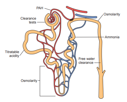
- These tests measure the ability of the tubules to remove essential salts and water using specific gravity and osmolarity.
- Osmolarity depends on the number of particles present.
- Its basic unit is an milliosmole (mOsm), which is the molecular weight (g) of a substance divided by the number of particles into which it dissociates.
- Major clinical uses of osmolarity include evaluating renal performance, monitoring renal disease, monitoring fluid and electrolyte therapy, establishing salt deficiency and excessiveness, and evaluating the secretion of and renal response to ADH.
- Normal serum osmolarity values are from 275 to 300 mOsm, and a normal ratio to urine osmolarity is 1:1 under random conditions, or 1:3 under controlled fluid intake.
- ADH is injected to test if diabetes insipidus or abnormal urination and thirst is caused by decreased ADH production or inability of the renal tubules to respond to ADH.
- The failure of urine osmolarity to be concentrated on a 1:3 ratio indicates that ADH receptors are not functioning.
- If it reaches the ratio, it indicates the body is unable to produce ADH.
- The osmolarity of a solution can be determined by measuring the colligative property or a property dependent on the number of particles in the solution, and comparing this value with the value obtained from the pure solvent.
- In a freezing point osmometer, the sample is supercooled and then vibrated it to produce crystallization of water in the solution.
- The heat of fusion produced by the crystallizing water temporarily raises the temperature of the solution to its freezing point; when the temperature of the sample remains constant, this represents the true freezing point of the sample.
- One mole of a non-ionizing substance dissolved in 1 kg of water is known to lower the freezing point 1.86°C, and it is equivalent to 1000 mOsm. By comparing it with a known molal solution (usually NaCl), the osmolarity of the unknown solution can be calculated.
- In a vapor pressure osmometer, the sample is evaporated and then condensed on a thermocoupler which determines te dew point temperature proportional to the vapor pressure.
- It is converted into milliomole and compared to NaCl standards; however, there is more variation as samples tend to evaporate before testing and are only 0.01 mL.
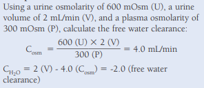
- The ratio of urine to serum osmolarity can be further expanded by performing the analyses using water deprivation and a timed urine specimen and calculating the free water clearance to test the ability of the kidney to respond to the state of body hydration.
- Calculate the osmolar clearance using the standard clearance formula and then subtract the osmolar clearance value from the urine volume in mL/min.
- The osmolar clearance indicates how much water must be cleared each minute to produce a urine with the same osmolarity as the plasma.
- This indicates whether the water being excreted is more or less than the amount needed to maintain an osmolarity the same as that of the ultrafiltrate.
- Factors to consider because of their influence on true osmolarity readings include lipemic serum, lactic acid, and volatile substances, such as ethanol, in the specimen.
- Insoluble lipids displace serum water and produce erroneous results.
- If serum samples are not separated or refrigerated within 20 minutes, lactic acid may form and cause false increases in the result.
- Volatile substances become part of the solvent; they cause false elevations in freezing point osmometry but are not detected in vapor pressure osmometry.
Tubular Secretion and Renal Blood Flow Tests
- It is measured by a substance that is secreted rather than filtered through the peritubular capillaries as it aims to test the ability of the bloodstream and tubules to exchange substances.
- Phenolsulfonphthalein (PSP) dye was formerly used, but standardization and interpretation of results was difficult because of interference by medications and elevated waste products in patients’ serum, and difficult because several very accurately timed urine specimens are needed.
- Currently, p-aminohippuric acid is used.
- It is completely removed once in contact with renal tissue as it is loosely bound to plasma proteins, and it is removed in the peritubular capillaries rather than by the glomerulus.
- The standard clearance formula is used, and normal values for the effective renal plasma flow range from 600 to 700 mL/min, making the average renal blood flow about 1200 mL/min.
- PAH is only contained within the plasma portion, and approximately 8% of the renal blood flow does not come into contact with the functional renal tissue.
- Nuclear medicine procedures using radioactive hippurate can also be used to determine renal blood flow by measuring the plasma disappearance of a single radioactive injection; it also provides a visualization of the blood flowing through the kidneys.
- Titratable acidity can also measure renal functioning in hydrogen ion and ammonia secretion by calculating the difference of free hydrogen ions (titratable acidity) and the total acidity of the specimen.
- The tests can be run simultaneously on either fresh or toluene-preserved urine specimens collected at 2-hour intervals from patients who have been primed with an acid load consisting of oral ammonium chloride.