Respiratory system
THE HEAD
Nasal Cavity
Function/structure: Warming and moistening Air – prevents epithelial damage
Large cavity that connects the external environment with the pharynx. - This allows air from the external environment to be warmed and moistened; Respiratory tissue is delicate so this prevents damage from microbes and air from dehydrating the epithelia
Sensory organ - sense of smell
Physical first line of defense against pathogens/ debris
Hair and mucus trap dirt particles and microbes before they enter the lungs
Projections (conchae) create turbulence in the air which helps the mucus trap dust and particles
Each lateral wall of the nasal cavity has three bony projections called the superior, middle and inferior nasal conchae
Nasal conchae increases the surface area of the nasal cavity to maximize warming and moistening of the air
EPITHELIUM IN THE RESPIRATORY SYSTEM
Nasal cavity
Pseudostratified ciliate columnar cells
Pseudostratified: looks like it has multiple layers but is one layer
Columnar: can specialize in secreting mucus
Ciliated: have small projections called cilia which move debris trapped in mucus out of the nose
THE PHARYNX
Function - Passage for food and air
Structure: 3 sections
Nasopharynx: Noso – closest to the nose
Oropharynx: Oro- (oral) closest to the mouth
Laryngopharynx: Laryngo – closest to the larynx
Epiglottis
Structure: flap of cartilage
Function: fits over the trachea when swallowing to prevent food from entering the trachea (and moving down into the lungs)
LARYNX
STRUCTURE: Vocal cords supported by cartilage
FUNCTION: Sound production as air moves out of the lungs
The bronchial tree
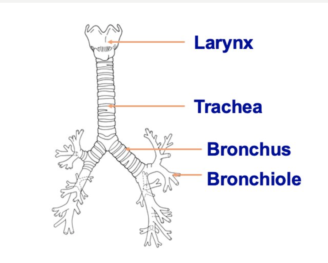 Trachea
STRUCTURE: Tube from pharynx to bronchi – supported by C-shaped rings of cartilage
FUNCTION:
- Cartilage holds the trachea open for air movement
- Ciliated epithelium along its length cleans the air with mucus
Bronchi
STRUCTURE:
-Division of the trachea into the right and left lungs
–further divide into secondary and tertiary bronchi
FUNCTION - Air passage
BRONCHIOLES
Structure
-very fine tubes with walls of smooth muscle
-end in groups of air sacs (alveoli)
Function -air passage
Alveoli
Structure:
Occur in clusters and very thin moist walls
a large capillary network covers the sacs
Function - Allow air to into very close contact with the blood so gas exchange can occur
Pleura
STRUCTURE: Membrane covering the lung and the internal thoracic cavity
FUNCTION: Membrane produces pleural fluid which acts to reduce friction as the lung moves within the thoracic cavity
Diaphragm
STRUCTURE: Sheet of muscle which separates the thoracic cavity from the abdominal cavity
FUNCTION: Aids in the ventilation of the lung
INTERCOSTAL MUSCLES
STRUCTURE: Muscles found between the ribs
FUNCTION: Aids in the movement of the ribs for the ventilation of the lungs