WK 3 Skull - Facial bones and sinuses
Major Bony features of the Facial bones
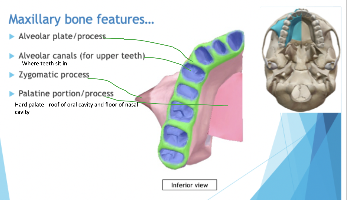
Maxillae (R and L Maxilla bones)
Maxillae: creates part of the orbital floor, medial wall of orbits, lateral wall of the nose and roof of the mouth
Maxillary sinus - biggest air sinuses and fractures can be seen
Maxillae had canine eminence, incisor fossa and canine fossa
Alveolar plate has teeth and roots embedded into it - raised up portion above the incisor tooth is the incisor fossa
floor of nose is the roof of the mouth
Anterior nasal spine helps to hold the nasal cartilage and septum in place - seen in lateral images, is also the acanthion
Zygomatic bones
Form the lateral wall and part of the floor of the orbit
Had temporal border and orbital border
Mandible
Symphysis Mentee - cartilage becomes ossified
Condyles form the TMJ
TMJ is a modified condylar hinge joint as it allows medial and lateral movement (grinding of food). Also protrudes and detrudes
Nasal bone
has internasal suture, frontonasal suture and frontomaxillary suture
Lacrimal
has fossa for the lacrimal gland which produces tears
Vomer
V shaped - pointy side pointing anterior and wider side is posterior
Palatine bone
3 portions
Palatine portion of the palatine bone
Nasal portion of the palatine bone: going up the lateral aspect of each nasal cavity
Orbital part of palatine bone
Forms part of posterior orbital floor
Inferior nasal conchae
covers lower part of the opening into the maxillary sinus
Orbital margins
Roof: frontal bone
Lateral wall: zygomatic bone
Medial wall: maxilla, lacrimal, ethmoid and palatine bone (orbital part)
Floor: zygomatic bone and the maxilla
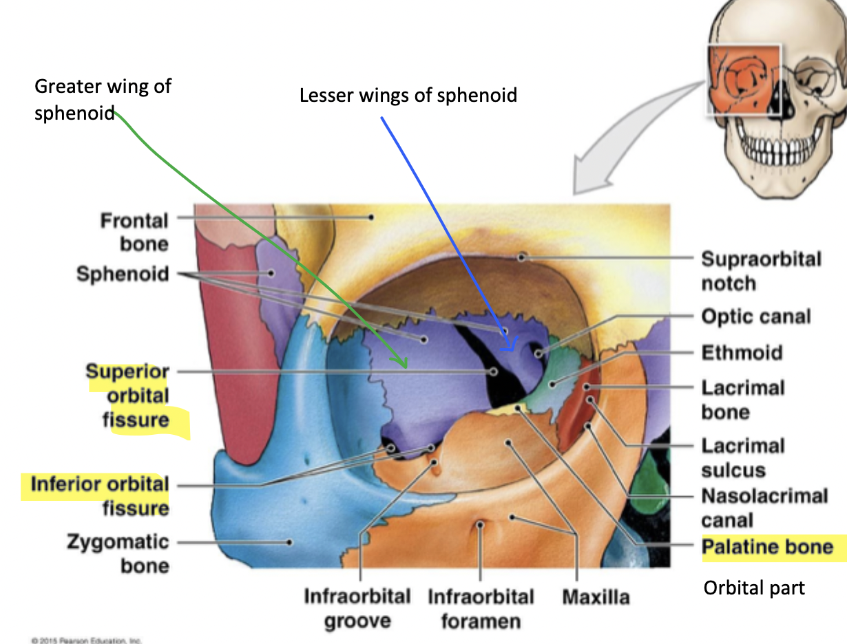
Optical canal of the sphenoid bone and optic nerve comes through there to connect to the back of the eyeball
Superior orbital fissure between the greater and lesser wing of sphenoid
Inferior orbital fissure between the maxilla and greater wing of sphenoid - allows things to pass down from the lower face region and not through the orbit
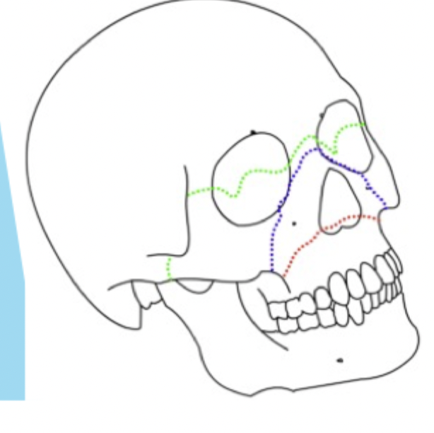
Le fort fractures
Type 1
fracture through the maxillae plate and removing the alveolar plate from the rest of the face - bottom of teeth and maxillae become disassociated with the face
Type 2
Full removal of maxillae from the zygomatic, nasal and frontal bones
Type 3
Same as type 2 but the zygomas are gone too - entire facial bones removed from cranium