System Neurobiology
Organization of the nervous systems
Major divisions of the vertebrate nervous system
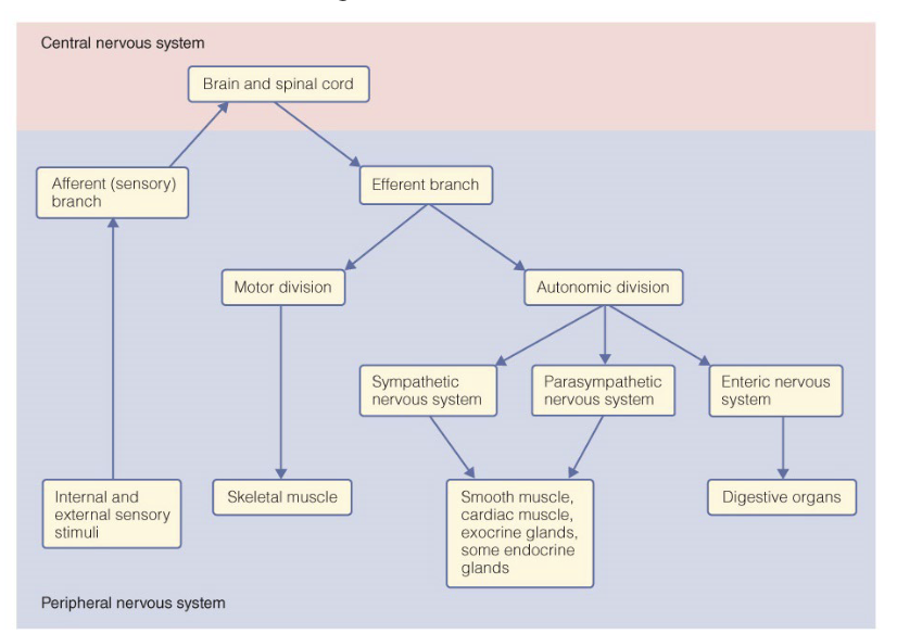
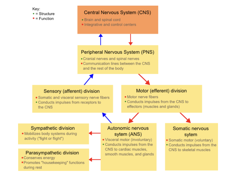
central nervous system
Brain
higher function
complex task initiation
perception
emotions
Spinal cord
reflexes
carries sensory and motor signals
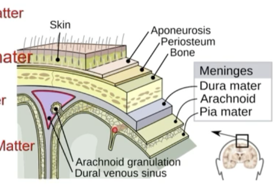
Protected by meninges and cerebrospinal fluid (CSF)
Meninges
Dura mater
tough matter
outermost
Arachnoid matter
weblike
middle layer
Pia mater
delicate matter
thin
inner layer
Blood brain barrier
prevents the wrong molecules from going to the brain
prevents glucose
does not prevent fats
White matter
myelinated
Brain: in the inner part
Spinal cord: on the outside
Gray matter
unmyelinated
Brain: on the outside
Spinal cord: on the inside
Functions of the spinal cord
mediates spinal reflexes
pathway for impulses to and from the brain
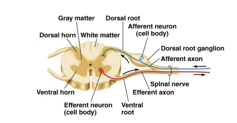
Axons of the peripheral neurons bundles into nerves
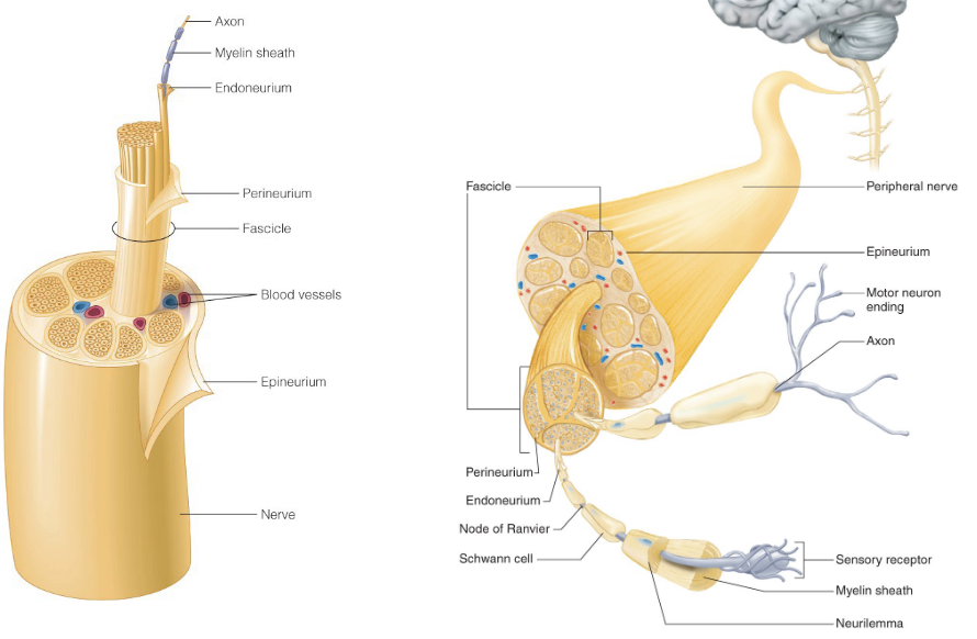
Nerve- a collection of nerve fibers in the PNS-connect sensory organs to CNS. May or may not be myelinated
Tract- collection of nerve fibers in the CNS (usually myelinated-white matter)
Peripheral neuron cell bodies cluster in or near the CNS
if neuron cell bodies are clustered in the CNS- nuclei
if neuron cells are clustered outside the CNS- ganglia
Nerves may contain sensory pr motor signals or both
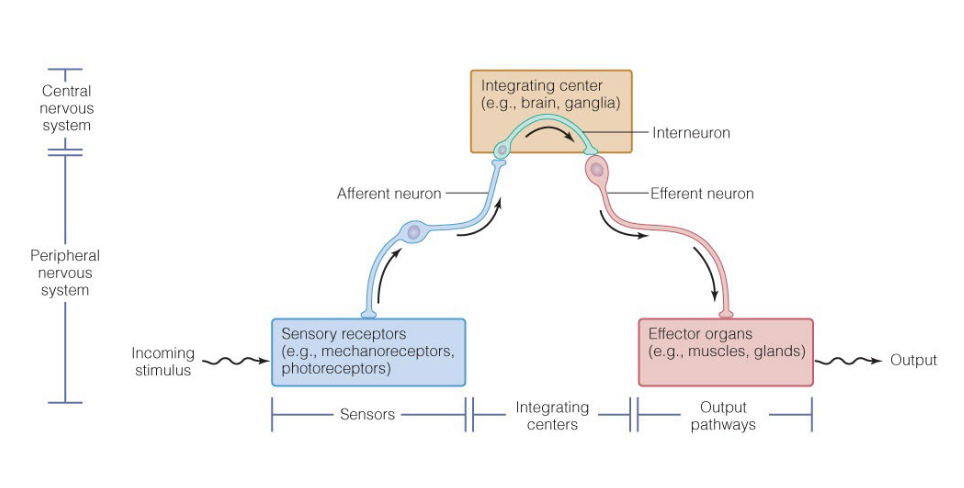
Reflex arc control many involuntary behaviors
automatic, subconscious response to stimuli within or outside the body
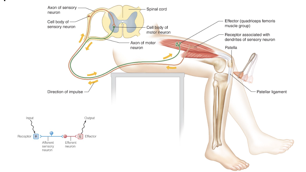
simple reflex arc(sensory-motor)
most common (sensory -assoication-motor)
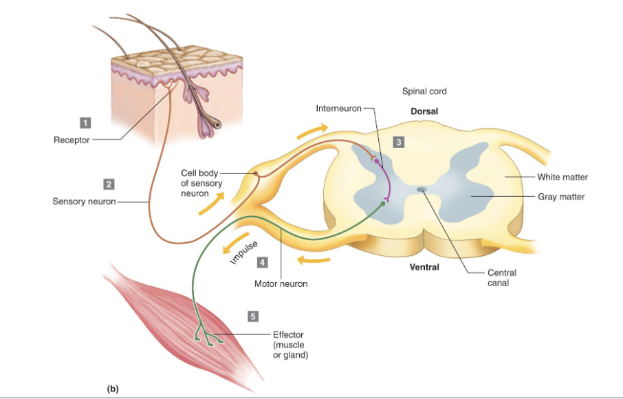
Simple reflex arc involve only 2 neurons
Most reflexes are mediated by more neurons
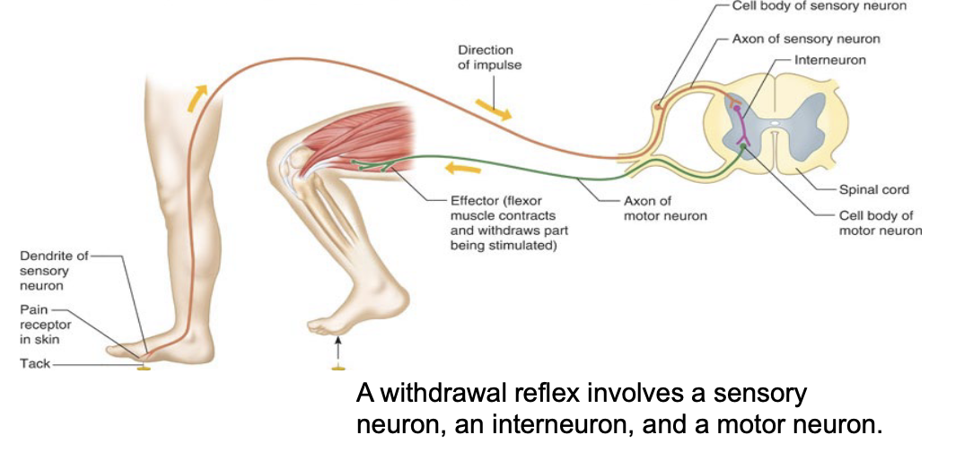
a withdrawal reflex involves a sensory neuron, an interneuron, and a motor neuron
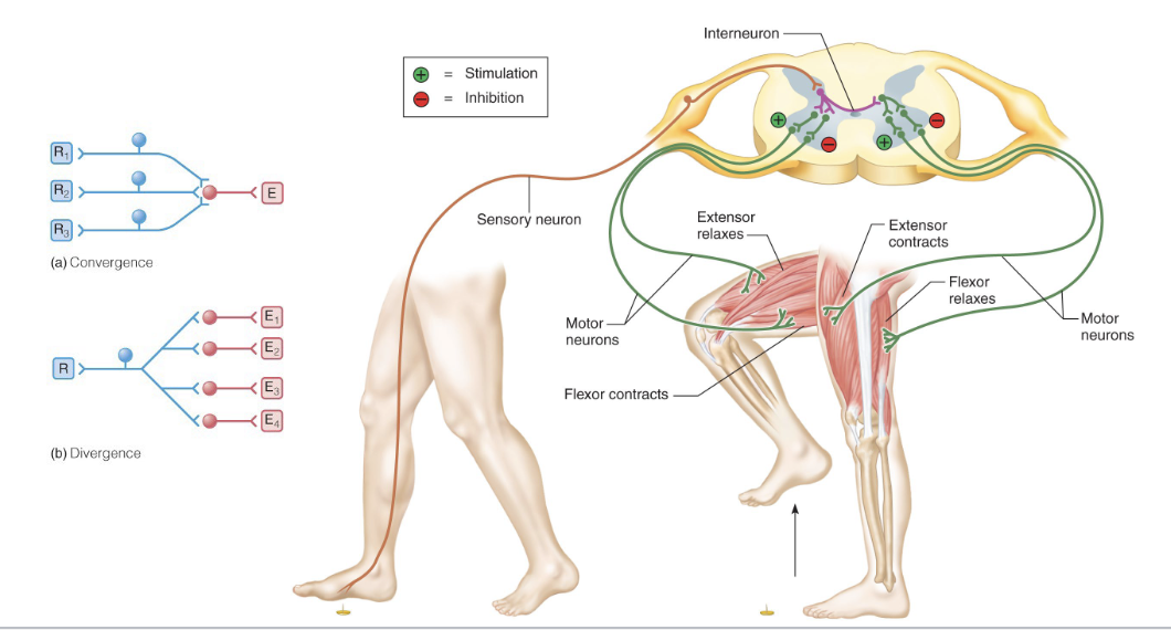
Reflex arcs employ divergence to ensure coordinated responses
Bundles of axons in the CNS are called tracts
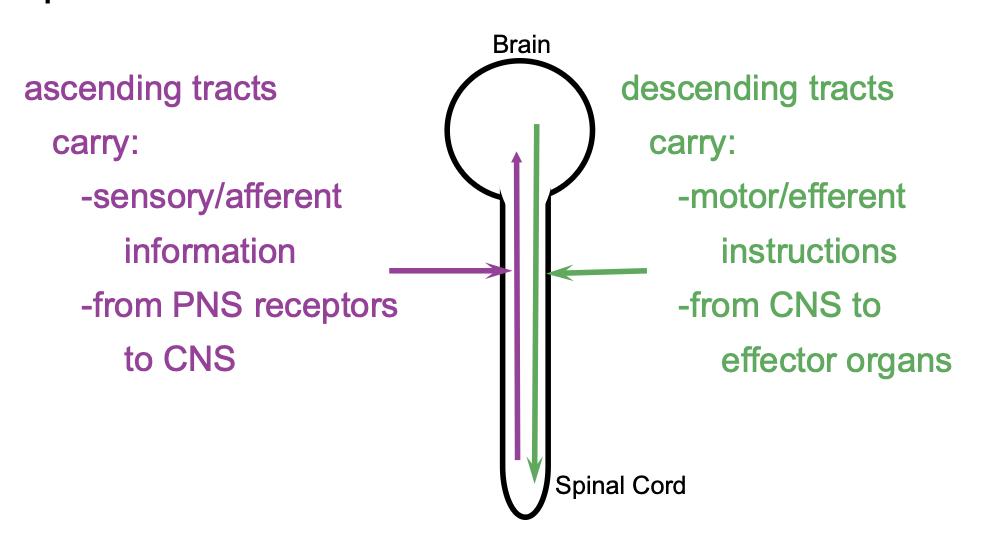
Different information carried in ascending and descending tracts
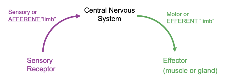
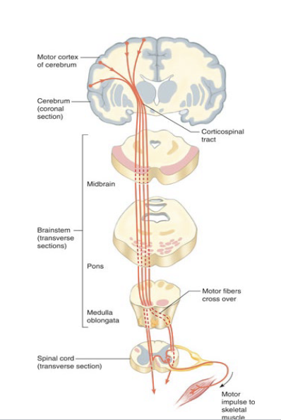
Efferent outputs
originate in the brain
ipsilateral
same side
contralateral
opposite side
mostly muscle neurons
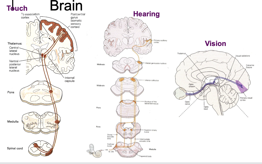
Sorting sensory inputs in the brain
Touch
contralateral
Hearing
both
eyesight
both
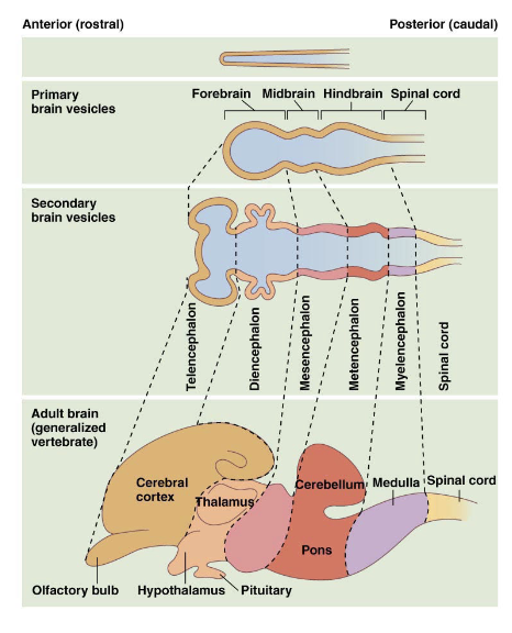
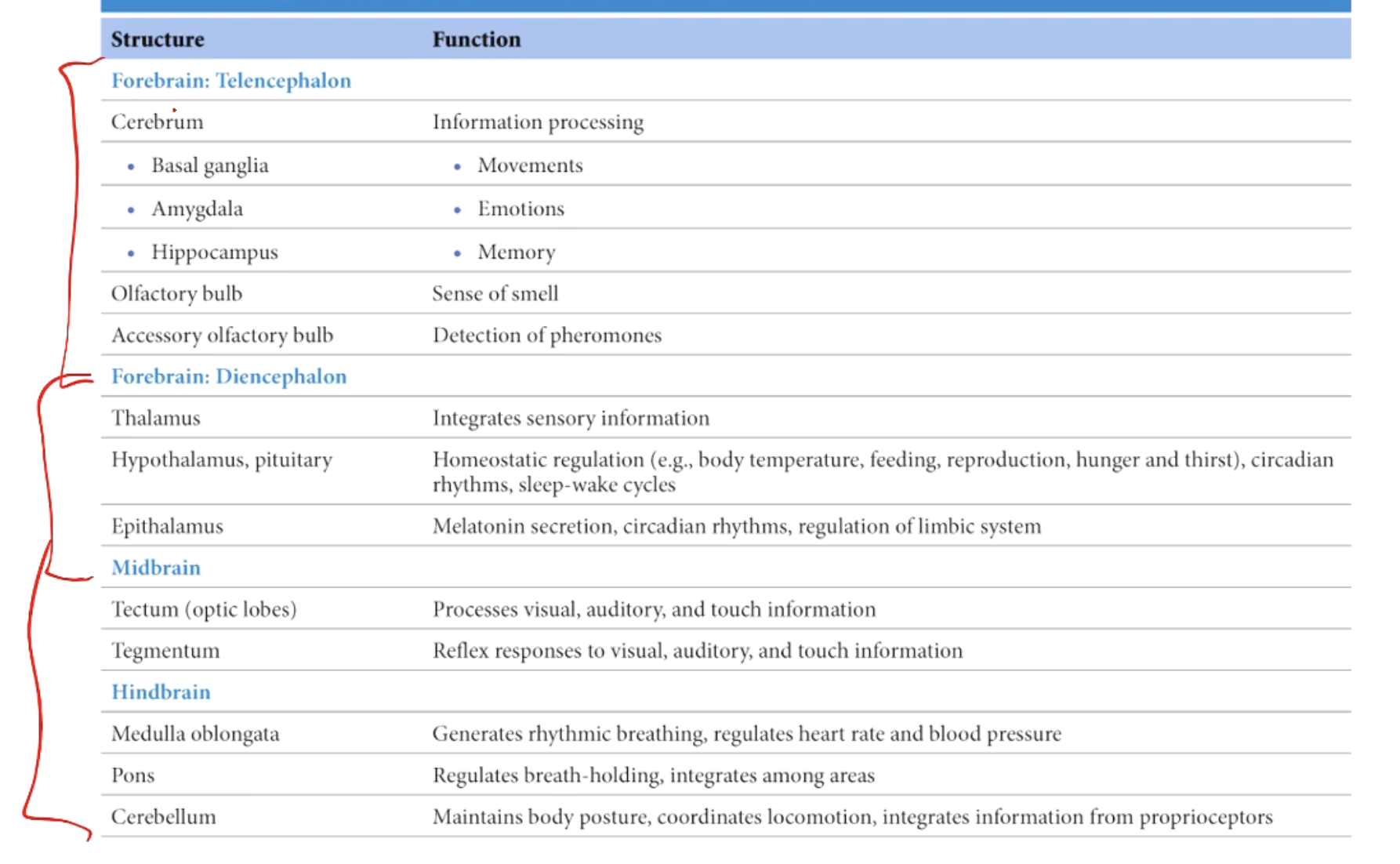
3 fundamental division of the vertebrate brain
Hindbrain
reflexes and involuntary behavior
metencephelon and myelencephalon
medulla oblongata
breathing, heart rate, blood pressure
pons
cerebellum
motor behaviors
midbrain
mesencephalon
forebrain
telencephelon and diencephalon
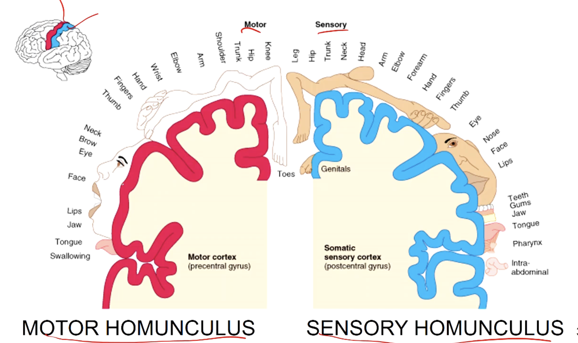
Frontal
primary motor cortex
controls voluntary movements in skeletal muscle
stimulation of different regions f PMC leads to movement
Damage to the PMC leads to paralysis and loss of voluntary movement
damage to the PMC usually results in permanent loss of these movements
pre motor cortex
coordinates movements of groups of muscles
damage:
loss of skill
can be relearned
parietal
primary somatosensory cortex
behind the central sulcis
primary input from sensory receptors in skin and muscle
damage leads to loss of sensation
somatosensory association area
adjacent to primary somatosensory cortex
interpretation of sensation
integration sensation with memory
damage: loss of identification of sensations
temporal
occipital
primary visual cortex
message goes to the cortex
visual association area
interepreting the message
Homunculus
motor
sensory
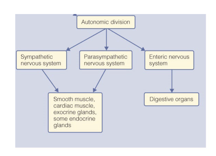
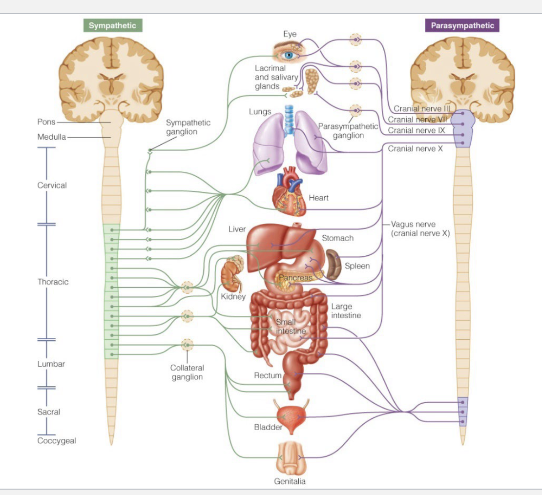
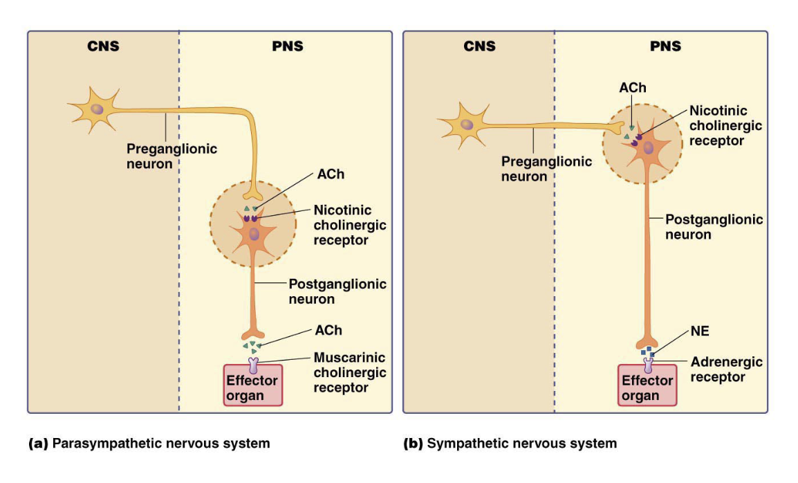
Autonomic nervous system
3 branches
sympathetic
most active during stress excitement, or physical activity
fight or flight
increases HR, breathing rate, directs blood to working muscles
parasympathetic
most active during rest
resting and digesting
redirects energy toward maintenance activated like digestion
enteric
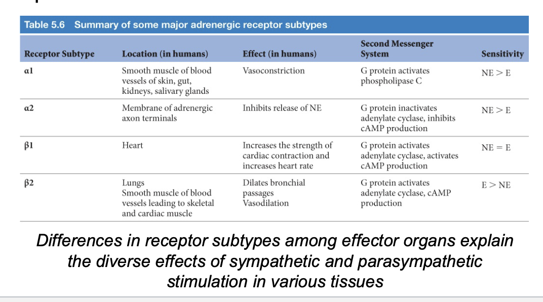
Effector organs express different metabotropic adrenergic receptors
Most organs have dual innervation from the parasympathetic and sympathetic branches
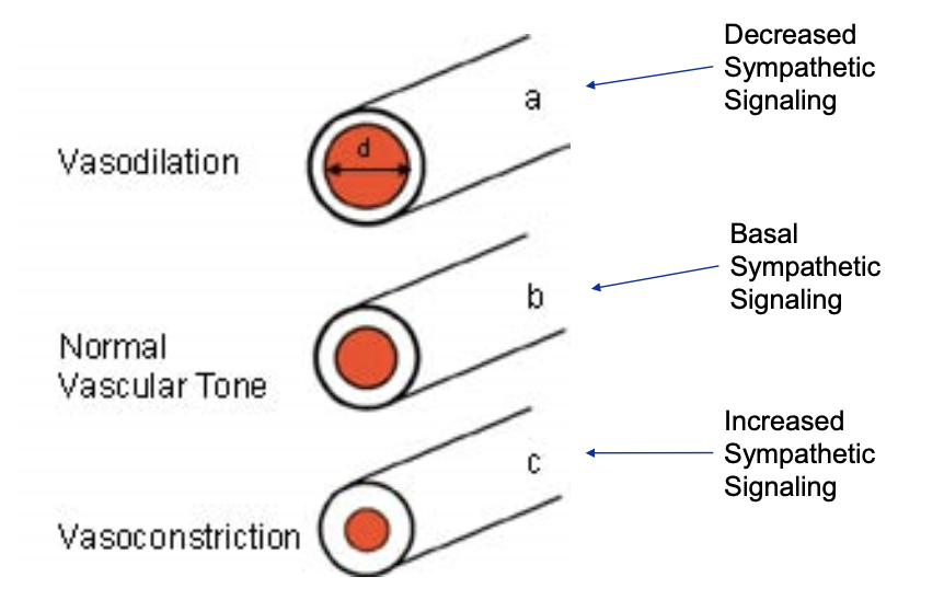
How can sympathetic input alone control dilation and constriction?
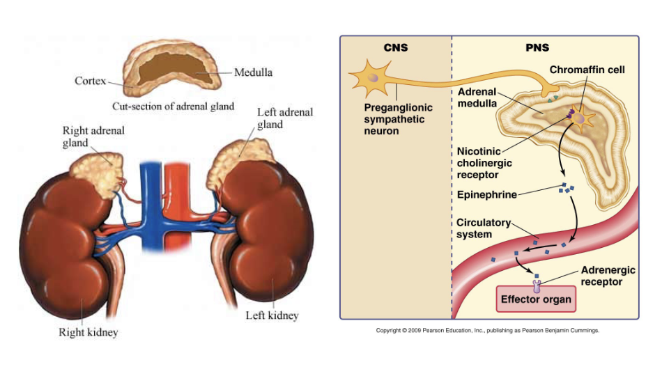
The adrenal medulla is innervated by the sympathetic nervous system only
The CNS regulates both voluntary and involuntary efferent branches
somatic motor neurons innervate skeletal muscle for voluntary movements
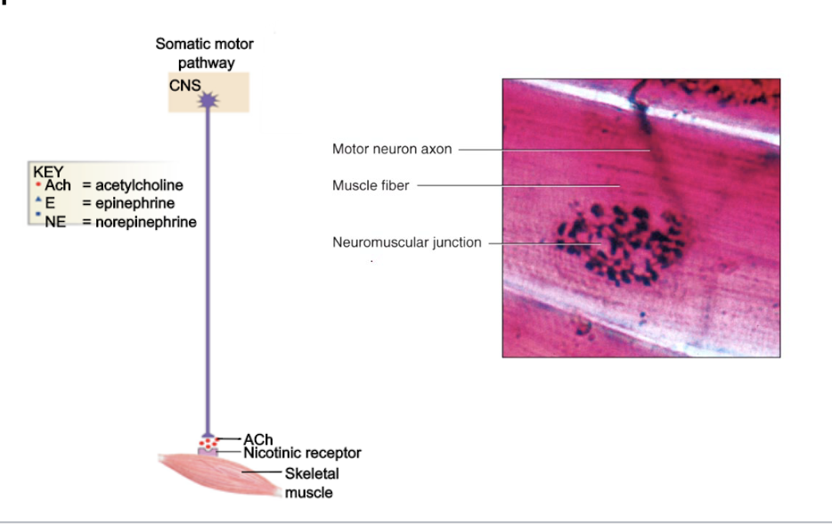
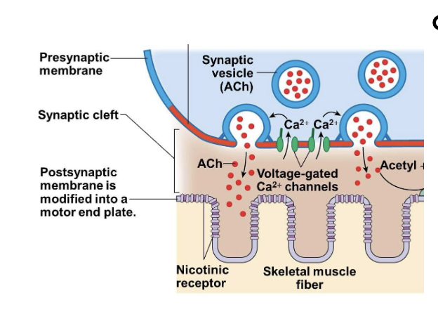
Vertebrate motor neurons release ACh at the neuromuscular junction
Acetylcholine (ACh)
released from synaptic vesicles
binds to nicotinic ACh receptors
causes skeletal muscle depolarization via entry of sodium ions into the cell