Cytoskeleton
Cells have a default shape, a sphere. Cells modify this shape in order to fit its their function. Bacteria do this by having a cell wall, animals cells do this via the cytoskeleton
Bacteria have a cell wall. A rigid wall made of polysaccharide cross-linked with peptides. Penicillin blocks the transpeptidase disrupting cell wall synthesis. Lysozyme digests the polysaccharide. Protects the bacteria but prevents it from changing shape
Animal and plant cells have an internal cytoskeleton. This give them (especially animal cells who don’t have a cell wall) much more dynamic control over their shape. However proteins are very small compared to the diameter of a cell, so in order to synthesise the cytoskeleton, proteins must be polymerised
The cytoskeleton is made of protein polymers
Intermediate filaments are rope-like bundles of proteins. Key use: rigid cell shape.

Actin filaments are helical polymers of G-actin. Key use: dynamic cell shape
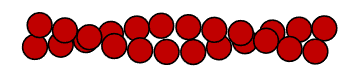
Microtubules are hollow polymers of tubulin. Key use: a framework for moving objects within the cell

Intermediate filament proteins are elongated with a central -helical region. Dimers form as head-to-head coiled coil structures. Tetramers form head-to-tail leaving overhangs, making it very easy for tetramers to come together and assemble into rope-like filaments. Very strong, very stable, very insoluble.


Resist mechanical force on the cell (like a rope resists pulling).
Keratin is the major intermediate filament protein in skin. It supports epithelial cells. Desmosomes are connections between epithelial cells. Hemidesmosomes connect cells to the underlying extracellular matrix. Keratin filaments span the epithelial cells forming a framework that supports the cells and links it mechanically to its neighbours. The cells are mechanically coupled together. The mechanical force from pulling on one keratin filament will be distributed to not only the other keratin filaments in the cells, but also the keratin filaments in other cells.

Defects in keratin cause epidermolysis bullosa simplex. Keratin filaments are weakened so instead of the skin resisting mechanical force the cells tear causing blisters. Blisters can form just from simple tasks that stretch the skin such as bending a finger

Actin polymerises to form filaments
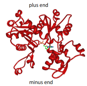
G-actin (globular actin) is the basic subunit. Actin is highly conserved across eukaryotic evolution. Actin binds a central ADP/ATP.
F-actin (filamentous actin) is a polymer of actin. It is a right handed helix of two protofilaments. F-actin is the building block of the actin cytoskeleton. Actin filaments are asymmetric. Growth is by addition of actin monomers to the plus end.

The first stage of actin polymerisation is nucleation. Actin nucleation factors stimulate polymerisation by catalysing this Rate limiting step (formation of the trimer)

Net actin polymerisation occurs above Cc
At the critical concentration of G-actin (Cc) the rates of addition and loss are balanced and there is no net polymerisation.
C = [G-actin]
Rate of addition = konC
Rate of loss = koff
At the critical concentration konC = koff
ATP-actin is added and hydrolyses to ADP-actin
kon is the same for ATP- and ADP-bound actin. koff is higher for ADP-actin so Cc (minus end) > Cc (plus end). So at steady state, actin is preferentially lost from the minus end - treadmilling.
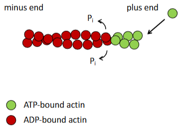
At the critical concentration, actin is lost at the minus end while at the plus end, actin is added so overall the filament remains the same length. Steady state actin filaments are dynamic and are a bit like a conveyor belt.


Phalloidin is an actin cytoskeleton toxin that comes from the mushroom Amanita phalloides. It is used to visualise the actin cytoskeleton as it binds to F-actin and stabilises the actin filaments. This makes the actin filaments no longer dynamic.
Actin cross-linking proteins connect actin filaments
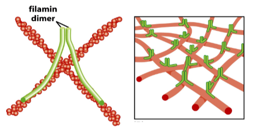
Actin filaments themselves are not very strong. Actin filaments are much more useful in cells when they are joined together to make bigger structures. Actin filaments are joined together by actin cross linking proteins such as filamin. Filamin is a dimer and each subunit has an F-actin binding domain. The filamin subunits join together at right angles. The actin filaments are therefore also joined at right angles. This allows actin filaments to form mesh-like structures.
The actin cytoskeleton braces the cell membrane.
Actin filaments form a cortical cytoskeleton beneath the plasma membrane. This is anchored by transmembrane proteins to give strength. In RBCs, actin filaments form a network with spectrin filaments to support the RBC shape and integrity.

This is another version of mechanical coupling. Force is transmitted between the cortical actin cytoskeleton and plasma membrane. Allows the plasma membrane to withstand mechanical stress as the force is spread out through the actin cytoskeleton.
Genetic defects lead to spherocytosis. Healthy RBCs have a biconcave disc shape and it is the actin cytoskeleton that gives it this shape. If the actin cytoskeleton is defected, then the RBCs will have a spherical shape and are more fragile
Actin polymerisation drives membrane protrusion
Actin cross-linking proteins bundle actin filaments together to make them more rigid

If actin filament polymerises on the plus end facing the plasma membrane, it can push against the plasma membrane and potentially push it out, forming a protrusion. However the force from 1 actin filament is not enough. At least 20 actin filaments bundled together are required to produce the force required to form a protrusion (20 pN)
Filopodia are used by cells to sense their environment. They are formed by bundles of actin filaments pushing on a single area of membrane.
Lamellipodia are used by cells to move. They are formed by multiple bundles of actin filaments pushing on a larger area of plasma membrane
Actin filaments interact with myosin motors
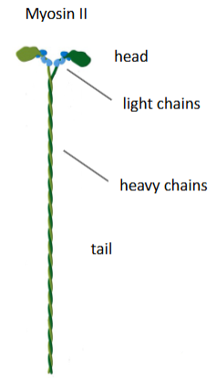
Two heavy chains intertwined forming a long coiled coil tail region with 2 globular heads. Two regulatory light chains bound to the head region. Myosins can interact tail-to-tail to form bipolar filaments. Mechanical movements of the head domain is linked to ATP hydrolysis

The actin cytoskeleton is contractile

Contractile actin is arranged in a sarcomeric array

In cell movement, the lamellipodia form in the direction in which the cell wants to go and then the back is pulled up by contractile actin.