Untitled
The Muscular System Study Notes
Overview of Muscle Tissue
Muscle Tissue Characteristics
One of the four primary tissue types.
Composed of elongated muscle cells specialized for contraction.
There are three types of muscle tissue: skeletal, cardiac, and smooth.
Skeletal Muscles
Definition and Composition
Composed of skeletal muscle tissue along with connective tissues, nerves, and blood vessels.
Skeletal muscles are directly or indirectly attached to bones.
The muscular system comprises about 700 skeletal muscles, which facilitate movement and stability.
Functions of Skeletal Muscles
Move the Skeleton
Pulls on tendons to move bones.
Maintain Posture and Body Position
Support Soft Tissues
Abdominal wall and pelvic cavity floor are made up of skeletal muscles.
Guard Entrances and Exits
Encircle openings of the digestive and urinary tracts.
Maintain Body Temperature
Muscle contractions generate heat.
Organization of Skeletal Muscle Tissue
Components of Skeletal Muscles
Contains muscle fibers, connective tissues, blood vessels, and nerves.
Each skeletal muscle cell is referred to as a muscle fiber.
Connective Tissue Organization
Epimysium: Covers the entire muscle.
Perimysium: Divides the muscle into fascicles (bundles of muscle fibers) and contains blood vessels and nerves.
Endomysium: Covers each muscle fiber and ties fibers together; contains capillaries and nerve fibers.
Contain myosatelite cells, which are stem cells that repair damaged skeletal muscles.
Connective Tissue Attachments
Tendon: A bundle of fibers that attaches the muscle to bone (formed by collagen fibers from endomysium, perimysium, and epimysium).
Aponeurosis: A broad sheet of fibers connecting muscles to each other.
Blood Vessels and Nerves
Skeletal muscles have an extensive network of blood vessels necessary for high energy use.
They are under voluntary control and require stimulation from the central nervous system.
Axons run through connective tissue layers to innervate individual muscle fibers.
Features of Skeletal Muscle Fibers
Muscle fibers can be extremely long (up to 60 cm).
Each fiber is multinucleate, containing hundreds of nuclei.
Skeletal muscle fibers exhibit a striated appearance due to the repeating pattern of thick (Myosin) and thin (Actin) filaments.
Sarcolemma and Transverse Tubules
Sarcolemma: The muscle cell's plasma membrane surrounding the sarcoplasm (cytoplasm).
T Tubules: Extensions of the sarcolemma that allow electrical impulses to reach the cell’s interior, facilitating a unified contraction of the entire fiber.
Myofibrils
Structure of Myofibrils
Cylinder-shaped structures running the length of the muscle fiber.
Each fiber contains hundreds to thousands of myofibrils encircled by T tubules, comprising thick and thin myofilaments whose sliding causes contraction.
Sarcoplasmic Reticulum (SR)
Specialized smooth endoplasmic reticulum forming a network around myofibrils.
Contains terminal cisternae with high concentrations of calcium ions, forming a triad with T tubules.
Sarcomeres
Functional Units
Repeating units of myofilaments that represent the smallest functional unit of skeletal muscle fibers.
Each myofibril contains about 10,000 sarcomeres arranged sequentially, giving muscle fibers a banded or striated appearance.
Sarcomere Lines and Bands
Z Lines: Mark the boundaries of each sarcomere; connected to thick (Myosin) filaments via titin.
M Line: Center of each sarcomere with proteins connecting thick (Myosin) filaments.
A Band: Darker region with thick filaments and zone of overlap.
I Band: Lighter region containing thin filaments, includes the Z line.
H Band: Area containing only thick filaments when muscle is relaxed.
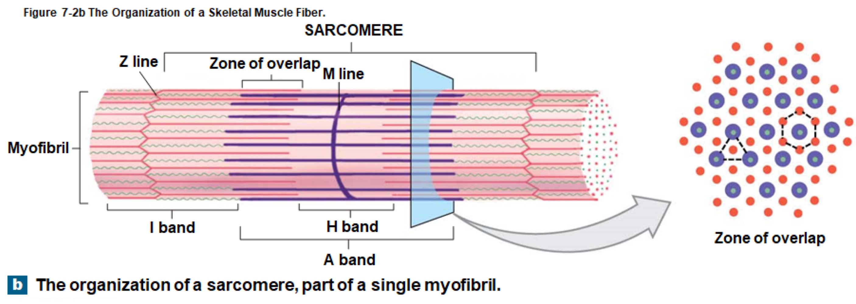
Thin and Thick Filaments
Thin Filaments: Composed of actin, containing active sites for myosin binding, covered by tropomyosin strands that are held in place by troponin.
Thick Filaments: Composed of myosin molecules, structured with tails pointing inward and heads projecting outward for binding to actin during contractions.
Sliding Filament Theory
Concept: Thin filaments slide towards the center of the sarcomere while thick filaments remain stationary during contraction.
Mechanism:
Myosin heads bind to active sites on thin filaments forming cross-bridges.
Heads pivot towards the M line (power stroke) and then detach.
Cross-bridge cycling repeats as long as ATP and calcium ions are present.
Neuromuscular Junction
Specialized connection between a motor neuron and a skeletal muscle fiber, comprised of an axon terminal and a motor end plate, which contains acetylcholine (ACh), a neurotransmitter.
Action potentials generated lead to muscle contractions, relying on the movement of ions across membranes and the neuromuscular signaling process.
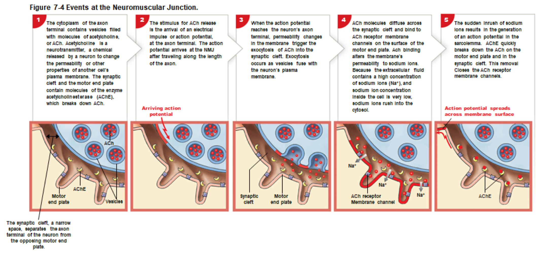
Contraction Cycle
Contraction Initiation: Begins with calcium ions arriving at the zone of overlap.
Active-Site Exposure: Calcium binds to troponin, exposing active sites on actin by moving tropomyosin.
Cross-Bridge Formation: Energized myosin heads bind to exposed active sites on actin.
Head Pivoting: Stored energy is used to pivot myosin heads toward the M line (power stroke), releasing ADP and phosphate.
Cross-Bridge Detachment: When ATP binds to the myosin head, it releases from actin.
Myosin Reactivation: Myosin head splits ATP into ADP and P, re-cocking the head for another cycle.
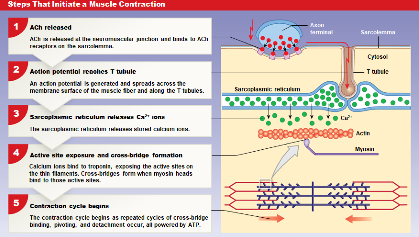
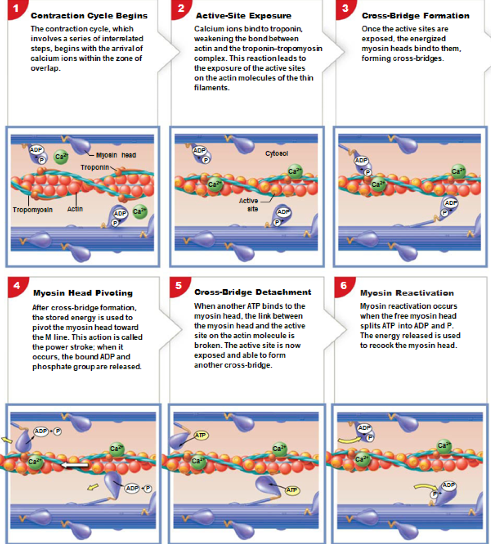
Muscle Contraction and Relaxation
Muscle Contraction: Involves ATP usage and is an active process.
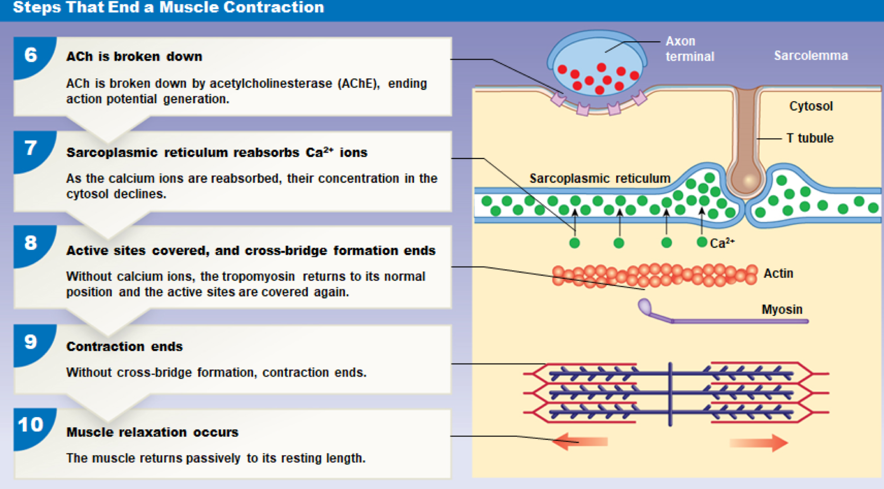
Muscle Relaxation:
ACh breakdown by acetylcholinesterase (AChE) ends action potential generation.
Calcium ions reabsorbed by the sarcoplasmic reticulum.
Cross-bridge formation stops as tropomyosin covers active sites again, resulting in muscle returning to resting length.
Sarcomere Shortening Produces Tension
Sarcomere shortening causes muscle fibers to contract, pulling on tendons and generating tension, which results in movement only if the tension exceeds the load's resistance.
Muscle Tension Factors
Amount of Tension: Depends on cross-bridges formed in muscle fibers; fibers can be fully contracted or relaxed, and contraction intensity cannot vary by altering individual sarcomere states.
Factors Affecting Tension:
Individual muscle fiber's resting length, frequency of stimulation, and overall muscle fiber recruitment in contraction.
Muscle Twitch
Definition: A single stimulus-contraction-relaxation cycle in a muscle fiber, lasting a brief period.
Phases of Muscle Twitch:
Latent Period: No tension is produced; it begins with stimulation.
Contraction Phase: Maximum tension is reached; involves cross-bridge interactions.
Relaxation Phase: Tension falls as calcium levels decrease and active sites are covered.
Summation and Muscle Tension
Summation: Addition of one muscle twitch to another, leading to increased tension when a second stimulus occurs before relaxation completes.
Incomplete Tetanus: Peaks tension during rapid contractions.
Complete Tetanus: Sustained muscle tension without relaxation due to overly high stimulation rates leading to maximal tension.
Motor Units and Muscle Control
Motor Unit Definition: A single motor neuron and all muscle fibers it controls.
Control Scale: Precise movements in eye muscles have few fibers per neuron, while gross movements in leg muscles have up to 2000 fibers per neuron.
Recruitment: Activation of more motor units to increase tension and produce smooth muscle contractions.
Peak tension occurs when all motor units are activated.
Muscle Tone vs. Atrophy
Muscle Tone: Resting tension stabilizes positions of bones and joints, maintaining posture.
Atrophy: Reduction in muscle size and strength due to lack of stimulation, can be reversible if caused by inactivity.
Isotonic vs. Isometric Contraction
Isotonic Contraction: Muscle length changes with constant tension; ex: lifting a book.
Isometric Contraction: Muscle length remains unchanged with tension not exceeding the load; ex: pushing against a wall.
Energy Storage and Metabolism in Muscle
Creatine Phosphate: Stores energy during rest; used to replenish ATP during contraction.
ATP Breakdown: Energy is released when ATP is broken down to ADP and phosphate.
Aerobic Metabolism: Requires oxygen and produces ATP efficiently under normal activities; relies on other substrates for energy conversion.
Glycolysis: Anaerobic process breaking glucose to pyruvate, yielding 2 ATP per glucose molecule, occurring in low oxygen conditions.
Muscle Fatigue and Recovery
Muscle Fatigue: Indicates inability to perform at required levels due to energy depletion or pH decline from lactic acid.
Recovery Period: Time taken to restore normal conditions, including reestablishing oxygen levels and eliminating lactic acid.
Muscle Performance Factors
Performance Measurement: Tension produced and duration of activity.
Controlled by: Muscle fiber types (fast-twitch and slow-twitch) and physical conditioning.
Fast-Twitch Fibers: Develop tension quickly; fatigue rapidly; rely on anaerobic glycolysis.
Slow-Twitch Fibers: Contract slowly, resistant to fatigue; supported by aerobic metabolism.
Aging and Muscular System Changes
Muscle fibers decrease in size, elasticity drops, fibrous tissue increases, and the capacity for recovery decreases with age.
Exercise Effects on Multiple Systems
Engages musculoskeletal, cardiovascular, respiratory, and nervous systems enhancing overall performance and efficiency.
Comparison of Muscle Types
Table 7-1: Compares properties among skeletal, cardiac, and smooth muscle.
Skeletal Muscle: Voluntary control; rapid fatigue; uses aerobic and anaerobic metabolism.
Cardiac Muscle: Involuntary; cannot undergo tetanus; relies on aerobic metabolism.
Smooth Muscle: Involuntary; continuous contractions; uses aerobic metabolism.
Major Skeletal Muscles Description
Axial Muscles: Muscles of head, neck, spine, trunk.
Appendicular Muscles: Muscles of the shoulder, upper/lower limbs.
Muscle Actions: Notations of origin, insertion, and action based on muscle functions.
Summary of Muscle Actions
Agonist: Prime mover of muscle action.
Antagonist: Opposes action of agonist muscle.
Synergist: Assists prime mover; stabilizes connections.
Fixator: A synergist that stabilizes the origin of a prime mover.