Unit 10 Obstructive Disorders
Obstructive Airway Disorders→ getting air OOOut
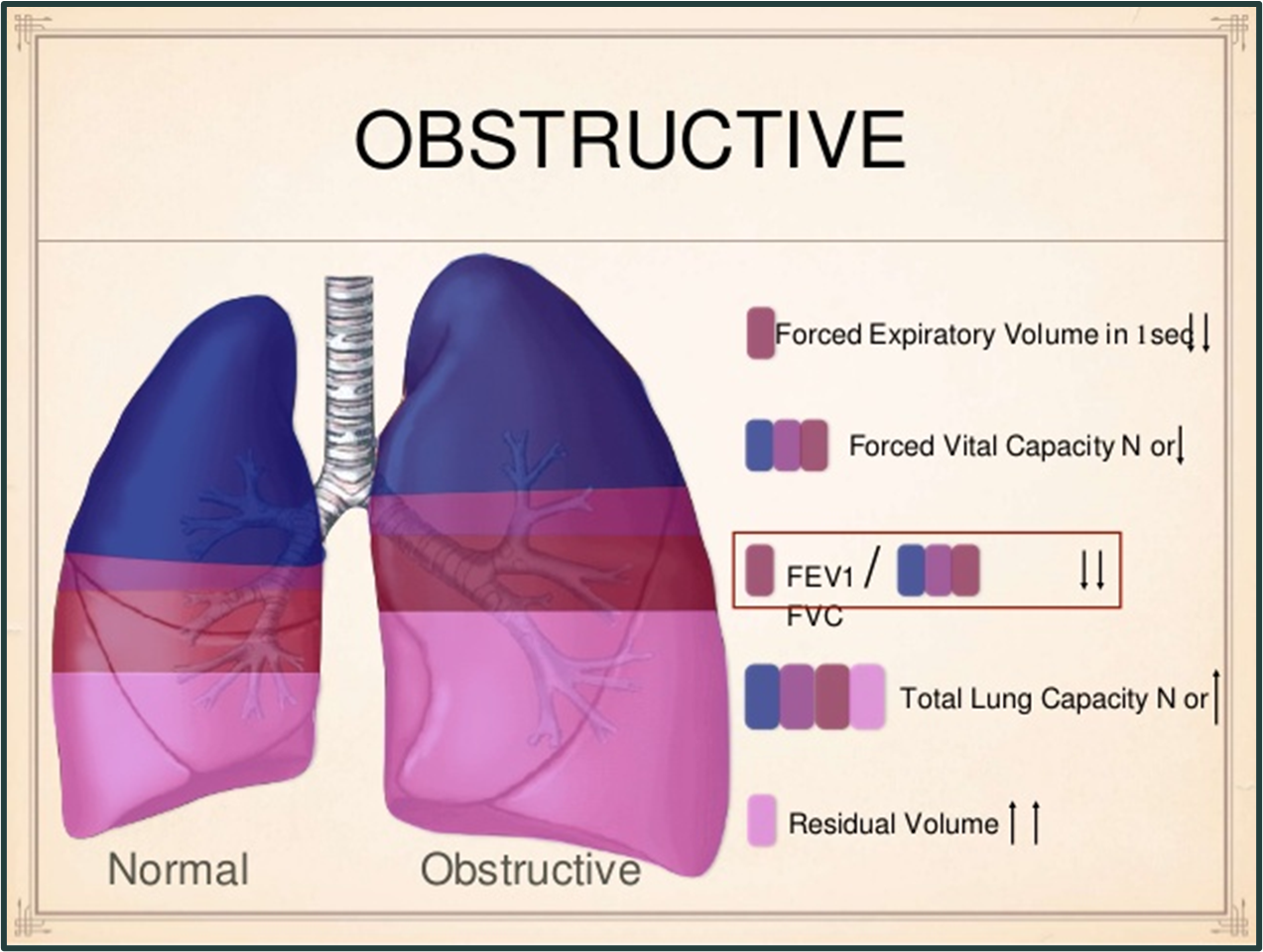
Definition: Obstructive airway disorders are conditions that limit expiratory airflow.
Characteristics:
The airway obstruction primarily causes difficulties during expiration, with a requirement for increased force or time to expel air.
Dyspnea & Wheezing → universal to all OAD
Affected individuals experience increased work of breathing, ventilation-perfusion mismatching, and reduced forced expiratory volume in one second (FEV1).
Common Disorders:
Asthma
Chronic Bronchitis
Emphysema
Coexisting conditions of chronic bronchitis and emphysema are categorized as Chronic Obstructive Pulmonary Disease (COPD).
Physiology of Airway Disease
Airway Anatomy: Air travels from upper airways (trachea and major bronchi) to lower airways (bronchi and alveoli).
Airway Structure:
In pulmonary airways, cartilage transitions into smooth muscle strips the closer you get the to alveoli
Smooth Muscle Control:
Controlled by the autonomic nervous system (ANS).
Parasympathetic stimulation (via the vagus nerve) causes bronchoconstriction.
Sympathetic stimulation (via beta-2 adrenergic receptors) promotes dilation
Asthma
Definition and Risk Factors
Asthma: A chronic obstructive airway disorder characterized by episodes of airway obstruction, bronchial hyper-responsiveness, airway inflammation, and occasionally airway remodeling.
Major Risk Factor: Genetic predisposition identified (over 100 genes linked to asthma susceptibility and IgE-mediated responses to allergens).
Onset: Can occur at any age; predominantly in childhood or early adulthood.
Approximately 50% of asthma cases begin in childhood; 30% before age 40.
Childhood Risk Factors: Family history, allergies, and prenatal exposure to tobacco smoke and pollution.
Classification
Types of Asthma:
Acute (single episode) vs. Chronic (long-term condition).
Extrinsic: Triggered by Type I hypersensitivity (allergens).
Intrinsic: Adult-onset asthma.
Exacerbations: Recurrent episodes
Severity Factors: Genetics, allergens, pollution exposure, and environmental triggers (e.g., tobacco smoke).
Increased risks associated with viral infections (trigger exacerbations) and obesity.
Emotional factors and hormonal changes may exacerbate symptoms.
Current trends show decreased mortality rates but increased morbidity, particularly among Blacks and older adults (>85 years).
Etiology and Pathogenesis
Underpinning Factor: Exaggerated IgE mediated hyper-responsiveness to stimuli, leading to:
Inflammation with eosinophils, lymphocytes, mast cells.
Damage to bronchial epithelium.
Chronic Inflammation:
Causes bronchial hyper-responsiveness, mucosal edema, and airflow obstruction, often reversible.
Episodes include bronchospasm, inflammation, edema, and increased mucus production.
Cytokine Role: Involved in inflammatory responses and asthma complications.
Exacerbation:
1)Inflammation of the mucosa with noted edema;
2)↑ contraction of respiratory-associated smooth muscles (bronchoconstriction); and
3)Secretions of thick mucus in the respiratory passages
vInflammatory process → recurrent episodes (i.e., exacerbations) of airway obstruction and episodic bronchospasms
§Characterized by:
oInspiratory and expiratory wheezing, breathlessness or dyspnea, tachypnea, chest tightness, and a non-productive cough that is often worse at night and in the early morning
Clinical Manifestations
Asthma Attacks: Can occur spontaneously or due to triggers (e.g., infections, emotional stress).
Nocturnal asthma is common due to circadian variations and hormone changes.
Symptoms:
Episodic bronchospasms, expiratory and inspiratory wheezing.
Dyspnea, nonproductive cough, tachypnea.
Asymptomatic between exacerbations with normal pulmonary function tests.
Additional Symptoms:
Tightness in the chest, agitation, inability to talk, accessory muscle use, potential hypoxia.
Initial respiratory alkalosis leads to respiratory acidosis over time due to air trapping.
Diagnosis and Treatment Goals
Diagnosis: Based on history, physical exams, lab findings, and pulmonary function studies.
Treatment Goals:
Minimize frequency and severity of acute attacks to avoid lung damage.
Reduce risks for chronic obstructive lung disease.
Most effective strategy: Avoidance of triggers
Severe or Refractory Asthma
Definition: Refers to asthma individuals requiring high medication dosages for symptom control or who experience persistent symptoms despite treatment.
Risks: Increased likelihood of fatal asthma outcomes.
Etiologies: Include genetic factors, infections, continued allergen exposure, and comorbidities such as gastroesophageal reflux.
Acute Attack Risk Factors: Cardiac arrhythmia and asphyxia may cause death during attacks.
Asthma in Older Adults
Immunologic Considerations: Decreased immunity with age heightens asthma control difficulties.
Comorbidities: Older adults are more likely to have conditions affecting asthma management.
Management Challenges: Complexity of treatment regimens and potential physical or cognitive impairments impede control.
Asthma in Children
Prevalence: Leading cause of chronic illness in children; more frequent admissions in Black children.
Pathogenesis: IgE-mediated responses to respiratory viruses often precede asthma onset.
Contributing Factors: Environmental allergens and tobacco smoke increase risk.
Clinical Manifestations: Include symptoms of prolonged cough without cold symptoms in infants.
Potential rapid progression with irritability, cough, tachypnea, and severe respiratory distress indicated by cyanosis and tachycardia.
Chronic Obstructive Pulmonary Disease (COPD)
Definition and Epidemiology
Definition: Chronic obstructive pulmonary disease is marked by persistent and progressive airflow obstruction due to inflammatory responses to harmful particles and gases.
Epidemiology: Fourth leading cause of mortality in the US; disproportionately affects assigned-at-birth females (56%) compared to males (44%).
§COPD is a leading etiology of worldwide morbidity and mortality
Leading Etiology: Smoking (80% of COPD deaths attributed to smoking); hereditary deficiency of alpha-1 antitrypsin is rarer.
Risk Factors and Diagnosis
At-Risk Populations: Older adults with recurrent respiratory infections, asthma, and those with chronic productive cough are at higher risk.
Recognition: Symptoms are often advanced by the time they are noted, with many smokers being undiagnosed.
Potential Indicators: Chronic cough, sputum production, and dyspnea should prompt suspicion of COPD.
Etiology and Pathogenesis
Mechanisms: COPD involves inflammation and fibrosis of bronchial walls, mucus hypersecretion, and loss of lung elasticity.
Consequences:
Inflammation obstructs airflow, leads to mismatching ventilation/perfusion, and reduces gas exchange surface area.
Loss of elastic recoil exacerbates airflow limitation and increases air trapping, resulting in airway collapse.
Emphysema
Definition and Pathology
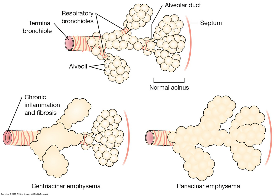
Definition: Emphysema is characterized by loss of lung elasticity and abnormal enlargement of airspaces distal to terminal bronchioles and alveolar wall destruction.
Mechanism: Caused by protease activity that breaks down elastin and alveolar structures.
Physiological Changes: Results in hyperinflation of the lungs and increased total lung capacity.
Clinical Manifestations:
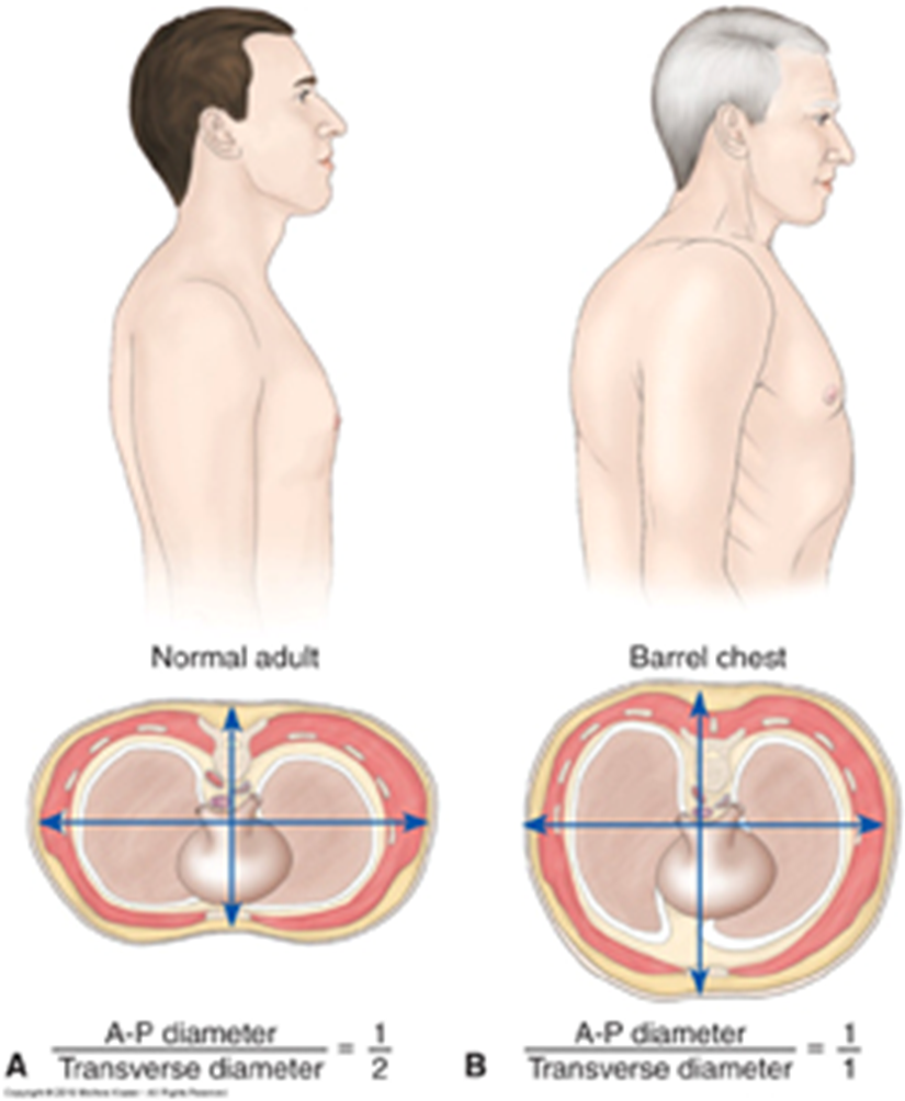
Presents with barrel chest (increased anterior-posterior diameter), circumoral cyanosis, and pursed-lip breathing during prolonged expiration.
Types of Emphysema
Centriacinar (Centrilobular):
Most common form, predominantly in smoking males, affecting bronchioles while preserving alveolar ducts initially.
Panacinar:
Involves alveoli and typically occurs in alpha-1 antitrypsin deficiency cases; can occur with centriacinar features in smoking.
Chronic Bronchitis
Pathogenesis and Definition
Definition: Chronic bronchitis is marked by inflammation leading to airway obstruction, characterized by hypersecretion of mucus and chronic productive cough.
Pathological Changes:
Initiated by inhaled irritants that induce neutrophilic infiltration, bronchial edema, and thick mucus production due to increased mucous gland size.
This compromises lung defense mechanisms, enhancing susceptibility to infections.
Diagnosis
Clinical Criteria: Diagnosis requires a chronic productive cough lasting at least three consecutive months for two consecutive years, with typical findings of purulent sputum during exacerbations.
Clinical Findings: Most common in middle-aged individuals with chronic irritative smoke exposure and recurrent infections.
Irreversibility: Unlike acute bronchitis, chronic bronchitis leads to irreversible airway changes.
COPD Clinical Manifestations
Symptoms and Progression
Symptom Onset: Progressive and insidious; typically features morning productive cough with worsening dyspnea.
Exacerbation: Frequent respiratory infections and failure are common in advanced COPD.
Presentation: Clinical attention is usually sought in the fifth or sixth decade of life due to symptoms such as fatigue, intolerance, and cough.
Mortality: Often occurs during infectious exacerbations leading to respiratory failure.
Emphysema vs. Chronic Bronchitis Differences
Emphysema:
Presentation: “Pink puffers”, accessory muscle use, breathlessness, barrel chest due to air trapping.
Chronic Bronchitis:
Presentation: “Blue bloaters”, cyanosis indicating hypoxia, potential heart failure.
Despite differences, patients often exhibit symptoms of both conditions.
Diagnosis and Management of COPD
Diagnosis: Based on history, physical examination, pulmonary function testing, imaging, and lab tests.
Management: No cure exists; however, treatment can modify disease progression with smoking cessation being crucial in managing COPD and emphysema.
Bronchiectasis
Definition and Pathophysiology
Definition: Bronchiectasis is an uncommon type of COPD characterized by permanent dilation of bronchi and bronchioles due to elastic tissue destruction.
Etiology: Not a primary disease; often occurs secondary to frequent pulmonary infections.
Associated Conditions: Seen in individuals with cystic fibrosis, tuberculosis, and previously after bacterial pneumonia.
Etiology and Pathogenesis
Mechanism: Mucus obstruction and persistent infection weaken and dilate bronchial walls.
Forms:
Localized: Affects specific lung segments, usually due to obstruction of airways.
Generalized: Bilateral and mainly affects lower lobes, associated with hereditary or acquired disorders.
Clinical Manifestations and Diagnosis
Symptoms: Include recurrent infections, copious purulent sputum, and hemoptysis. Weight loss and anemia are potential complications.
Additional Signs: May show signs of chronic bronchitis and emphysema leading to significant dyspnea and cyanosis, with digital clubbing commonly seen.
Diagnosis: Based on history, imaging studies, and high-resolution CT scans.
Cystic Fibrosis (CF)
Definition and Pathogenesis
Definition: Cystic fibrosis is an autosomal recessive disorder with defective chloride ion transport, causing thick mucus that obstructs airways and various ducts.
Pathophysiology: Abnormal protein production leads to respiratory, digestive, and reproductive system complications.
Common Manifestations:§Respiratory manifestations: persistent cough or wheeze, sputum production (most often characterized as “sticky”—difficult for infants and young children to expectorate), and recurrent or severe pneumonia
§Gastrointestinal manifestations: meconium ileus, constipation, gastroparesis, biliary cirrhosis, cholangiectasis, pancreatic insufficiency, distal intestinal obstruction syndrome (DIOS)
CF children get these infections→ Pseudomonas aeruginosa; Staphylococcus aureus; Haemophilus influenza; Burkholderia cepacia→ KNOW THIS
Diagnosis and Treatment
Diagnosis: Now involves universal newborn metabolic screening; the sweat test (sweat chloride concentration > 60 mEq/L) remains the gold standard.
Treatment Goals: Focus on limiting symptoms and managing complications; mortality is mainly due to respiratory disease.