The Axial Skeleton Pt. 2 (The Spine, Ribcage, and Sacrum)
Lesson Goals:
Identify the bones of the spine, ribcage, and sacrum.
Identify true, false and floating ribs.
Identify bony landmarks on the spine, ribcage, and sacrum.
Vocabulary
Lordosis: Spinal curve with the convexity of the curve anterior.
Kyphosis: Spinal curve with the concavity of the curve anterior.
Hyperlordosis: An increased lordotic curve.
Hyperkyphosis: An increased kyphotic curve.
Introduction
Besides the skull, the axial skeleton includes the bones of the spine, ribcage and the sacrum. In this section, you will learn about these bones and their structures.
The Spine
The spine consists of 24 vertebra “stacked” one on the other forming a column. There are regions of the spine which include the cervical, thoracic and lumbar.
The cervical spine consists of 7 vertebrae and has 2 very unique vertebrae called the atlas and axis.
The thoracic spine consists of 12 vertebrae that articulate with ribs.
The lumbar spine consists of 5 large vertebrae.
The vertebrae are numbered according to their location from top to bottom. For example C2 is the second cervical vertebra, T5 is the fifth thoracic vertebra and L5 is the fifth lumbar vertebra.
There are 4 spinal curves. These include cervical, thoracic, lumbar and pelvic curves.
The cervical and lumbar curves are both known as lordotic curves or lordoses. A lordotic curve is characterized by having its convexity anterior. Lordotic curves are considered secondary curves because they develop after birth when humans begin to hold their heads up, sit up and walk.
The thoracic and pelvic curves are called kyphotic curves. Kyphotic curves or kyphoses are characterized as being concave anteriorly. The kyphotic curves are considered primary curves because they are present at birth.
An increased curvature of the cervical or lumbar spine is called a hyperlorosis. A decreased curvature is called a hypolordosis.
Study the spine and the spinal curves in the following images.

Study the individual bones of the spine in the following diagrams.

C1 (Atlas): Image by Dr. Bruce Forciea

C2 (Axis): Image by Dr. Bruce Forciea

Typical Cervical Vertebra: Image by Dr. Bruce Forciea
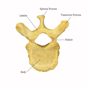
Thoracic vertebra: Image by Dr. Bruce Forciea
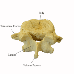
Lumbar Vertebra: Image by Dr. Bruce Forciea
Ribcage
The ribs attach to the 12 thoracic vertebra in the back and some ribs attach to the sternum in the front.
There are 3 types of ribs.
Ribs 1-7 are called true ribs because they attach directly to the sternum by way of their cartilage called costochondral cartilage.
Ribs 8-10 are called false ribs because their cartilage attaches to the cartilage of the true ribs.
Ribs 11-12 are called floating ribs because they have no connection to the sternum.
The sternum has 3 parts. The superior portion is called manubrium, the middle is called the body and the inferior portion is called the xiphoid process.
Study the ribcage in the following image.

Ribcage: Image by Dr. Bruce Forciea
Sacrum
The sacrum is a triangular bone that consists of a series of 5 fused vertebrae. At the inferior end is a small bone called the coccyx which consists of another series of 3-5 very small fused vertebrae.
View the sacrum and coccyx in the following image.