Small & Large Intestines
Describe the process of digestion, absorption, secretion, and motility of the small intestine and colon.
PART 1: SMALL INTESTINE
1. Digestion in the Small Intestine
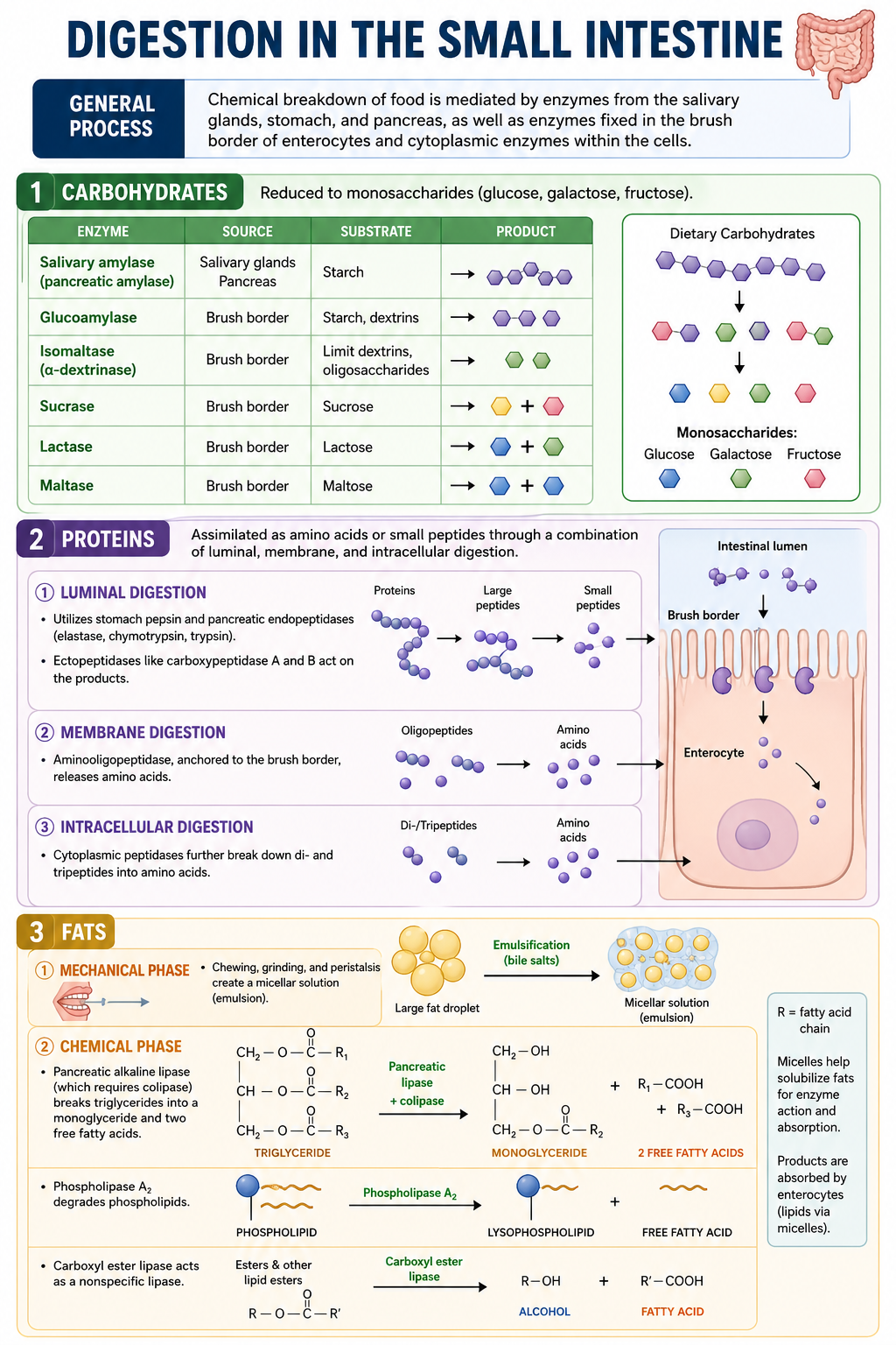
General Process: Chemical breakdown of food is mediated by enzymes from the salivary glands, stomach, and pancreas, as well as enzymes fixed in the brush border of enterocytes and cytoplasmic enzymes within the cells.
Carbohydrates: Reduced to monosaccharides (glucose, galactose, fructose).
Enzymes used: Salivary amylase and pancreatic lipase (for starch), glucoamylase and isomaltase (for oligosaccharides), sucrase (for sucrose), lactase (for lactose), and maltase (for maltose).
Proteins: Assimilated as amino acids or small peptides through a combination of luminal, membrane, and intracellular digestion.
Luminal digestion: Utilizes stomach pepsin and pancreatic endopeptidases (elastase, chymotrypsin, trypsin). Ectopeptidases like carboxypeptidase A and B act on the products.
Membrane digestion: Aminooligopeptidase, anchored to the brush border, releases amino acids.
Fats:
Mechanical phase: Chewing, grinding, and peristalsis create a micellar solution (emulsion).
Chemical phase: Pancreatic alkaline lipase (which requires colipase) breaks triglycerides into a monoglyceride and two free fatty acids. Phospholipase A2 degrades phospholipids, and carboxyl ester lipase acts as a nonspecific lipase.
2. Absorption in the Small Intestine
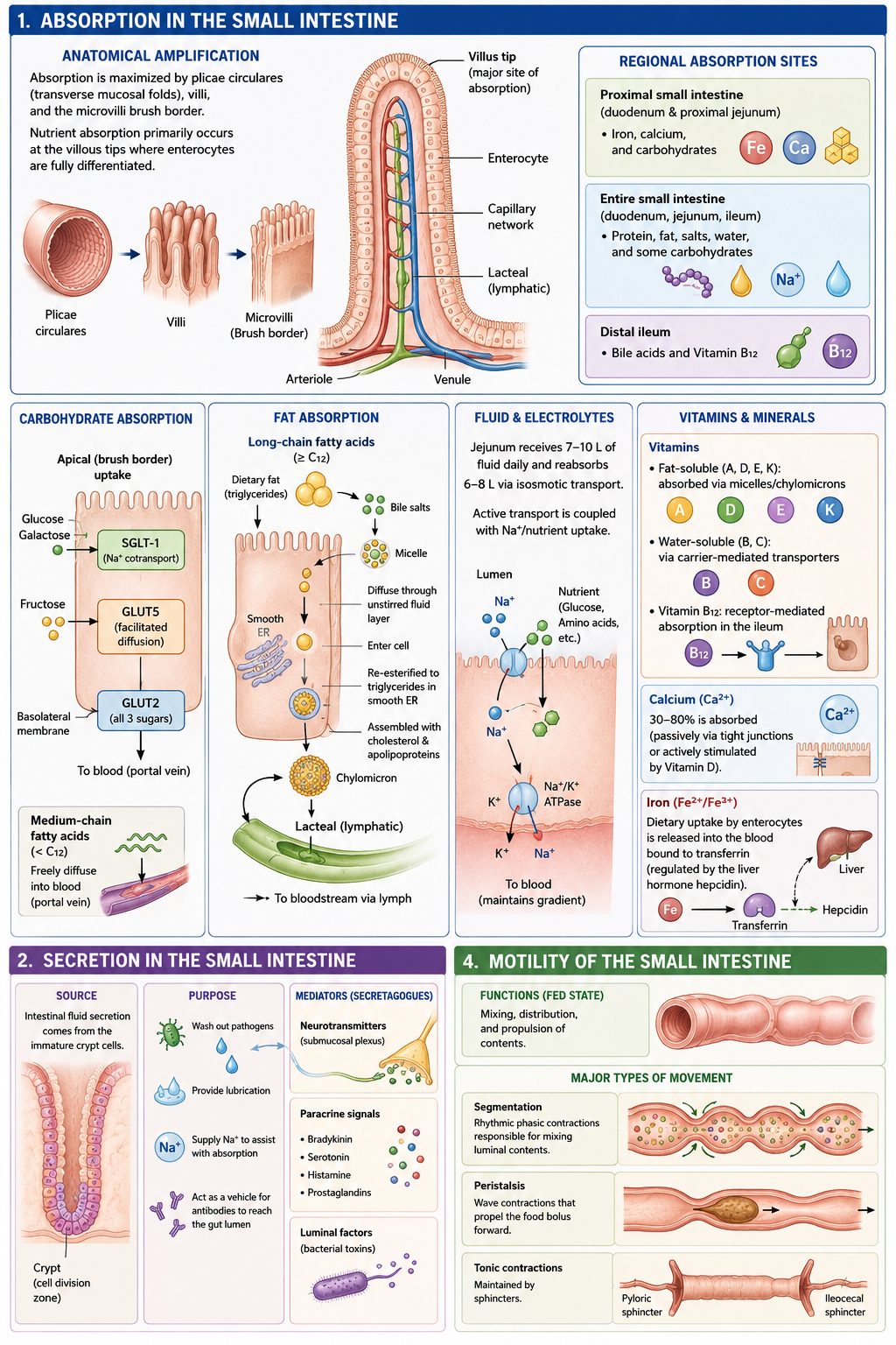
Anatomical Amplification: Absorption is maximized by plicae circulares (transverse mucosal folds), villi, and the microvilli brush border. Nutrient absorption primarily occurs at the villous tips where enterocytes are fully differentiated.
Regional Absorption Sites:
Proximal small intestine: Iron, calcium, and carbohydrates.
Entire small intestine: Protein, fat, salts, water, and some carbohydrates.
Distal ileum: Bile acids and Vitamin B12.
Carbohydrate Absorption: Uses SGLT-1 (for glucose and galactose) and GLUT5 (for facilitated diffusion of fructose). GLUT2 transports all three.
Fat Absorption:
Medium-chain fatty acids: Freely diffuse into the blood.
Long-chain fatty acids: Dissolve into micelles to diffuse through the unstirred fluid layer. They enter the cell, are converted back into triglycerides in the smooth ER, and form chylomicrons. Chylomicrons are exocytosed into lymphatic lacteals before entering the blood.
Fluid & Electrolytes: The jejunum receives 7-10L of fluid daily and reabsorbs 6-8L via isosmotic transport. Active transport is coupled with Na+/nutrient uptake.
Vitamins & Minerals:
Vitamins: Fat-soluble (A, D, E, K) are absorbed via micelles/chylomicrons; water-soluble (B, C) via carrier-mediated transporters; B12 is receptor-mediated in the ileum.
Calcium: 30-80% is absorbed (passively via tight junctions or actively stimulated by Vitamin D).
Iron: Dietary uptake by enterocytes is released into the blood bound to transferrin (regulated by the liver hormone hepcidin).
3. Secretion in the Small Intestine
Source: Intestinal fluid secretion comes from the immature crypt cells.
Purpose: To wash out pathogens, provide lubrication, supply Na+ to assist with absorption, and act as a vehicle for antibodies to reach the gut lumen.
Mediators (Secretagogues): Secretion is driven by neurotransmitters (submucosal plexus), paracrine signals (bradykinin, serotonin, histamine, prostaglandins), and luminal factors (bacterial toxins).
4. Motility of the Small Intestine
Functions (Fed State): Mixing, distribution, and propulsion of contents.
Major Types of Movement:
Segmentation: Rhythmic phasic contractions responsible for mixing luminal contents.
Peristalsis: Wave contractions that propel the food bolus forward.
Tonic contractions: Maintained by sphincters
PART 2: LARGE INTESTINE (COLON)
1. Digestion in the Colon
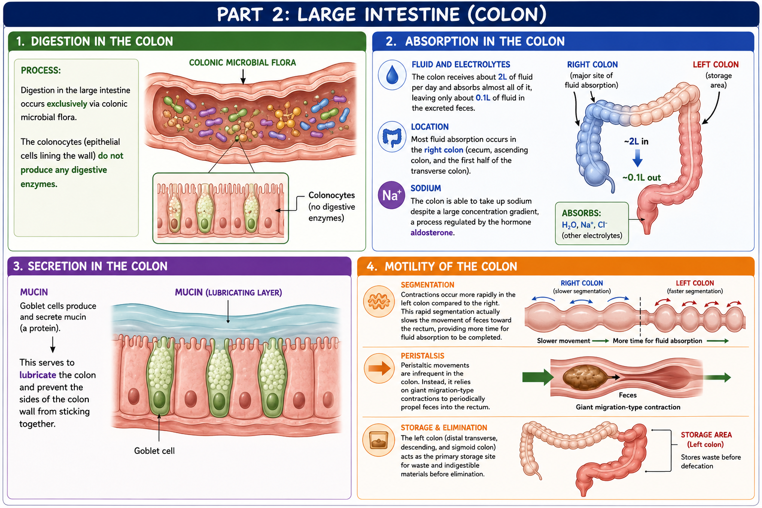
Process: Digestion in the large intestine occurs exclusively via colonic microbial flora. The colonocytes (epithelial cells lining the wall) do not produce any digestive enzymes.
2. Absorption in the Colon
Fluid and Electrolytes: The colon receives about 2L of fluid per day and absorbs almost all of it, leaving only about 0.1L of fluid in the excreted feces.
Location: Most fluid absorption occurs in the right colon (cecum, ascending colon, and the first half of the transverse colon).
Sodium: The colon is able to take up sodium despite a large concentration gradient, a process regulated by the hormone aldosterone.
3. Secretion in the Colon
Mucin: Goblet cells produce and secrete mucin (a protein). This serves to lubricate the colon and prevent the sides of the colon wall from sticking together.
4. Motility of the Colon
Segmentation: Contractions occur more rapidly in the left colon compared to the right. This rapid segmentation actually slows the movement of feces toward the rectum, providing more time for fluid absorption to be completed.
Peristalsis: Peristaltic movements are infrequent in the colon. Instead, it relies on giant migration-type contractions to periodically propel feces into the rectum.
Storage and Elimination: The left colon (distal transverse, descending, and sigmoid colon) acts as the primary storage site for waste and indigestible materials before elimination
Describe the pathophysiology including the clinical presentation, epidemiology and risk factors, pathogenesis, pathology and clinical manifestations of the following disorders of the small intestine and colon:
Diarrhea
Inflammatory bowel disease
Diverticular disease
Irritable bowel syndrome
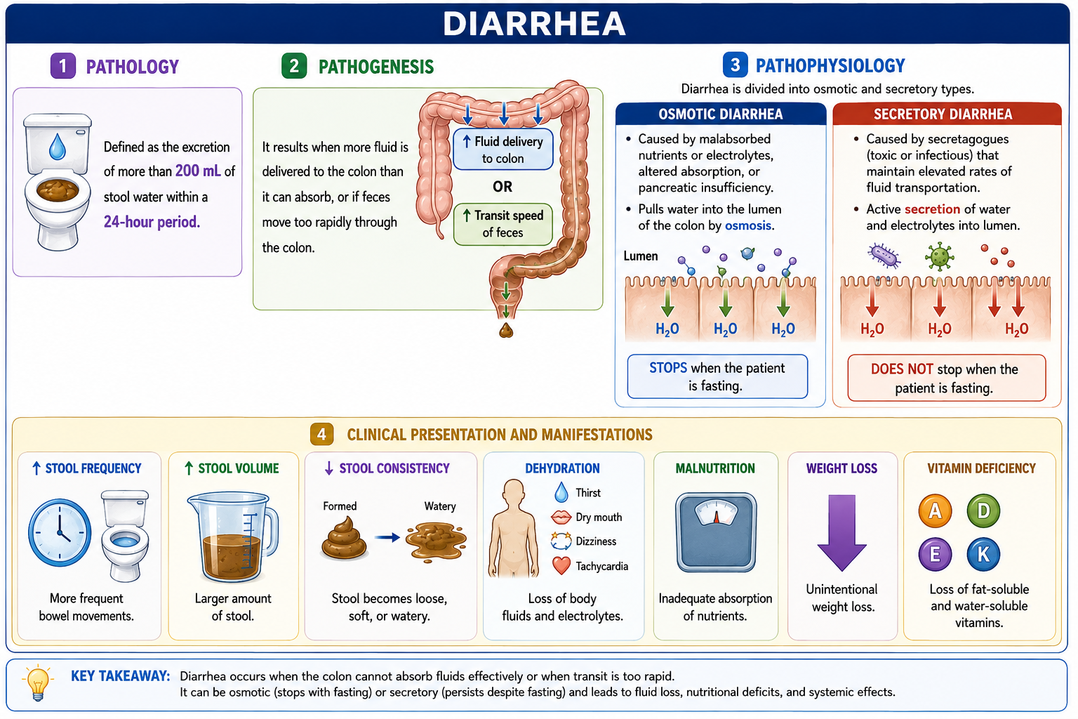
Diarrhea
Pathology: Defined as the excretion of more than 200 mL of stool water within a 24-hour period.’
Pathogenesis: It results when more fluid is delivered to the colon than it can absorb, or if feces move too rapidly through the colon.
Pathophysiology: Diarrhea is divided into osmotic and secretory types.
Osmotic diarrhea: Caused by malabsorbed nutrients or electrolytes, altered absorption, or pancreatic insufficiency. This type stops when the patient is fasting.
Secretory diarrhea: Caused by secretagogues (which can be toxic or infectious) that maintain elevated rates of fluid transportation. This type does NOT stop when the patient is fasting.
Clinical Presentation and Manifestations: Symptoms include an increase in stool frequency and volume, a decrease in stool consistency, dehydration, malnutrition, weight loss, and vitamin deficiency.
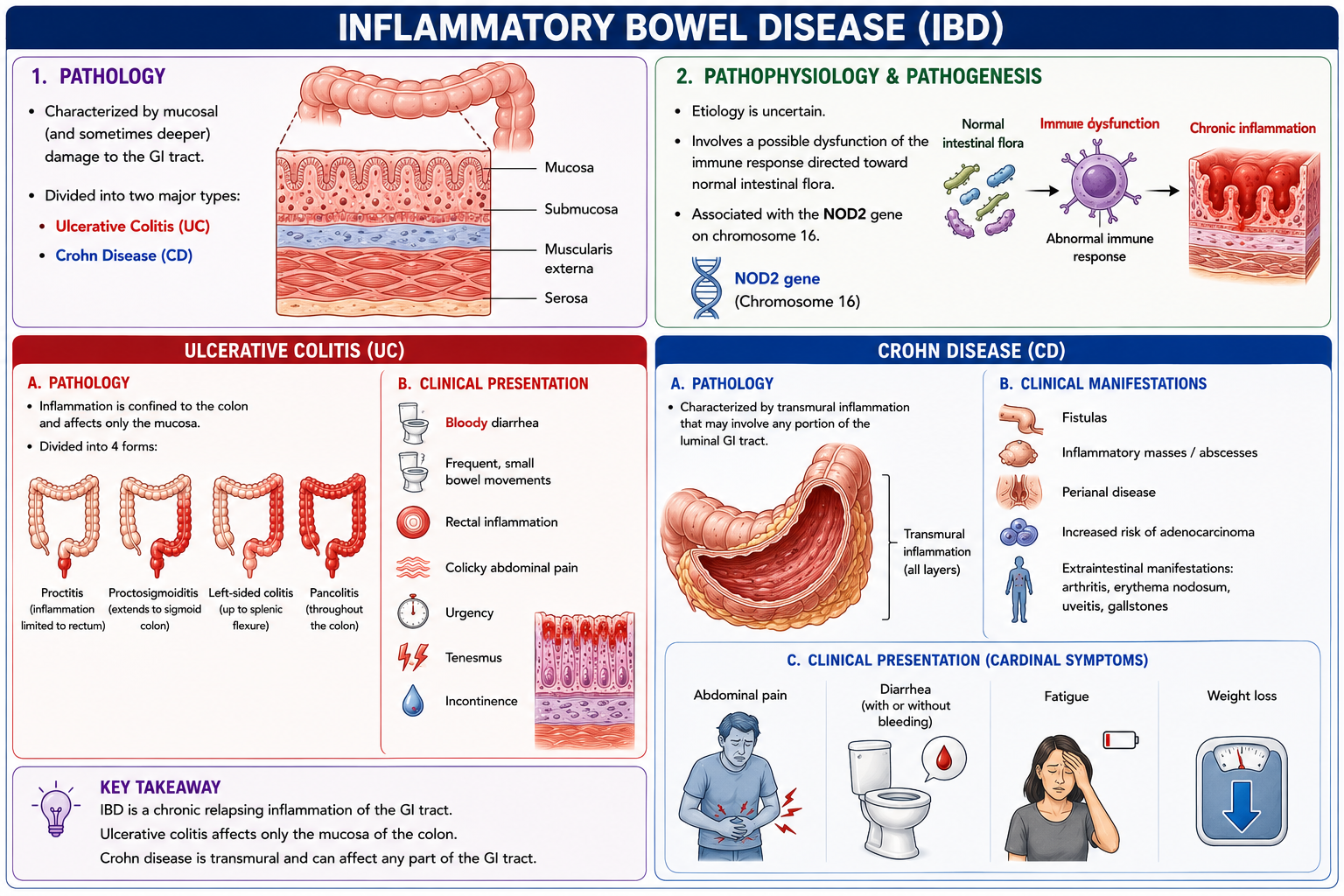
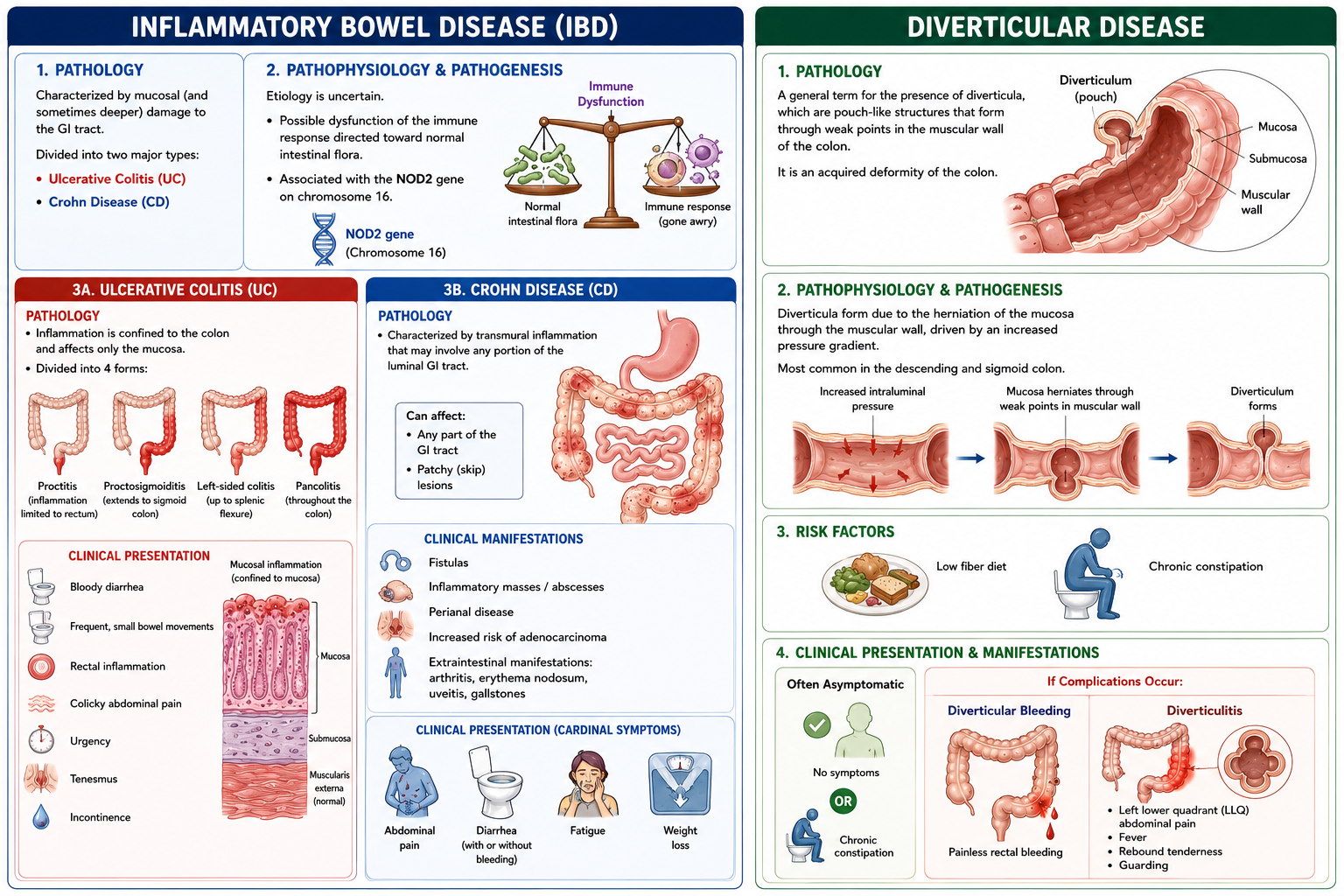
Inflammatory Bowel Disease (IBD)
Pathology: Characterized by mucosal (and sometimes deeper) damage to the GI tract. It is divided into two major types: Ulcerative Colitis and Crohn disease.
Pathophysiology and Pathogenesis: The etiology is uncertain, but it involves a possible dysfunction of the immune response directed toward normal intestinal flora. It is also associated with the NOD2 gene on chromosome 16.
Ulcerative Colitis:
Pathology: Inflammation is confined to the colon and affects only the mucosa. It is divided into 4 forms: proctitis, proctosigmoiditis, left-sided colitis, and pancolitis.
Clinical Presentation: Presents with bloody diarrhea, frequent and small bowel movements associated with rectal inflammation, colicky abdominal pain, urgency, tenesmus, and incontinence.
Crohn Disease:
Pathology: Characterized by transmural inflammation that may involve any portion of the luminal GI tract.
Clinical Manifestations: The transmural inflammation can lead to fistulas, inflammatory masses/abscesses, and perianal disease. There is also an increased risk of adenocarcinoma. It is associated with numerous extraintestinal manifestations, such as arthritis, erythema nodosum, uveitis, and gallstones.
Clinical Presentation: Cardinal symptoms include abdominal pain, diarrhea (with or without bleeding), fatigue, and weight loss.
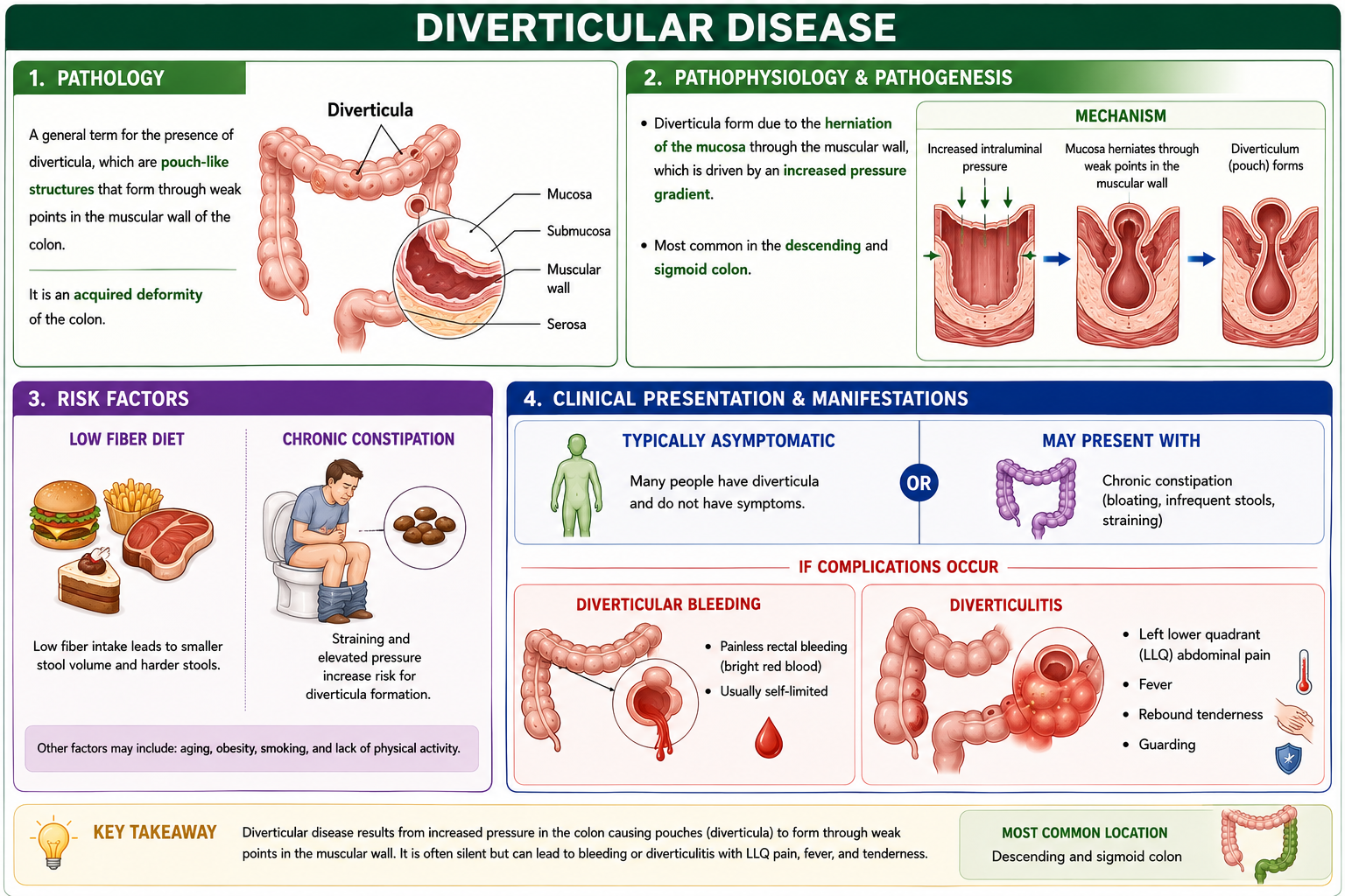
Diverticular Disease
Pathology: A general term for the presence of diverticula, which are pouch-like structures that form through weak points in the muscular wall of the colon. It is an acquired deformity of the colon.
Pathophysiology and Pathogenesis: Diverticula form due to the herniation of the mucosa through the muscular wall, which is driven by an increased pressure gradient. It is most common in the descending and sigmoid colon.
Risk Factors: Includes a low fiber diet and chronic constipation.
Clinical Presentation and Manifestations: It is typically asymptomatic or presents with chronic constipation. If complications occur, they manifest as diverticular bleeding or diverticulitis (which presents with left lower quadrant/LLQ abdominal pain, fever, rebound tenderness, and guarding).
Irritable Bowel Syndrome (IBS)
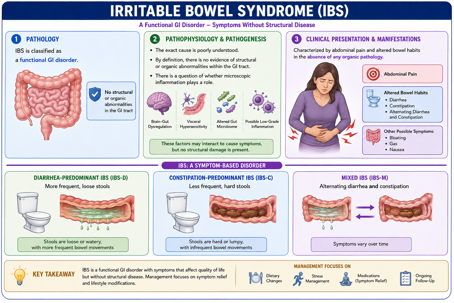
Pathology: Classified as a functional GI disorder.
Pathophysiology and Pathogenesis: The exact cause is poorly understood. By definition, there is no evidence of structural or organic abnormalities within the GI tract, though there is a question of whether microscopic inflammation plays a role.
Clinical Presentation and Manifestations: Characterized by abdominal pain and altered bowel habits in the absence of any organic pathology. The altered bowel habits can present as diarrhea, constipation, or an alternating pattern of both diarrhea and constipation.