Stem cells in drug development
4: Theoretically, pluripotent stem cells can give rise to every cell type in the animal body via fate determination and differentiation.
In the foetus, give rise to the mesoderm (→ cardiac muscle, skeletal muscle, kidney tubule cells, RBCs and gut smooth muscle), endoderm (→ alveolar cells, thyroid cells and pancreatic cells) and ectoderm (→ epidermal skin cells, neurons and pigment cells)
6: Drug development is expensive and has a high, costly failure rate. Pharma companies want most valid screening assays and preclinical models (most accurate to actual human situation) to ensure drug candidates are tuned to humans and human disease, and lack side effects.
7: One of the ways in which stem cells are used in drug development is to create valid in vitro screening processes. Somatic cells are sampled from human patient and made into induced pluripotent stem cells which can then be made to differentiate into 2D cultures of target cell types or 3D organoid systems eg mini brain, optic cup etc. These organoid systems or target cell types are then used in drug discovery/screening - focused screening and creating high content libraries
8: Stem cells can then be used to create valid pre-clinical (animal) models of the disease that the new drug is being designed to treat
9: iPS cells in the screening process have better validity and provide an ethical way to test human tissue
10: iPSCs in screening:
Somatic cells are taken from the diseased donor. These may be squamous epithelial cells, keratinocytes or fibroblasts.
Somatic cells exposed to Yamanaka factors/reprogramming which strip cells of all epigenetic modifications etc, reversing them back to iPSC state. These iPSCs are then able to differentiate into relevant screening cells - ‘disease in a dish’
11: OSKM (Yamanaka) factors induce pluripotency in differentiated somatic cells. They are a set of 4 transcription factors (Kif4, Myc, Sox2 and Oct4) which aid in upregulation of embryonic stem cell genes, upregulation of cell proliferation, downregulation of differentiation genes and loosening of chromatin structure (aids in epigenetic removal?)
12: One case in which iPSCs were used in screening is for the condition ‘Fibrodysplasia ossificans progressiva’ which is a rare condition in which soft tissue turns to bone.
Researchers took FOP patient iPS cells and differentiated them into pre-somite cells, as after this stage is where the cell would either differentiate to bone or dermal cells. These pre-somite cells were then used to screen for new drugs. 4892 compounds were screened, of which 7 were selected as result of high throughput screening. 3 out of the 7 were selected through use of FOP iPSC screening and two candidate drugs were taken on to FOP model preclinical testing.
The two candidate drugs were AZD and TAK - both reduced bone volume from approx 100mm³ to 40 mm³ - look into progression of these drugs maybe?
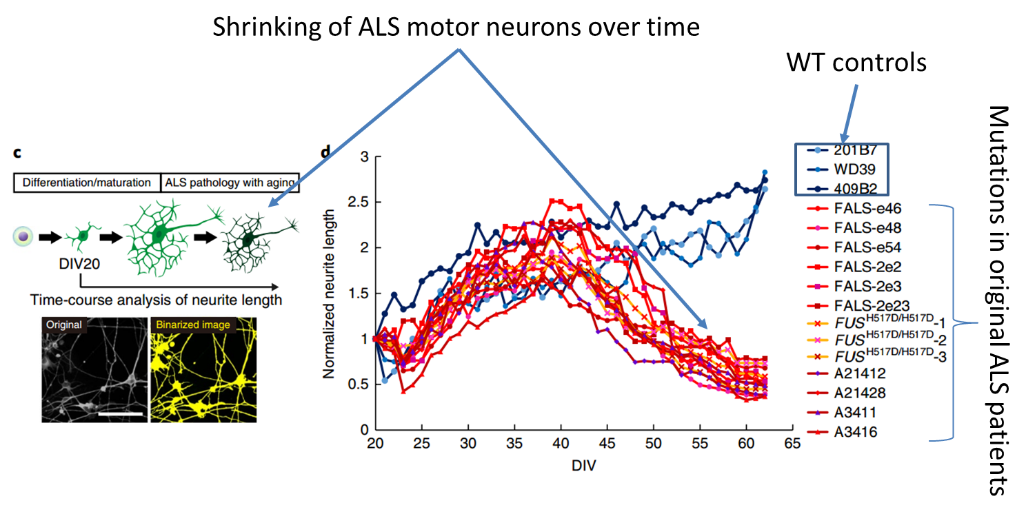
13: iPSC derived motor neurons have been used to model sporadic ALS (motor neuron disease) and this also identified a potential therapeutic target
14: Shown is a phenotypic screen of neurons derived from ALS patients. The branching of neurons is observed following by a degradation in neuron structure which would be typical of actual patient MND cases
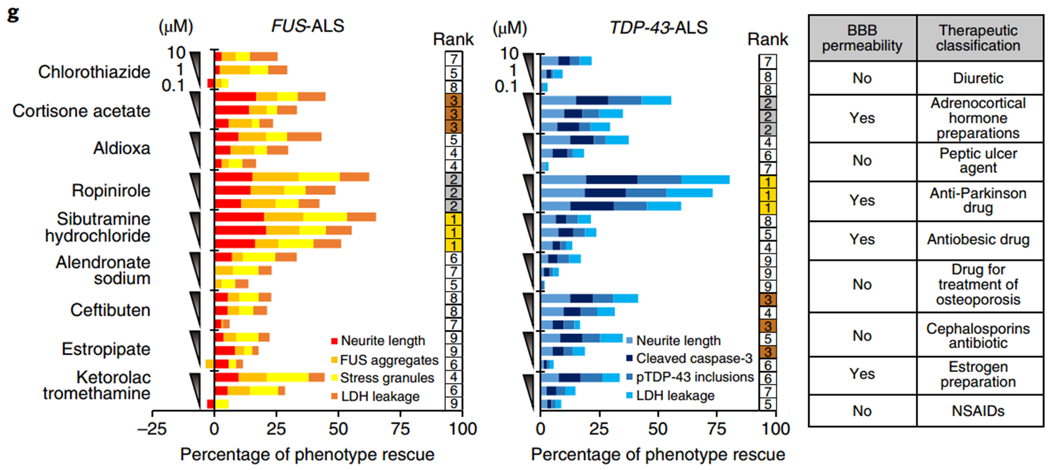
15: ALS model iPSCs were used to screen 1232 compounds in order to find compounds which both preserved neurite length and had minimal/no side effects. Drug efficacy was evaluated via ALS specific phenotypes of neurite length, FUS (fused in sarcoma, an RNA binding protein) aggregates, G3BP aggregates, LDH leakage, cleaved caspase 3 and pTDP 43 inclusions
16: Ropinirole, a drug already used for Parkinsons and restless leg syndrome, showed high phenotypic rescue and as it’s already in use for a neurodegenerative disorder is able to cross the blood brain barrier and was much easier to take to clinical trials
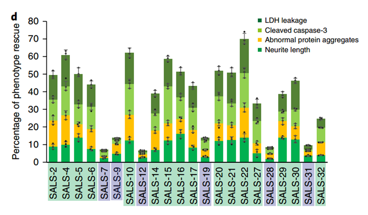
17: Ropinirole targets both familial and sporadic ALS cases where there isn’t a defined gene mutation
19: Gene knockout can be carried out in mice embryonic stem cells to create mice lacking that gene.
electroporation (electrical field is applied to cells in order to increase the permeability of the cell membrane)
homologous recombination to knock gene out in cells
selection of knockout cells with resistance marker
knockout cells injected into mouse cells
20: The knocked out cells are injected into the blastocyst/blastocoel cavity of embryo and implanted into the pseudo-pregnant female producing chimaeric mice (single organism composed of cells with more than one distinct genotype). Chimeric mice bred to produce litter with 25% homozygous KO mice, 25% WT controls.
22: Mice are often much more disease-resistant than humans, however ‘humanised’ mice have been created with the use of stem cells which more commonly mimic human disease
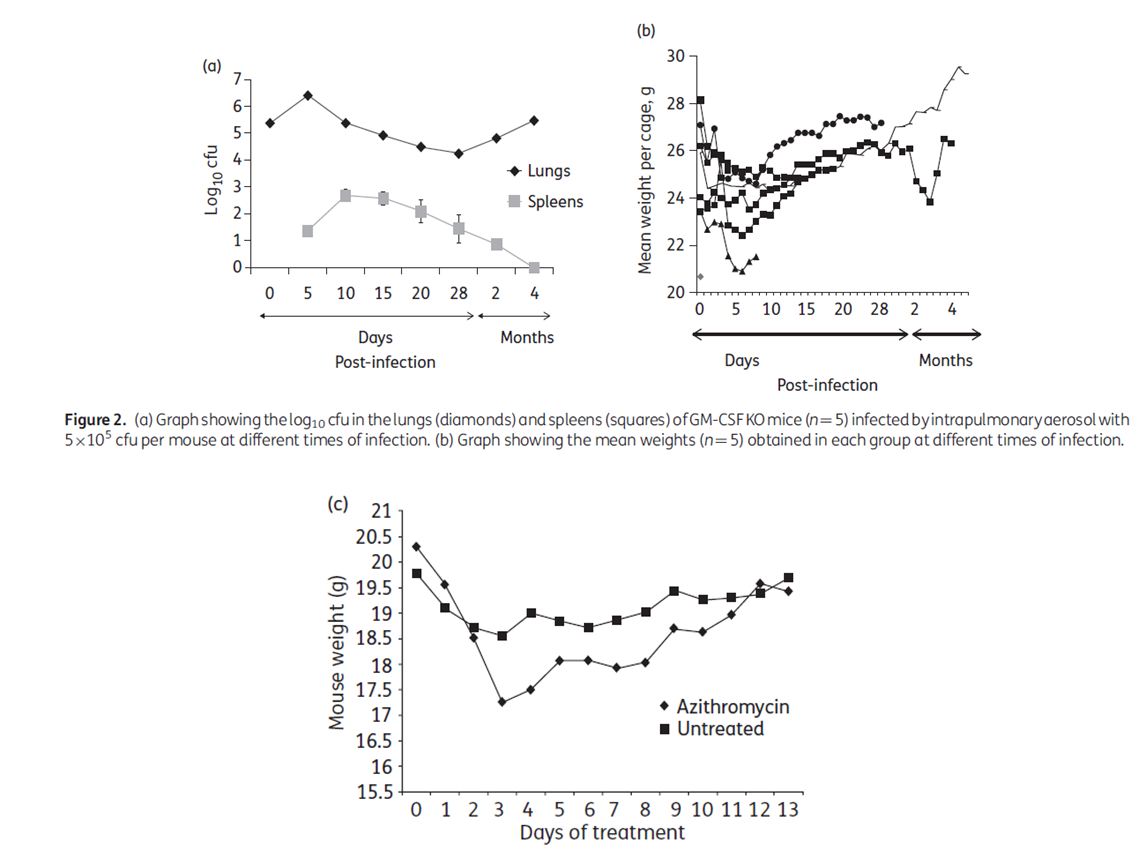
23: GM-CSF knockout mice have been created for preclinical testing of antimicrobial agents against Mycobacterium abscessus. Normally mice are able to quickly clear the infection which is not the same in humans.
24: Response to M. abscessus in mice uses immune cells and cytokines eg granulocyte-macrophage colony-stimulating factor (GM-CSF) so KO created a model of chronic pulmonary M. abscessus infection.
25: KO mice showed a week ish of acute infection which could be cleared using antibiotics