Osteology
Axial skeleton=skull vertebral column, ribs and sternum
Appendicular skeleton=pectoral girdle, upper limbs, pelvic girdle, and lower limbs
Functions of the skeletal system
Protection
ribs protect heart and lungs
skull protects brain
vertebrae protect the spinal cord
pelvic bones protect the reproductive organs
Support
provides framework for attachment of organs
Leverage
muscles pull on bones to produce movement
Blood cell production
bone marrow produces erthyocytes, leukocytes, and platelets
Storage of minerals
calcium Ions: 98% of bodies
phosphate ions
Classification of bones
Long bones= Typically longer than they are wide. Consists of a shaft and two ends
Flat bones=Thin and flatten, often a bit curved
Round/Short bones= Cube shaped bones (wrist and ankle)
Sesamoid bones=special short bones enclosed in tendons
Irregular bones= Complicated shape that do not fit into other classifications
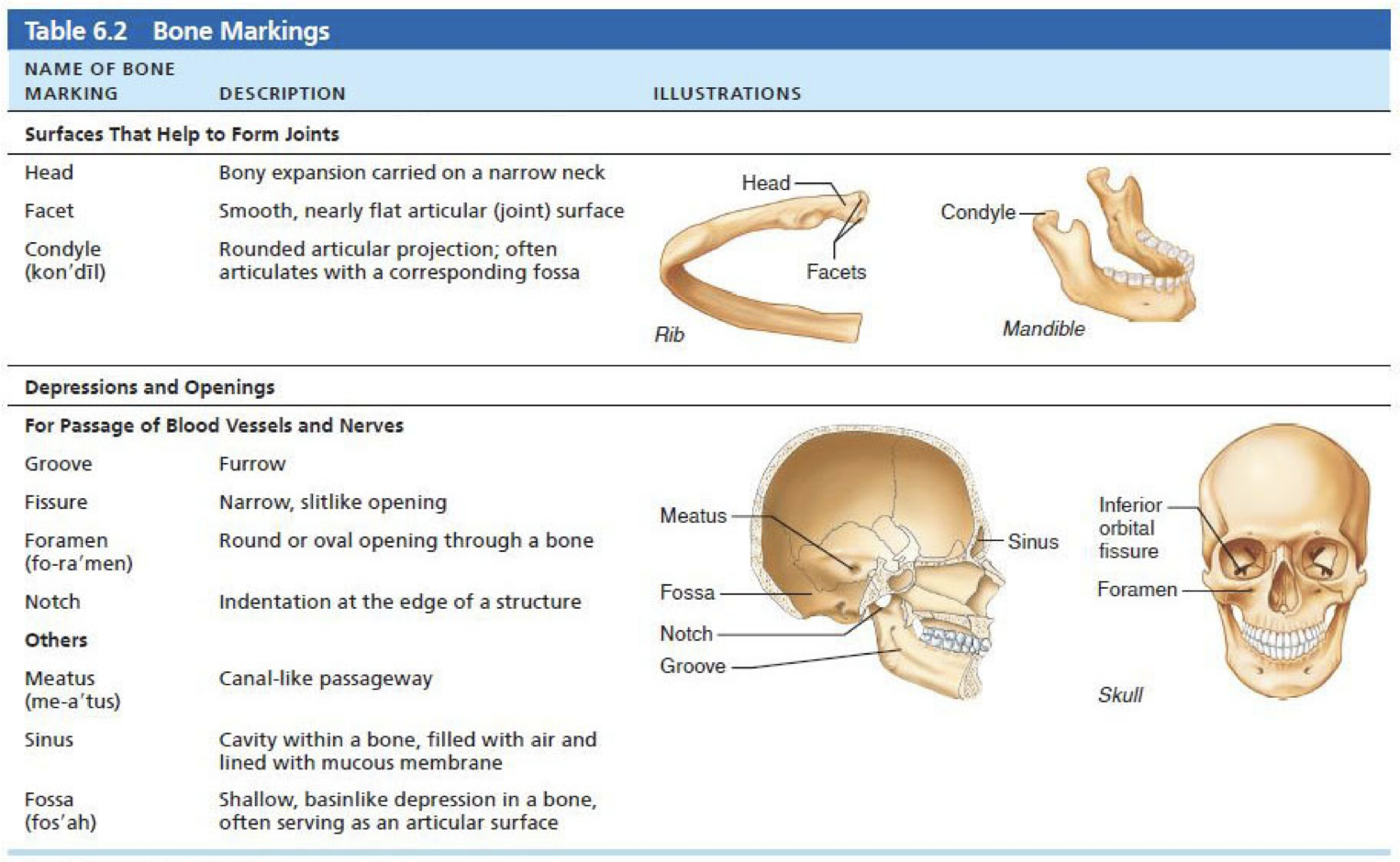
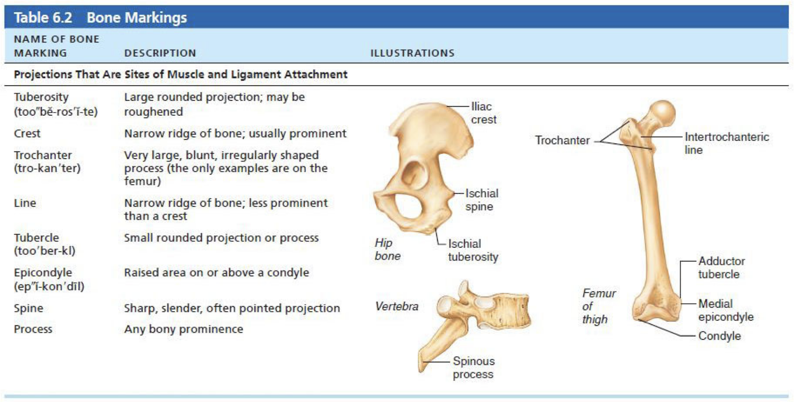
Bone Markings
Learning bone markings is part of the anatomical language
Sites of muscle, ligament, and tendon attachment on external surfacces
Areas involved in joint formation or conduits for blood vessels and nerves
Histological Organization of Bone
Osseous tissue is a supportive connective tissue
bone matrix: calcifed
collagen fibers-contribute to tensile strength of bones
bone cells
Composition of bone matrix
osteoid: organic component
ground substance
produced y the osteoblasts
consists mostly of collagen protein fibers-contirbutes to flexibility
Hydroxyapatite: inorganic component
calcium phosphate combines with calcium hydroxide
forms crystals of appetite
crystals then deposit around fiber to harder the matrix
BONE CELLS
Osteoprogenitor cells
Derived from mesenchymal cells
produce cells that the mature to become osteoblasts
located in the periosteum and endosteum in adults
initiate ossification in developing bone
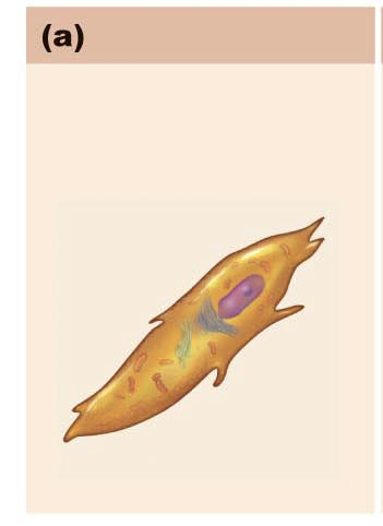
osteoblasts
Often positioned side by side on bone surfaces
synthesize and secrete osteoid
INITIAL SEMISOLID FORM OF MATRIC
later calcifies
osteocytes
mature bone cells derived from osterovlasts
have lost bone forming ability
maintain protein and mineral content of the matrix
stimulate the release of calcium ions form the bone to the blod
can detect mechanical stress on bone
osteoclasts
Breakdown bone through acidic secretions and lysomal enzymes
EAT BONE. Pac man
plays role in bone remodeling
reabsorb bone minerals such as calcium into blood stream
parathyroid hormone indirectly stimulates osteoclasts to reabsorb calcium from bone when blood calcium drops
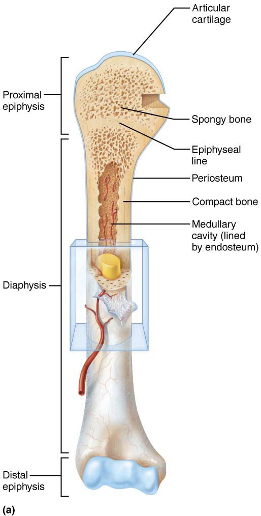
LONG BONE
Compact bone: dense outer layer that appears smooth and solid
spongy boen: moade up of a bhonoeycpmb of small needle liek pieces of bone called trabeculae
open spaces r filled with red or ywllo boen marrow
surronds the medullary cavity
Sturcture:
Diaphysis=tubular shaft
consists of compact bone surroning central meduallry caivety thatis fille dwith yellow marrow in adults
epiphyses=ends of long boens
articular cartilage covers articlar joint surfaces
between disphysis and epiphysis is epiphyseal line
remnant of childhood epiphyseal plate where bone grwon occurs
Short, irregular, and flat bones= consist of thin plates of spongy bone covered by compact bone
compact bone sanwiched between connective tissue membrnaces
hyaline carilage conver area of bone that is part of a movable joint
BONE MEMBRANES
periosteum= white double layered mem brane that covers external surfaces except joints
fibrous layer=dense irregular connective tissue
contians many nerve fibers and blood vessels
perforating fibers= anchoring points for tendons and liagments (collagen projecting into bone)
inner layer (osteogenic) =contains osteoprogeniotr cells, osteoblasts
endosteum
delicate connective tissue membrane covering internal bone surface
covers trabecular of spongy bon
lines canals that pass thorugh compact bone
COMPACT-LAMELLAR BONE
consists of
osteon
canals and canaliculi
intersitial and circymferential lamellae
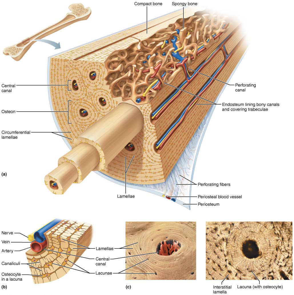
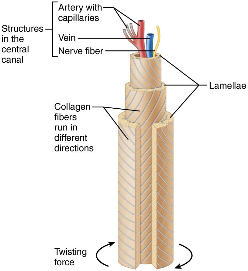
Central (Haversian) canal runs through core of osteon
Contains blood vessels and nerve fibers
Perforating (Volkmann’s) canals:
Canals connect blood vessels and nerves of periosteum, medullary cavity, and central canal
lacunae=small caivites that c ontian osteocytes
canaliculi=hairlike canals that connect to lacuasne to each other and to central canal
osterobalsts that secrete bone matric maitain contact with each other and osteocytes via cell projection siwth gap juntion
when matric harderns and cells r traped the canaliculi form
allow communication between all osteocytes or osteon and permit nutrients and wastes to be realted from one cell to antoher
BONE DEVELOPMENT
ossification (osteognesises) is the process of bone tissue fromation
formation of bony skelton begins in month 2 of developemtn
postanatl bone growth occurs until early adulhoot
always bone remodeling
UP to about week 8, fibrous mebrane and hyaline carilage o ffetal skelton are replaced with bone tissue
endochondral ossifciatoion
bone forms by replacing hyaline carilage
called cartilage (endochondral bones)
form most of skeleton
intrambranous ossificaiton