Muscles
Types of Muscle
Cardiac muscle
Smooth muscle
Skeletal muscle
Skeletal Muscle
essential for voluntary movement
Constantly used. to maintain posture
Covers the skeleton, allowing bones to be moved relative to one another
Attached to bones via a tendon (An inelastic tendon)
Voluntary nervous system controls skeletal muscles
Structure
Myofibrils - the muscle fibres which muscles are made up of
Sarcoplasm - the nuclei and cytoplasm shared by myofibrils
Within the sarcoplasm is a large concentration of mitochondria and endoplasmic reticulum
Each myofibril is mainly made up of two types of protein filaments (Actin and Myosin)

Muscle function
Muscles cannot push, only pull
Muscles work in antagonistic pairs
Two muscles acting in opposing directions
Ligaments = Bone to bone
Tendons = Muscle to bone
Ligaments and tendons cannot contract
Muscle Contraction Evidence
WHen a muscle contracts, there are changes which occur to the sarcomere
I band becomes narrower
Z lines move closer together
Sarcomere shortens
H zone becomes narrower
A band remains the same width
In a contracted muscle there is more overlap of the acting and myosin, caused by the actin filaments being pulled over the myosin filaments towards the centre of the sarcomere, therefore shortening the length of the sarcomere
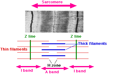
Muscle contraction
Sliding filament mechanism
An action potential reaches the neuromuscular junction
Acetylcholine is released and depolarises the sarcolemma
The action potential travels into the muscle fibre via the T tubules (A system of tubules) That branch throughout the sarcoplasm
The tubules are in contact with the sarcoplasmic reticulum which has absorbed Ca2+ from the sarcoplasm
The action potential opens Ca2+ channels and the calcium ions rush into the sarcoplasm down a diffusion gradient
The Ca2+ ions cause the tropomyosin molecules that were blocking the myosin binding sites on the actin filament to pull away
A cros-bridge forms between actin and myosin without ATP
The myosin heads change their angle pulling the actin filament along and release and ADP
A new ATP attaches to the myosin head causing it to detach from the actin
The Ca2+ ions activate the ATPase enzymes which hydrolyse ATP to ADP.
The energy released makes the myosin head return to its original position
The myosin head, once more with an attached ADP can reattach itself further along the actin filament and the cycle is repeated as long as Ca2+ ion concentrations
in the myofibril are high
Summary of mechanism
Action potential reaches neuromuscular junction
Acetylcholine is released
Sarcolemma is depolarised
Action potential travels via T tubules
Sarcoplasmic reticulum has absorbed Ca2+ from sarcoplasm
Action potential opens Ca2+ channels and Ca2+ rushes into the sarcoplasm down a diffusion graient
Presence of Ca2+ causes tropomyosin molecules to pull away
Myosin binding sites are no longer blocked
Crossbridge forms between actin and myosin
Myosin heads pull the actin filament along
Myosin heads release ADP
ATP attaches to the myosin head causing it to detach from the actin
Ca2+ ions activate ATPase
ATPase hydrolyse ATP to ADP
Myosin head returns to original position as a result of energy released
Myosin head is able to reattach itself further along the actin filament
Cycle is repeated as long as Ca2+ ion concentrations in the myofibril are high
Energy supply for muscle contraction
The hydrolysis of ATP to ADP + Pi provides the energy for movement of the myosin heads and active transport of caalcium ions back into the sarcoplasmic reticullum
ATP cannot be stored and aerobic respiration is slow, so muscles need another way of regenerating ATP
A method of rapidly regenerating ATp anaerobically can be achieved party by glycolysis and party by using pyosphocreatine
Phosphocreatine is stored in muscle and acts as a reserve supply of phosphate which can combine which ADP to form ATP under anaerobic conditions
Muscle Relaxation
Ca2+ ions are reabsorbed using energy from ATP hydrolysis
Tropomyosin therefore blocks the myosin binding sites on the actin filaments
Mosin heads are unable to bind and the muscle relaxes