Sensing the World flashcards
3: reception → amplification via enzymes/ion channel action → transmission via cellular response → response eg nerve conduction, brain circuitry, behavioural response
Slide 4: GPCRs act as sensory receptors. Alpha beta and gamma subunits. Different alphas shown in diagram represent different areas the alpha subunit affects. GDP to GTP causes dissociation.
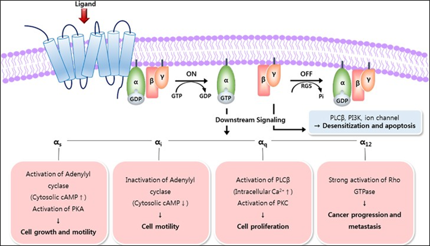
Slide 5: Different classes of GPCRs are associated with different senses - smell, taste and vision etc. We have over 800 GPCRs
Slide 7: The 2004 Nobel prize for physiology/medicine was awarded for the discovery of hundreds of genes which encode odorant receptors and the organisation of the olfactory system.
Slide 8: The olfactory system consists of exposed, highly ciliated sensory neurones. Humans have approximately 400 olfactory receptors expressed on cilia. Different organisms have different amounts.
Bowman’s gland = contains enzymes, helps produce mucus
Lamina propria = connective fibroblast tissue
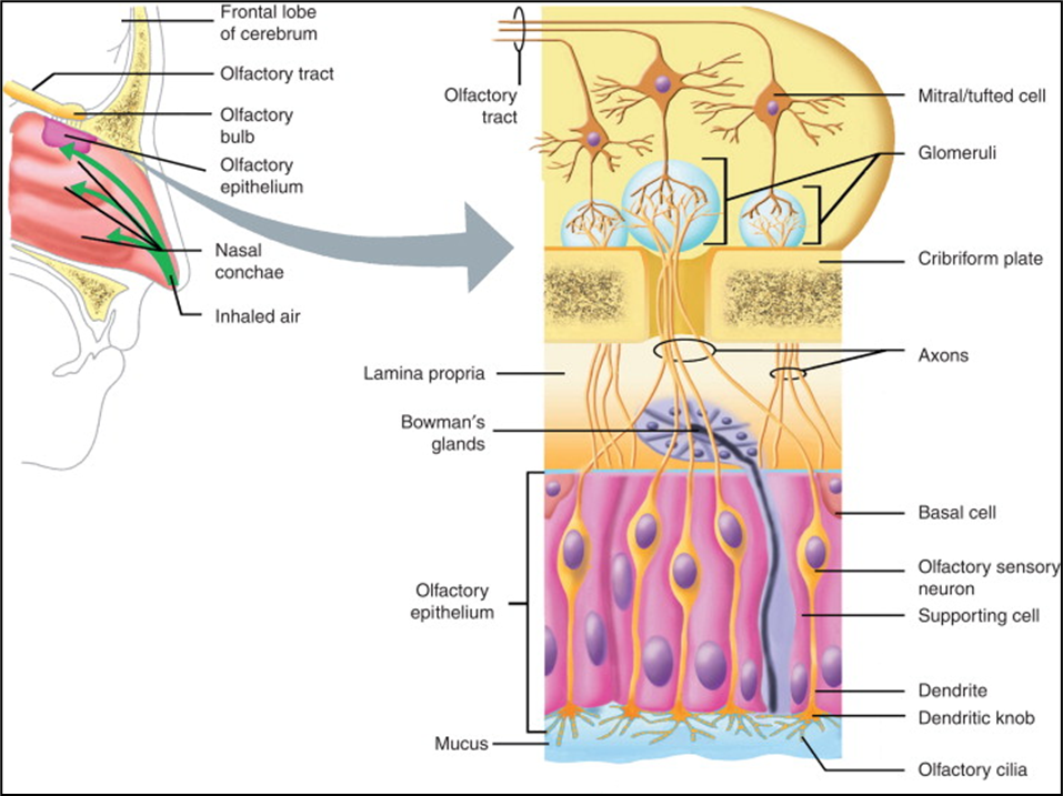
Slide 9: Each neurone expresses only 1 type of olfactory receptor - how does that happen in development? Genetic, random gene exchange on chromosomal level.
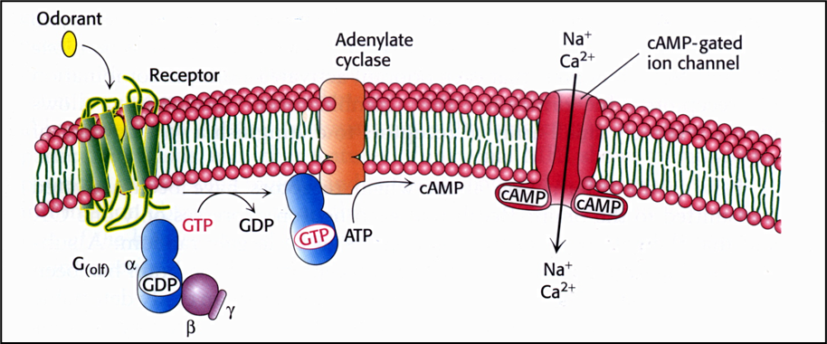
Golf is the alpha subunit of the heterotrimeric complex linked to Olf receptors which stimulates adenylyl cyclase (ATP → cAMP). cAMP opens cation channels → depolarisation. Action potential propagates into olfactory bulb in brain.
10: (summary) There are thousands of sensory neurones but only 400 olfactory receptors. All neurones expressing one particular olfactory receptor converge on their own discrete area of the olfactory bulb. How this wiring doesn’t get confused is another mystery we’re yet to uncover. We know there must be amplitude sensing at neuronal level to detect how strong a smell is.
11: Different smells or ligands are recognised by specific groups or ensembles of receptors meaning similar ligand structures can elicit very different smell responses - olfactory code.
12: Different ‘senses of smell’ is found in other parts of the body.
Odorant receptor hOR 17-4 which interacts with a particular floral odour also has a role in guiding the sperm to the egg.
similar heart muscle cell receptors may metabolically regulate heart function
immune receptors activated promote death of certain leukaemia cells
skin receptors increase skin cell regeneration
digestive tract receptors cause chronic diarrhoea/constipation but also may contribute to improved digestion
14: gustation senses components in your food triggering response to brain about whether good or bad/safe. This was helpful back when our ancestors were hunter gatherers as taste allows us to detect if something could be poisonous eg if bitter or sour. Umami is also a savoury taste from some L-amino acids eg MSG. Hedonic (pleasure) or homeostatic (good for you) eating interacts with taste (palatable/aversive) and state (hunger). Smell also can play a role.
15: soft palate = muscle fibres and connective tissue covered by membrane and contains secondary salivary glands. Epiglottis = cartilage, helps swallow.
NST = major sensory nucleus
LOOK INTO PARTS OF MOUTH
Gust cortex distinguishes taste
16: Taste receptors are mainly GPCRs. GPCRs work as dimers for umami, sweet and bitter.
V different receptors for sodium, sour and carbonation
ENaC = epithelial sodium channel
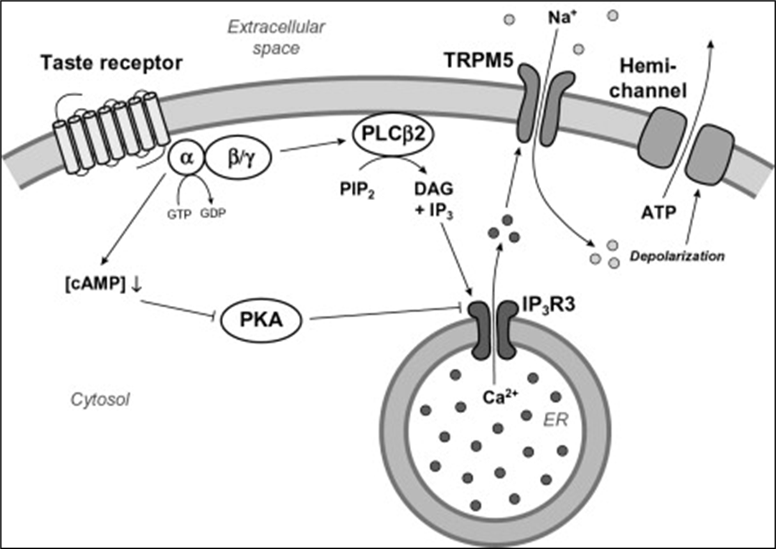
17: Taste receptor activation depolarises the taste cell eg TRPM5 is a calcium-activated non-selective cation channel which induces depolarisation upon intracellular calcium increase.
TRPM5 important in taste esp sweet.
Calcium channel shown can also be calcium/sodium
18: Taste responses are dependent on taste cell type, not the receptor. Artificial GPCR ‘Receptor activated solely by a synthetic ligand’ has a ligand, spiradoline, which is normally tasteeless. Artificially expressed RASSL in mouse taste cells caused different reactions depending on what cells they were expressed in.
19: hyposmia/anosmia/dysosmia = disease related to smell - decreased/loss/altered perception of smell.
Hypogeusia/ageusia = disease related to taste - reduced and total loss
These conditions are caused by ageing, COVID-19, mineral deficiencies, Parkinson’s and some psychiatric conditions.
21: Anatomy is as important to vision as signal detection. The lens, iris and retina can all be affected to cause degraded vision eg astigmatism, presbyopia, myopia, amblyopia, glaucoma and retinal detachment.
22: Anatomy of the retina
different types of cell which recognise stimuli
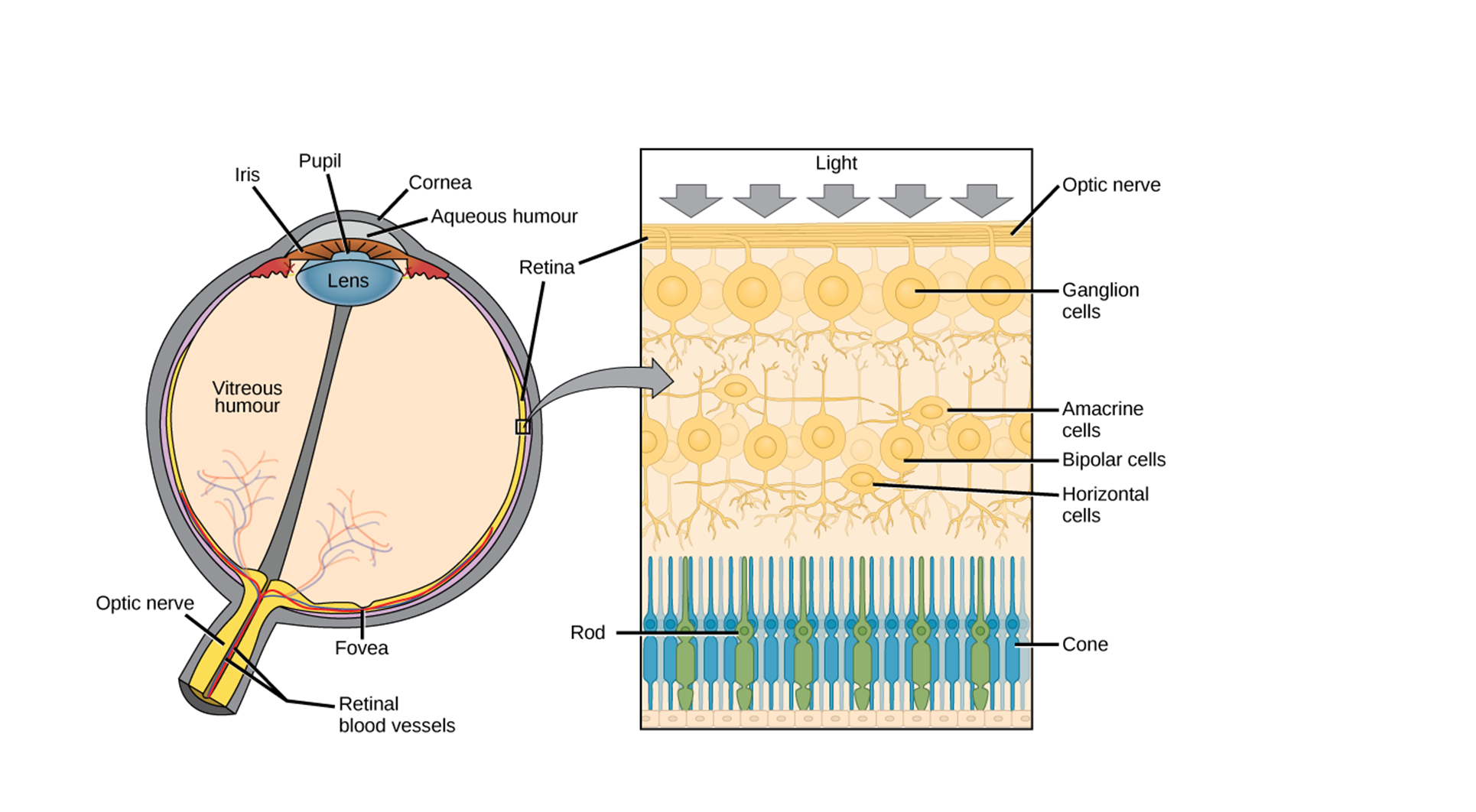
23: Rhodopsin (RHO gene) is the receptor. It’s a GPCR associated with photon ligands. Rhodopsin contains chromophore ‘retinal’ which is located within the TMDs (??) of rhodopsin.
Light induces cis → trans retinal inducing a cascade of conformational changes in rhodopsin alpha and beta subunits and triggering the heterotrimeric G protein signal transduction cascade. Trans retinal dissociates from the rhodopsin and undergoes enzymatic recycling to its cis form.
24: Normal nerve signalling works by depolarisation - action potentials. However for sight ending the inhibition of neighbouring bipolar cells is needed via hyperpolarisation so that glutamate isn’t released from photoreceptors.
25: ASK
26: Most photoreceptor cells are rods which deal with low light level and motion. Cones can differentiate colour and setail as they express different opsins. L(ong) cones = red, M(edium) cones = green and S(hort) cones = blue. Based on different wavelengths of light
27: Night blindness occurs due to vit A deficiency as this is converted into retinal. Macular degeneration may also occur which is either dry or wet. Dry is age related, due to thinning of macular and deposits of Drusen material. Wet = vasculature leakage problem.
28: diabetic retinopathy is a very common form of vision loss caused by retinal microvasculature damage.
Colour blindness (achromatopsia) is more common in males as cone genes are found on the X chromosome
29: OPN4 encodes melanopsin in the retina. Melanopsin is expressed in retinal ganglion cells and senses light to differentiate day and night for our body clocks. These cells signal to the suprachiasmatic nucleus in the brain.
Slides 30 to 38 not examinable
31: Outer ear = pinna, middle = incus, malleus, stapes and inner = cochlea and semicircular canals