Soft tissue exam
Appreciate why a thorough examination of soft tissues is essential
for accurate diagnosis and treatment planning, as it helps identify underlying pathologies that may not be apparent through imaging alone.
Also allows for a comprehensive understanding of the patient's condition, guiding clinicians in selecting the most effective therapeutic interventions.
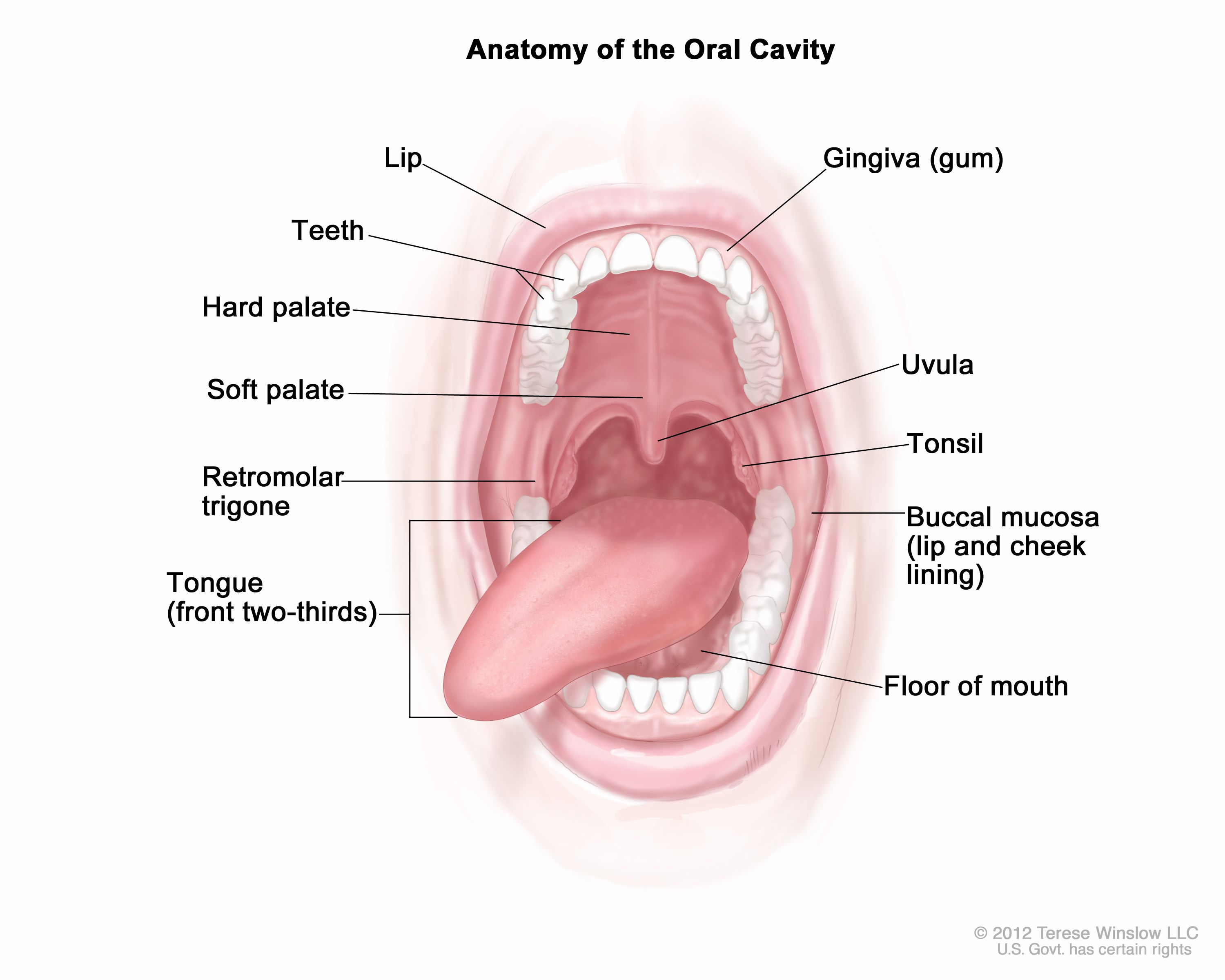
Recognise, describe and record surface anatomy of the healthy oral cavity, and identify features that deviate from health.
Soft tissue structures include:
The lips: Critical for function and aesthetics, their health can indicate overall oral hygiene.
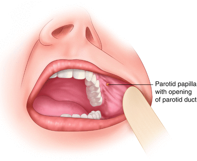
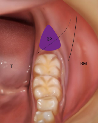
The cheeks (buccal mucosa): Important for protecting the oral cavity; any lesions could indicate systemic conditions.
The tongue: A muscle with a unique surface texture; changes in color or texture can signify underlying issues.
The gums: Healthy gums are pink and firm; signs of inflammation or recession should be documented.
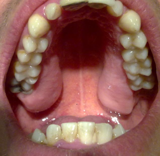
The palate: Includes hard and soft tissues; abnormalities may affect swallowing or speech.
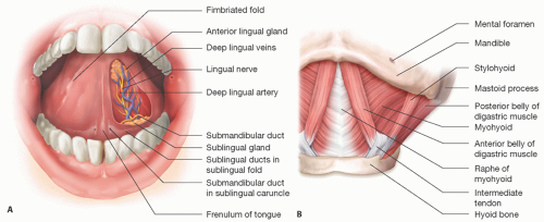
Floor of mouth: The area beneath the tongue; abnormalities, such as swelling or lesions, can indicate systemic conditions or infections.
Fauces: The region at the back of the mouth that connects the oral cavity to the oropharynx; any swelling, discoloration, or lesions may signal underlying health issues.
Describe and identify examples of structures that represent the range of normal variability
Uvula: The small, fleshy extension at the back of the soft palate; variations in size and shape are typical among individuals.
Tonsils: Lymphoid tissues located on either side of the fauces; they can vary in size, often being larger in children and smaller or even absent in adults.
Lingual frenum: The tissue connecting the underside of the tongue to the floor of the mouth; its length and thickness can differ significantly, affecting tongue mobility.