Lecture 20 - T Cell Activation
Route of circulation for mature naive T cells
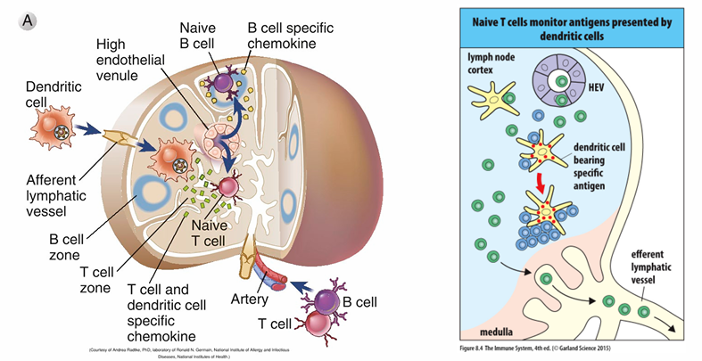
mature naive T cells exit the thymus into the bloodstream to secondary lymphoid organs like lymph nodes via HEVs
they migrate to the T cell zone and interact with dendritic cells
antigen recognition will activate the T cell
no antigen recognition prompts exit via the efferent lymphatic vessel to the next lymph node
naive T cells can enter a draining lymph node by two routes:
in the blood
in the afferent lymph coming from an upstream lymph node
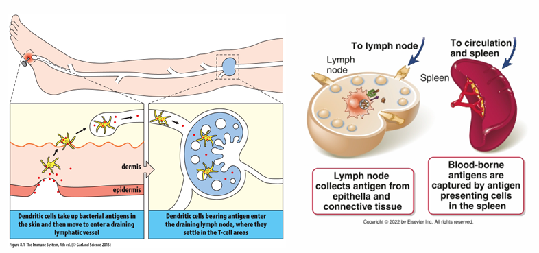
secondary lymphoid tissues collect antigens
Dendritic cells in the circulation of T cells
dendritic cells bring antigen to lymph nodes via lymphatics
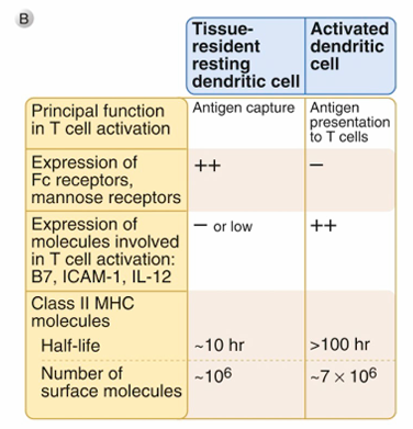
they phagocytose microbes and antigens at local sites
they are activated by PAMPs at local sites
express more MHC molecules
express co-stimulatory molecules for T cell activation (B7)
migrate into lymph nodes following chemokine gradient
these dendritic cells diff from follicular dendritic cells
T cells and dendritic cells meet in secondary lymphoid tissues
T cell Activation
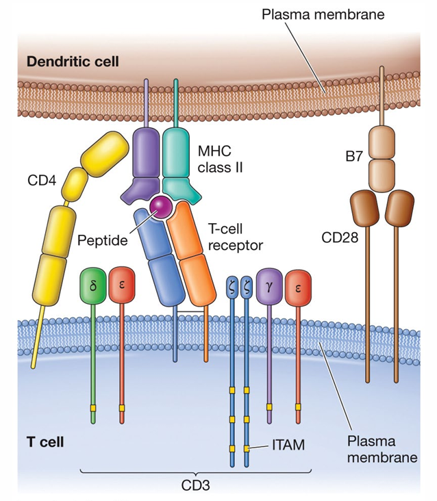
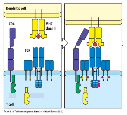
1) MHC - TCR with co-receptor (either CD4 or CD8)
dendritic cell — MHC molecule presenting antigen
T cell — TCR, CD3 complex, co-receptor
2) Co-stimulatory signal
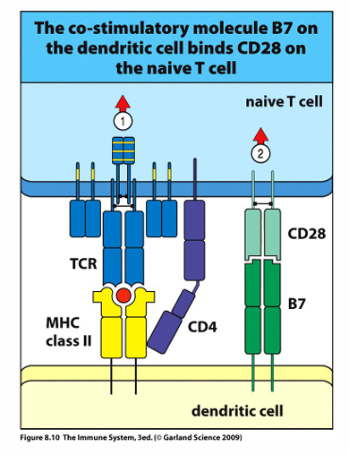
B7 - CD28
B7 is expressed on dendritic cells activated by PAMPs
the only other cells that can express B7 are macrophages and B cells, but these must be activated by T cells to express this molecule
dendritic cell — B7, expression induced when PRRs are activated by PAMPs, B cells express when activated so that they can establish the conjugate pair
T cell — CD28 & all naive T cells express CD28
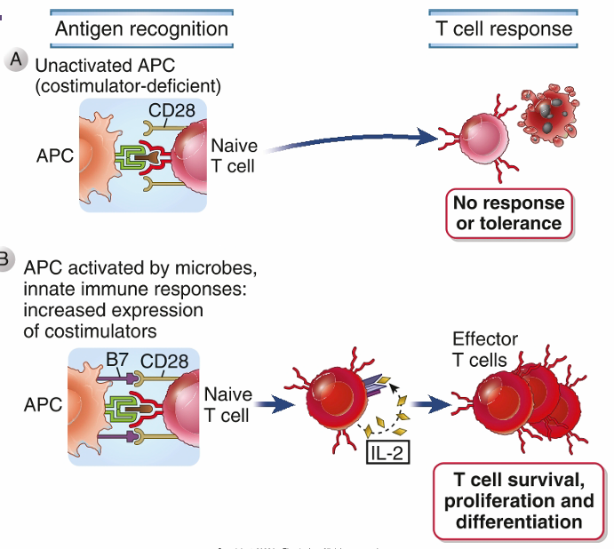
T cell activation requires co-stimulation
T cells always require co-stimulation to activate, regardless of MHC class
dendritic cells express B7 ligand only after recognizing a PAMP
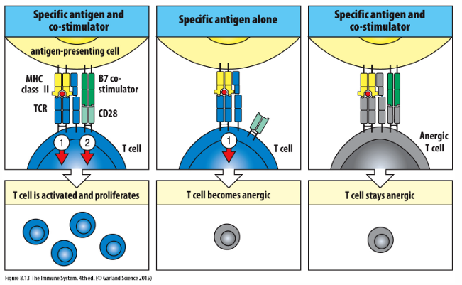
no co-stimulation = anergic T cell
peripheral tolerance — requirement of B7 co-stimulatory signal serves to negatively select T cells that bind to antigen presented by activated dendritic cells
only professional APCs can express B7 (macrophages, neutrophils)
3) Cytokine activation
IL-2
additional cytokine secretion defines T cell phenotype/functions
T cells — activated by MHC-antigen and co-stimulator
express high affinity receptor for IL-2 & secrete IL-2
IL-2 has autocrine signaling which induces T cell proliferation
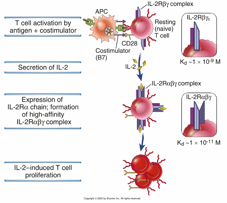
After signal 1 & 2 have been completed:
once activated by the first 2 signals, the T cell will secrete IL-2 and express high affinity receptor for IL-2