Spinal Cord Segment
Define, Describe, Differentiate
Central Nervous System (CNS) vs Peripheral Nervous System (PNS)
CNS: Brain and spinal cord; encased in bony structures.
PNS: Remainder of the nervous system; makes connections to peripheral structures.
Spinal cord and associated terms
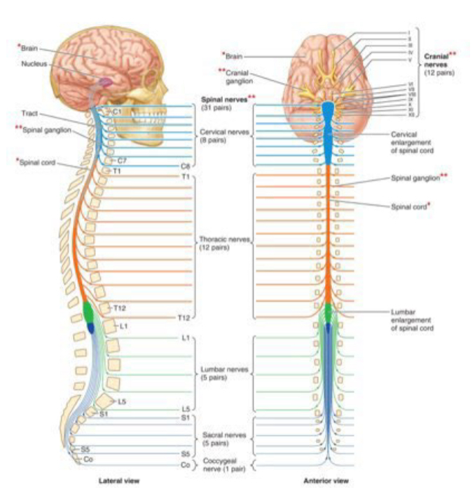
Spinal cord: continuation of the medulla oblongata of the brainstem; extends from the foramen magnum in the occiput to somewhere between the lower thoracic and upper lumbar vertebrae.
Spinal cord occupies only the superior 2/3 of the vertebral canal (because in fetal development, the vertebral column grows faster than the spinal cord).
Enlargements: cervical enlargement contributing to brachial plexus innervating the upper limbs; lumbar enlargement contributing to plexuses innervating the lower limbs.
Nerve anatomy terms (order is hierarchical)
Spinal cord segment
Spinal nerve
Ventral rootlets, ventral root, ventral ramus
Dorsal rootlets, dorsal root, dorsal ramus
Peripheral nerve
Mixed nerve (both sensory and motor)
Plexus
Dermatomes, myotomes, and cutaneous nerves
Dermotome: area of skin innervated by a single spinal nerve.
Myotome: group of muscles innervated by the fibers of a single spinal nerve.
Cutaneous nerve: nerves that supply the skin; may contain fibers from several spinal nerves; the cutaneous nerve distribution is often broader than a single dermatome.
Note: Plexuses are formed by ventral rami and distribute fibers to a region; cutaneous territories can be supplied by nerves that originate from plexuses.
Spinal Cord
Brain and spinal cord vs the remainder of the nervous system that connects to peripheral structures.
Spinal Cord specifics
Extends from the foramen magnum to between the lower thoracic and upper lumbar vertebrae.
Occupies the superior 2/3 of the vertebral canal due to differential growth rates during development.
Enlargements correlate with plexus formation:
Cervical enlargement contributes to innervation of the upper limbs via the brachial plexus.
Lumbar enlargement contributes to innervation of the lower limbs via the lumbosacral plexuses.
Pathway
Rootlet → Root → Spinal Nerve → Dorsal (posterior) ramus and Ventral (anterior) ramus
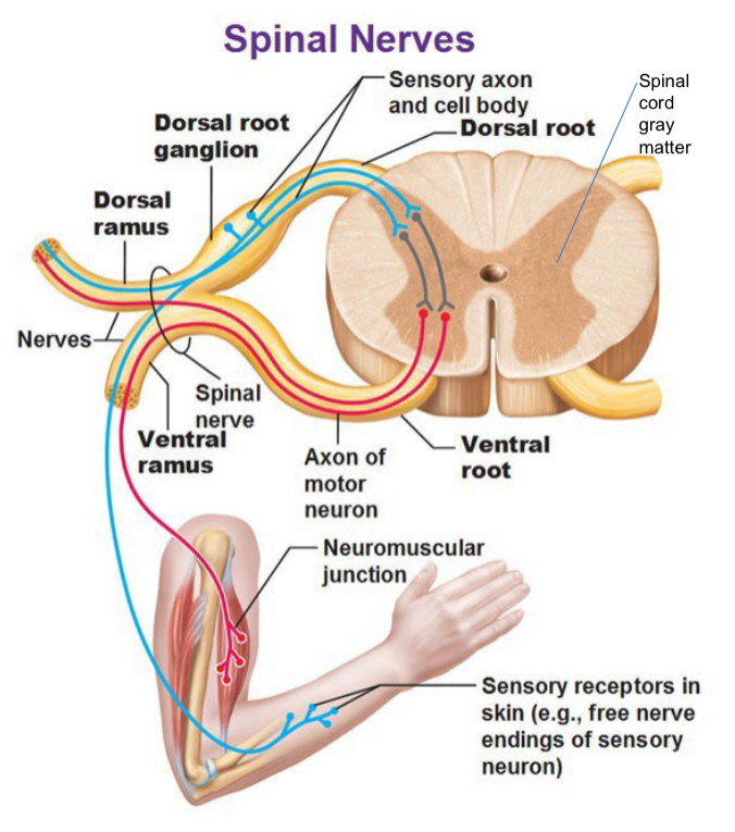
Ventral (anterior) root
Contains motor fibers from cell bodies in the anterior horn of spinal cord gray matter, traveling to effectors (e.g., muscles) peripherally.
Dorsal (posterior) root
Contains sensory fibers from cell bodies in the dorsal root ganglion (spinal sensory ganglion) that extend peripherally to sensory endings and centrally to the posterior horn of the spinal gray matter.
Spinal nerve
Contains both motor and sensory fibers; i.e., it is a mixed nerve.
Almost immediately divides into two rami (anterior and posterior) with each carrying both motor and sensory fibers.
Spinal cord gray matter
Contains neurons that contribute to motor output (anterior horn) and sensory processing (posterior horn).
The gray matter in the spinal cord is involved in reflexes and voluntary motor control.
Spinal Cord Segment
Spinal Cord Segment
The portion of the spinal cord forming a single pair of spinal nerves.
Spinal nerves
There are pairs of spinal nerves: cervical, thoracic, lumbar, sacral, coccygeal.
Additional notes
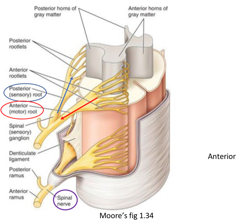
Anterior (ventral) roots carry motor fibers; posterior (dorsal) roots carry sensory fibers.
The rootlets coalesce to form the dorsal and ventral roots, which join to form the spinal nerve; the spinal nerve then divides into dorsal (posterior) and ventral (anterior) rami, each carrying mixed fibers.
The ventral ramus supplies anterior body wall and limbs; the dorsal ramus supplies the posterior trunk and muscles.
Lumbosacral Plexus
Lumbosacral Plexus overview
Lumbosacral plexus encompasses spinal nerve contributions roughly from to .
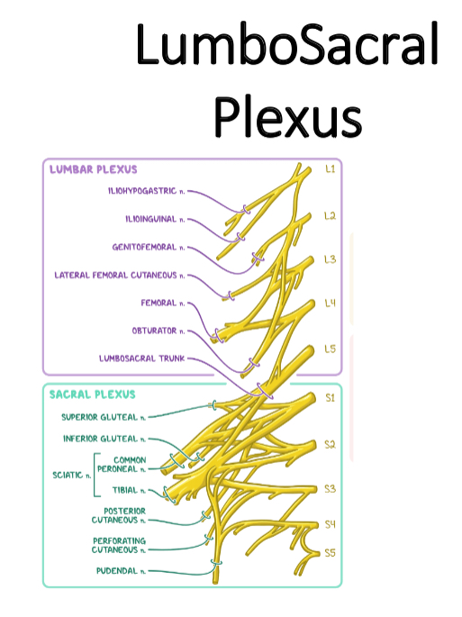
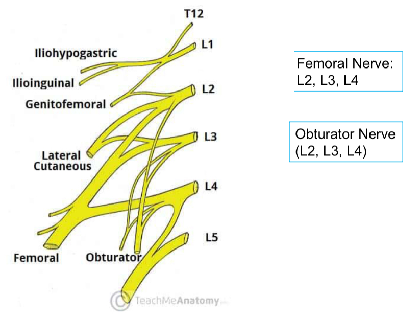
Lumbar plexus
Origin: ventral rami from .
Major nerves and targets:
Femoral Nerve: – innervates anterior thigh muscles and provides sensory innervation to parts of the thigh and leg.
Obturator Nerve: – innervates medial thigh muscles.
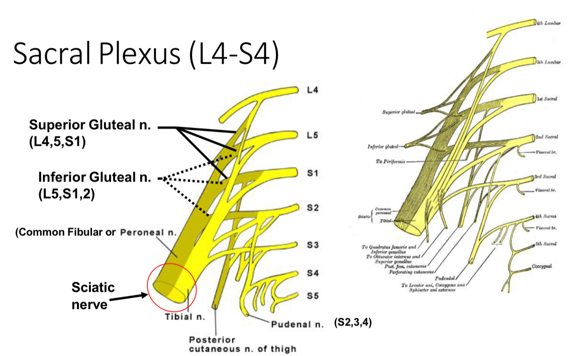
Sacral plexus
Origin: from .
Major nerves and targets:
Inferior Gluteal Nerve – gluteal muscles and posterior thigh.
Superior Gluteal Nerve – gluteal region.
Sciatic Nerve – main nerve of the posterior thigh; often divides into tibial and common fibular (peroneal) nerves distally, innervating most of the muscles below the knee.
Plexus, Plexuses (Plexi)
Plexus (plexuses, plexi) definition
A nerve plexus is a network of intersecting nerves formed by the merging and branching of spinal nerves.
Function: Plexuses sort and recombine spinal nerve fibers so that all fibers innervating a specific body part are bundled together.
The four major nerve plexuses
Cervical plexus (to head and neck; forms the phrenic nerve)
Brachial plexus (to chest, shoulders, and upper limbs)
Lumbar plexus (to back, abdomen, groin, thigh, knee, and calf)
Sacral plexus (to pelvis, buttock, genitals, thigh, calf, and foot)
Note on lumbar sacral plexus terminology
The combined LumboSacral Plexus covers contributions to the lower limb and pelvic region; it’s often visualized as a “box” or network near the lower spine/pelvis.
Lumber Plexus (ventral rami of T12-L4)
Lumbar Plexus specifics (ventral rami of )
Femoral Nerve:
Obturator Nerve:
Common references
Example nerves and their root values: Femoral Nerve (); Obturator Nerve ().
Sacral Plexus (L4-S4)
Sacral Plexus (approx. )
Major branches:
Superior Gluteal Nerve: roots
Inferior Gluteal Nerve: roots
Sciatic Nerve: main nerve of the posterior thigh; divisions into:
Common Fibular (Peroneal) Division
Tibial Division
Notes on the sciatic nerve divisions
Short head of biceps femoris is innervated by the common fibular division of the sciatic nerve.
Long head of the biceps femoris is innervated by the tibial division.
The sciatic nerve provides motor innervation to most of the posterior thigh and all lower leg and foot muscles via its branches.
Once in the Periphery…
Once in the periphery
Spinal nerves are composed of sensory fibers (information from a region of skin) and motor fibers (to muscle).
Dermatome
Definition: area of skin innervated or supplied by a single spinal nerve.
Example: anterior thigh is supplied by nerves corresponding to .
Myotome
Definition: group of muscles innervated by the fibers of a single spinal nerve.
Example: Quadriceps group corresponds to and is supplied by the Femoral nerve.
Practical implications
Mapping spinal nerves to sensory and motor areas helps identify intact or injured nerves.
Spinal nerves are formed by the joining of dorsal and ventral roots; testing dermatomes and myotomes helps assess nerve integrity.
Dermatomes
Dermatome maps
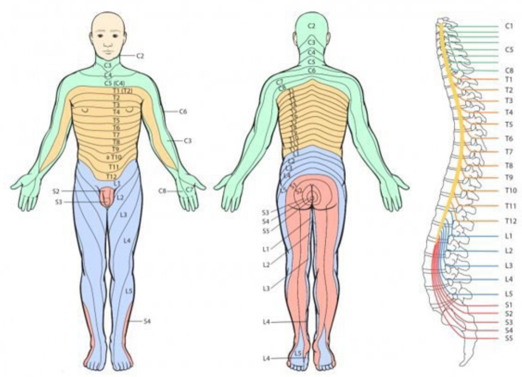
Dermatome maps depict skin areas innervated by specific spinal levels.
Example spinal levels typically shown on dermatome maps include , , , , , etc., illustrating regions such as the shoulder, chest/abdomen, groin, thigh, leg, and foot.
Note: Specific map details may vary by chart, but the overall concept remains: each skin region corresponds to a spinal level or a few adjacent levels.
Cutaneous Nerve
Cutaneous nerves
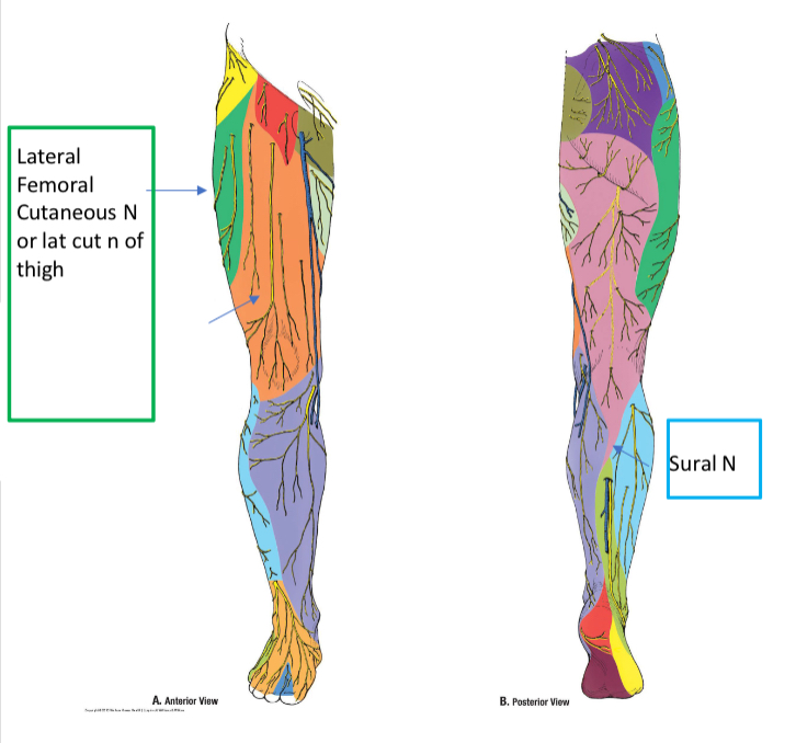
Function: supply the skin; they may carry fibers from several spinal nerves; their cutaneous territories are broader than a single dermatome.
Primarily sensory, but some cutaneous nerves carry autonomic (sympathetic) fibers that influence sweat glands and smooth muscles of blood vessels and hair follicles; they do NOT supply motor to skeletal muscle fibers.
Examples of cutaneous nerves related to the thigh region:
Lateral Femoral Cutaneous Nerve (lateral thigh)
Anterior Cutaneous Nerve of the thigh (anterior thigh)
Sural Nerve (posterior/lateral leg and foot region)
Spinal Cord Segment
Spinal cord segments and the lumbar/sacral plexus relationship
Lumbar/Sacral Plexus ranges and major nerves (T12-S4)
Femoral Nerve:
Obturator Nerve:
Sciatic Nerve:
Common Fibular (Fibular) division:
Tibial Division:
Summary mapping
Lumbar Plexus contributions: (to the anterior/medial thigh and associated muscles)
Sacral Plexus contributions: (to gluteal region, posterior thigh, leg, and foot)
Major peripheral nerves arising from these plexuses include the Femoral, Obturator, Sciatic (and its tibial and common fibular divisions).
Notes:
The organization of the nervous system emphasizes that many functional blocks (dermatomes and myotomes) map back to spinal nerve roots, which combine into spinal nerves and then diverge into dorsal/ventral rami and eventually plexuses that innervate specific regions of the body.
Numerical labels are given using spinal nerve root levels (e.g., ) and are presented in LaTeX format within double dollar signs as requested.