Anatomy Unit 4: Skeletal Systems
5.1 Skeletal basics
Main Functions
Support the body
Protect soft organs
Movement due to attached skeletal muscles
storage of minerals and fats
blood cell formation.
Skeletal system parts
Bones (skeleton)
Joints
Cartilages
Ligaments (connect bone to bone)
Divisions
Axial skeleton
Forms longitudinal axis of body
Appendicular Skeleton
Bones of limbs and girdles
Types of bones
Bone: connective tissue
2 basic types
Compact bone
homogeneous
Spongy Bone
Small needle-like pieces of bone with many open spaces
Classification of bones by shape
Long bones
Longer than wide
Shafts with heads at both ends
Contain mostly compact bone
Examples include: femur and humerus
Short bones
Cube-shaped
Contain mostly spongy bone
Examples include: carpals, tarsals
Flat bones
Thin and flattened
Usually curved
Thin layer compact bone around layer of spongy bones
Examples include: skull, ribs, and sternum
Irregular bones
Irregular shape
Usually curved
Do not fit in any other bone shape category
Examples include: vertebrae, pelvis
5.2 Bone Anatomy
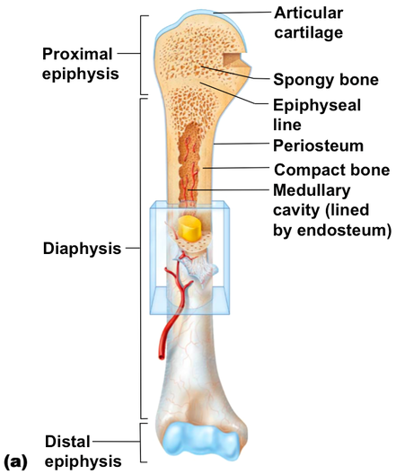
Gross Anatomy of long bones
Diaphysis
Shaft
Composed of compact bone
Epiphysis
Ends of the bone
Composed mostly of spongy bone
Long Bones
Periosteum
Outside covering of diaphysis
Fibrous connective tissue membrane
Sharpey’s fibers
Secure periosteum to underlying bone
Arteries
Supply bone cells with nutrients
Articular Cartilage
Covers external surface of epiphyses
Made of hyaline cartilage
Decreases friction at joint surfaces
Medullary cavity
Cavity of shaft
Contains yellow marrow(adults) or red marrow (infants)
Bone markings
Surface features of bones
Sites of attachments for muscles, tendons, and ligaments
Passage for nerves and blood vessels
Categories
Projections and processes
Grow out from bone surface
Depressions of cavities
Indentions
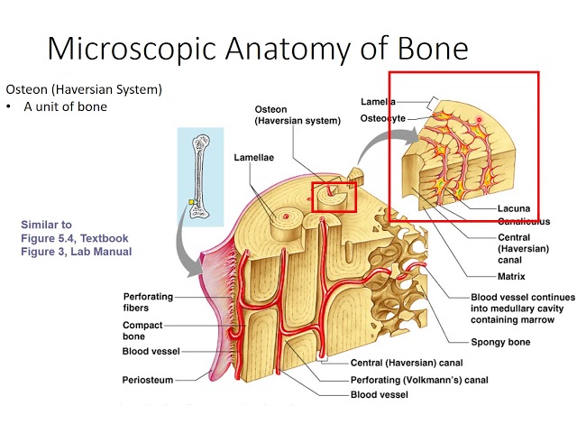
Bone anatomy (Microscopic)
Osteon
A unit of compact bone
Central (Haversian) canal
Opening in center of osteon
Carries blood vessels, nerves
Perforating (Volkmans) canal
Canal perpendicular to central canal
Carries blood vessels, nerves
Lacunae
Cavities containing bone cells (osteocytes)
arranged in concentric rings
Lamellae
Rings around central canal
Sites of lacunae
Canaliculi
Tiny canals
Radiate from central canal to lacunae
Form a transport system
Bone development: Childhood
Embryos: skeleton is primarily hyaline cartilage
During development, much of this cartilage replaced by bone
Cartilage remains in isolated areas
Bridge of nose
Parts of ribs
Joints
Epiphyseal plate allow for growth of long bone during childhood
New cartilage is continuously formed
Older cartilage becomes ossified
Bone grows in length because
Cartilage is broken down
Bone replaces cartilage.
Bones remodeled and lengthened until growth stops
Bone change shape somewhat
Bone grows in width
Remodeling
Bone resorbed in epiphyseal plate
Bone added by appositional growth
Bone is reabsorbed
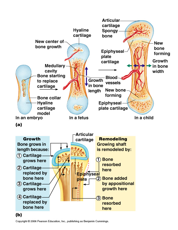
Remodeling
Osteocytes
Mature bone cells
Osteoblasts
Bone forming cells
Osteoclasts
Bone-destroying cells(break down bone matrix for remodeling and release of calcium)
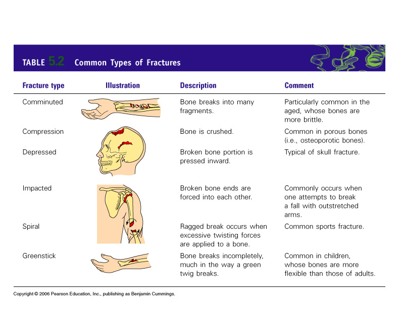
Bone Fractures
A break in a bone
Types of bone fractures
Closed(simple) fracture
does not penetrate the skin
Open(compound) fracture
broken bone penetrates through the skin
Treated by
Reduction(realignment) and immobilization
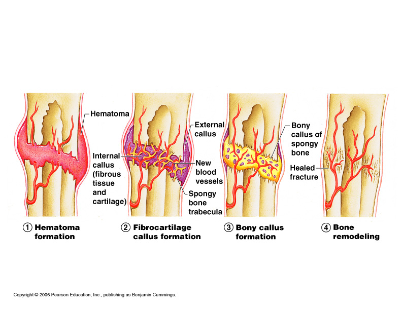
Fracture repair
Hematoma formation
Blood filled swelling is formed
Fibrocartilage callus formation
Break is splinted by fibrocartilage to form a callus
Bony callus formation
Fibrocartilage callus is replaced by a bony callus
Bone remodelling
Bony callus is remodeled to form a permanent patch
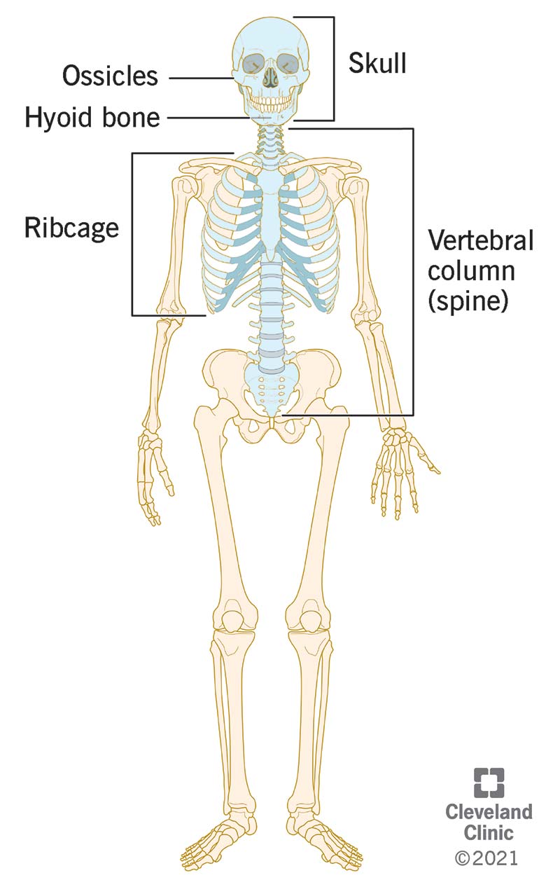
5.3 Axial Skeleton
Axial Skeleton Overview
Forms longitudinal part of body
Divided into 3 parts
Skull
Vertebral column
Bony thorax
Skull
Two sets of bones
Cranium
Facial bones
Bones are joined by sutures
Only mandible is attached by a freely movable joint
Paranasal sinuses
Hollow portion of bones surrounding nasal cavity
Functions to lighten the skull
Hyoid Bone
Only bone does not articulate with another bone
Serves as moveable base for tongue
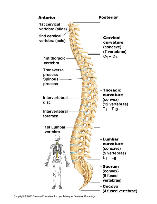
Vertebral Column
Vertebrae separated by intervertebral discs
SPine has normal curvature
Each vertebrae is given name according to location
1st cervical vertebra(atlas)
2nd cervical vertebra(axis)
Cervical curbature (C1-C7)
Thoracic curvature(T1-T12)
Lumbar curvature(L1-L5)
Sacrum and Coccyx
Bony Thorax
Forms a cage to protect major organs
The Skull
At birth, skull bones incomplete
Bones joined by fibrous membranes- fontanelles
Allow brain to grow
Convert to bone within 24 months after birth
Fontanelles are completely replaced with bone within two years after birth
Fetal skull large compared to infants total body length
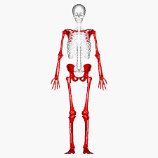
5.4 Appendicular Skeleton
General Parts
Three parts
Pectoral girdle
Two bones (per side)
Clavicle (collar bone)
Scapula(shoulder bone)
Allow upper limb to have exceptionally free movement
Pelvic girdle
Limbs
Bones of upper limb
Upper arm formed by single bone: Humerus
Forearm has two bones: Ulna and Radius
Hand: Carpals(wrist),metacarpals(palm),phalanges(fingers)
Pelvic gidgle
Hip Bones
Composed of three pair of fused bones
Ilium, Ischium, Pubic Bone
Total weight of upper body rests on pelvis
Protects several organs
Reproductive organs
urinary bladder
Part of large intestine
Gender Differences of the pelvis
Female
False pelvis is wider
Pubic arch is more than 90 degrees
Pelvis in general is larger and rounder
Male
Pubic arch is less than 90 degrees
Pelvis is taller and longer
Not meant for holding a child
Bones of lower limb
Thi
gh has one bone: Femur
Lower leg has two bones
Tibia and Fibula
Foot
Tarsus- ankle
Metatarsals- sole
Phalanges- toes
Arches of the foot
Bones of the foot are arranged to form three strong arches
Two longitudinal
One transverse
5.5 Joints
General Overview
Articulations of bones
Functions of joints
Hold together bones
Allow for mobility
Ways joints are classified
Functionally
Structurally
Functional classification
Synarthroses: immovable joints
Amphiarthrosis: slightly moveable joints
Diarthroses: freely moveable joints
Structural classification
Fibrous joints: generally immovable
Examples,
Sutures and syndesmoses(allow more movement than sutures)
Cartilaginous joints: immovable or slightly moveable
Examples
Pubic symphysis
Intervertebral joints
synovial joints: freely movable
Synovial fluid is found in joint cavity
Articular cartilage covers ends of boners
Joint surfaces enclosed by fibrous articular capsule
Have joint cavity filled with synovial fluid
Ligaments reinforce joint
Bursae- flattened fibrous sacs
Lined with synovial membranes
Filled with synovial fluid
Not actually part of the joint
Tendon sheath
Elongated bursa that wraps around tendon
Synovial Joint Types
Plane
Carpals
Gliding movement
Hinge joint
Elbows
Like how a door opens
Pivot joint
Rotation both internal and external
Turning skull around
Condyloid joint
Allows jaw, wrists, toes, and fingers to move up and down, from side to side, and around in circumduction.
Not a full rotation though!
Saddle Joint
Flexion, extension, etc
For example, if you were throwing objects from above the head in a sport.
Ball and socket
Full rotation
Ex. Pelvis
Inflammatory Conditions
Bursitis- inflammation of a bursa usually caused by blow or friction
Tendonitis- inflammation of tendon sheaths
Arthritis- inflammatory or degenerative diseases of joints
Osteoarthritis
Rheumatoid arthritis
autoimmune disease- immune system attacks joints
symptoms begin with bilateral inflammation of certain joints
often leads to deformities
Gouty arthritis
Inflammation of joints cause by deposition of urate crystals from the blood
can usually be controlled by diet