2. Electrical Signalling & Synaptic Transmission
Overview
Neurons are specialised, excitable cells designed for rapid, one-way communication.
Communicate by converting electrical signals (action potentials, APs) into chemical signals (neurotransmitters, NTs) and back again.
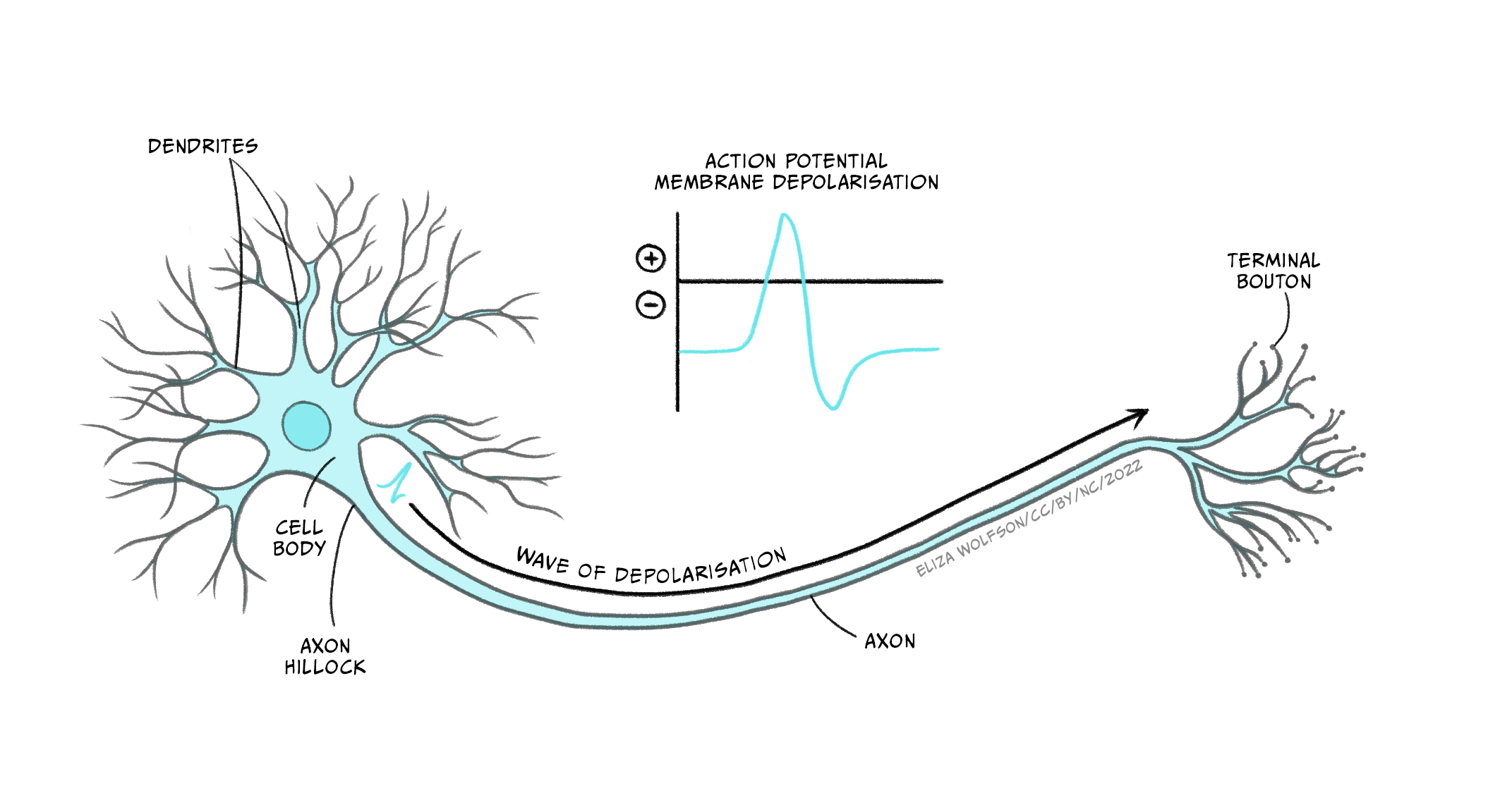
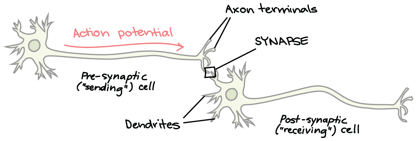
Functional polarity describes the unidirectional flow of information within a neuron and between neurons. Information is consistently received by the dendrites, which function as the receptive zone of the neuron. From the dendrites, the electrical signals are transmitted to the soma, or cell body, where integration of these signals occurs. Following processing in the soma, the integrated signal travels down the axon, which serves as the conductive zone. Upon reaching the axon terminals, specialized structures at the end of the axon, the neuron acts as a secretory zone, releasing neurotransmitters into the synapse. The synapse is the crucial junction where these chemical signals are transmitted to the next cell in the neural pathway. This ensures a consistent and directional flow of information throughout the nervous system.
Multiple neurons are chained together to span long distances (e.g. brain → spinal cord → foot).
Axon length & cell-body placement vary to minimise space (e.g. long peripheral axon with soma off to the side vs. short intracranial axon).
Electrical vs Chemical Signalling
Electrical: within a neuron (membrane potentials & APs).
Chemical: between cells at the synapse (NT release).
Region terms:
• Pre-synaptic = before synapse (sending cell).
• Post-synaptic = after synapse (receiving cell).
Three Key Events in Neuronal Communication
Generate the AP (depolarisation).
Conduct the AP along axon.
Synaptic transmission (chemical → electrical in next cell).
Membrane Potential (MP) Basics
Definition: charge difference across plasma membrane.
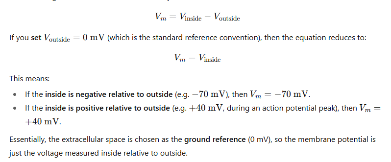
All cells have an MP; only neurons & muscle can change it rapidly.
At rest the inside is negative relative to outside (≈ in neurons).
Ionic Environment
Fluids are isotonic (equal total solute) but differ in individual ions.
• Inside (ICF): high , low , many negatively charged proteins ().
• Outside (ECF): high , low , plentiful.Ion movement requires membrane proteins because lipid bilayer blocks charged particles.
Ion Channels
Selective (e.g. channel, channel) or non-selective cation channels.
Opening → increases permeability for that ion, allowing diffusion down its concentration gradient.
Establishing Resting MP
At rest:
• Many leak channels are OPEN → diffuses out, taking + charge with it.
• Most channels are CLOSED → little Na⁺ influx.
• Result: net positive charge accumulates just outside membrane, leaving inside negative.Na⁺/K⁺ ATPase (pump) maintains gradients: pumps 3 out & 2 in using ATP (active transport) to reset concentrations long-term.
MP Changes & Electrical Signals
Depolarisation: MP becomes less negative/positive.
• Open channels ⇒ rushes in (↑ positivity) ⇒ MP moves toward & beyond.Repolarisation: return to resting negativity.
• Close channels, open channels ⇒ exits, removing + charge.Hyperpolarisation: MP becomes more negative than resting (e.g. ).
• Open extra channels or open channels (Cl⁻ enters) ⇒ inhibitory, harder to fire AP.
Action Potential (AP) Generation
Triggered when depolarisation reaches threshold (≈ ).
All-or-none: once begun, amplitude is constant.
Events:
Rapid influx (voltage-gated channels) ⇒ sharp depolarisation.
Inactivation of channels + opening of voltage-gated channels ⇒ repolarisation.
Brief overshoot ⇒ after-hyperpolarisation.
Na⁺/K⁺ pump restores ionic gradients.
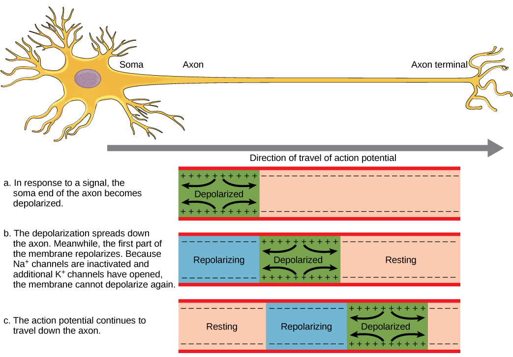
AP Conduction Along Axon
Opening of channels is sequential: depolarisation at one segment brings adjacent segment to threshold ("Mexican wave").
Unidirectional because channels behind the wave are transiently inactivated (refractory period).
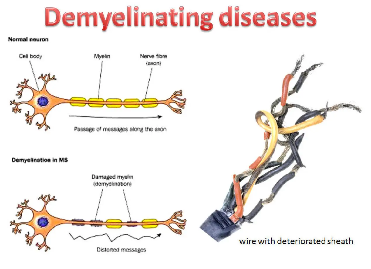
Myelination & Saltatory Conduction
Myelin (lipid sheath by oligodendrocytes CNS / Schwann cells PNS) prevents ion leakage.
Gaps = Nodes of Ranvier where voltage-gated channels cluster.
Local current leaps node-to-node (saltatory) ⇒ fewer openings, faster transmission.
Demyelination (e.g. Multiple Sclerosis):
• ↑ ion leak, ↓ conduction velocity, conduction block, neurological deficits.
Synaptic Transmission
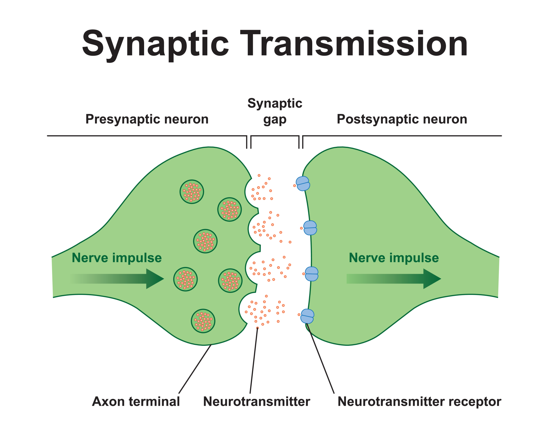
Synapse = junction between presynaptic axon terminal & postsynaptic membrane, separated by synaptic cleft (~20–40 nm).
Three Fundamental Steps
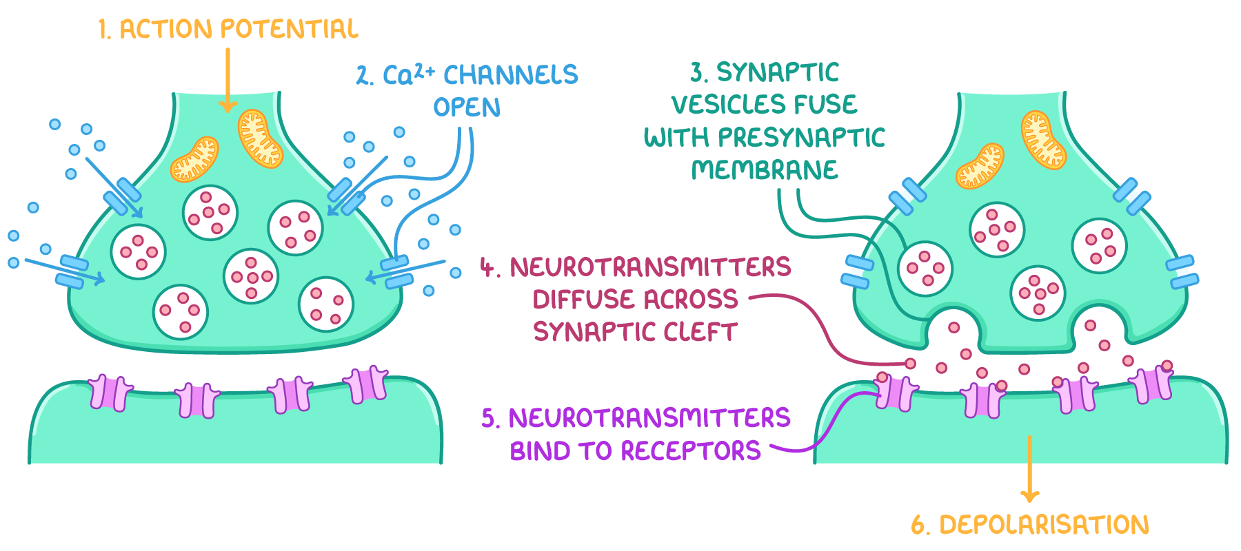
AP arrival & Ca²⁺ entry
AP depolarises axon terminal → voltage-gated channels open.
Extracellular (high outside) floods in.
NT release (exocytosis)
triggers synaptic vesicle fusion with membrane.
NT molecules released into cleft.
Postsynaptic effect
NT binds receptors → opens/closes ion channels.
EPSP (excitatory, usually entry) or IPSP (inhibitory, out or in).
If local depolarisation reaches threshold at axon hillock, new AP fires in postsynaptic neuron.
Terminating NT Action (3 ways)
Diffusion out of cleft.
Enzymatic degradation (e.g. acetylcholinesterase hydrolyses ACh).
Reuptake into presynaptic terminal or surrounding glia (astrocytes).
Clinical / Functional Connections
Hyperpolarisation as inhibition used for muscle relaxation & signal gating.
Many drugs/toxins target:
• Ion channels (local anaesthetics block channels).
• NT reuptake (SSRIs inhibit serotonin reuptake).
• Enzymes (neostigmine blocks AChE).Demyelinating diseases slow/stop conduction → motor & sensory deficits.
Study Tips
Master ion distributions first ("Na⁺ out, K⁺ in").
Use coloured counters/M&Ms to simulate charge movement for depolarisation vs hyperpolarisation.
Sketch graphs of MP vs time (label phases, ion movements).
Memorise the "rule of threes":
• 3 Na⁺ out / 2 K⁺ in (pump).
• 3 events for AP (generate, conduct, synaptic transmission).
• 3 steps of chemical synapse.
• 3 NT termination mechanisms.