Gene Mutations and DNA Repair Mechanisms
# Introduction to Gene Mutations and Molecular Biology
Course Context: This lecture is part of the 4BBY1070 Genetics and Molecular Biology course, delivered by Dr. Shirley Coomber. It builds upon knowledge from previous lectures including DNA replication (4BBY1013 L07), Transcription/Protein synthesis I (4BBY1013 L08), and Translation/Protein synthesis II (4BBY1013 L09).
Observations in Model Organisms: The study of mutations is exemplified by mutant colonies of the fungus Aspergillus nidulans and eight distinct morphological mutants of Drosophila (fruit flies).
Core Concept of Mutation: A mutation is defined as an alteration in the DNA sequence. Every organism carries mutant alleles, regardless of whether they manifest as distinct physical phenotypes. New mutations can arise in every generation.
Mutation
mutation is a change in DNA sequence
every organism carries mutant alleles, whether they manifest in distinct phenotypes or not
new mutations can occur in every generation
gene mutation occur as a result of change in nucleotide sequence → consequence in protein structure or gene expression
many causes:
DNA replication errors
spontaneous mutations
chemicals and irradiation (e.g. UV)
Classification of Point Mutations at the DNA Level
General Point Mutations: These occur within individual genes as changes in the nucleotide sequence, which can subsequently impact protein structure or gene expression.
somatic and germ-line mutations: somatic - ones that occur in any cell apart from germ cells, not inherited. Germ-line - occur in gametes, are inherited.
Base-Pair Substitution Mutations: The replacement of one nucleotide base pair by another. There are three primary types:
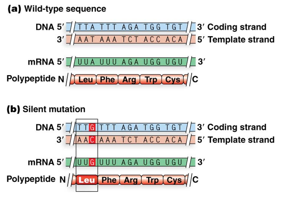
Silent Mutation (Synonymous): A base-pair change that does not result in a change to the amino acid sequence. Changes one codon for an amino acid to another codon for the same amino acid. This is possible due to the redundancy (degeneracy) of the genetic code.
Missense Mutation: changes codon for one amino acid to codon for another amino acid. The severity of this change depends on the specific role of the amino acid in the protein’s function.
conservative amino acid substitutes a chemically similar amino acid - less likely to alter function (neutral mutation)
non-conservative amino acid substitutes chemically different amino acid - more likely to alter function
consequences for protein depend on the function of the amino acid within the 3D structure
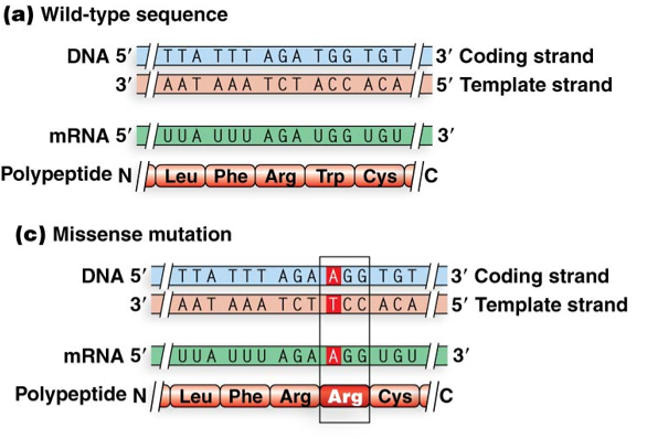
in three letter code a mutation in the first or second letter is more likely to produce a change in amino acid
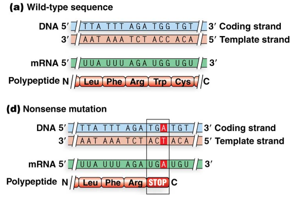
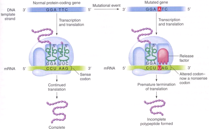
Nonsense Mutation: A base-pair change that converts a codon specifying an amino acid into a stop codon, leading to premature termination of translation. Truncated polypeptides are often nonfunctional
Transitions vs. Transversions:
Transition: A point mutation that replaces a purine with another purine (A ↔ G) or a pyrimidine with another pyrimidine (C ↔ T).
Transversion: A point mutation that replaces a purine with a pyrimidine or vice versa (e.g., A ↔ C, A ↔ T, G ↔ C, G ↔ T).
Frameshift and Regulatory Mutations
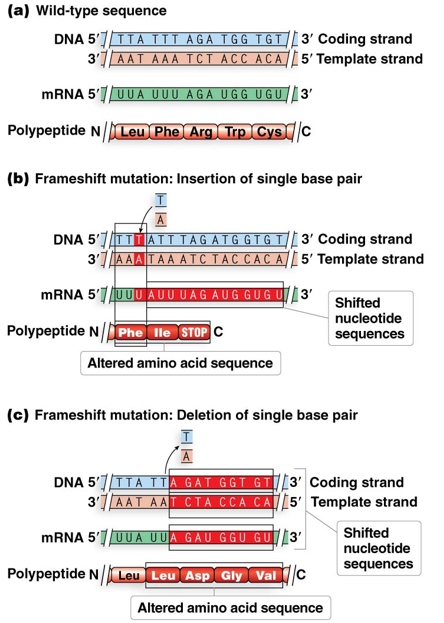
Frameshift Mutations: These are caused by the addition (insertion) or deletion of one or more base pairs (provided the number is not a multiple of three). This alters the reading frame and changes every amino acid downstream of the mutation site.
Regulatory Mutations: some point mutations alter the amount (not amino acid sequence) of protein product produced by a gene. affect regions such as promotors and other regulatory protein binding sites.
Promoter Mutations: These interfere with transcription initiation, often by affecting the binding of RNA polymerase or regulatory proteins.
Impact Levels: Some promoter mutations cause mild to moderate reductions in transcription, while others may abolish transcription entirely or even enhance it.
Case Study: Mutations in the RNA polymerase binding site or repressor protein binding site of the lac operon demonstrate how regulatory mutations affect gene expression.
Origin of mutation
Induced mutation
action of mutagen, an environmental agent that alters nucleotide sequence
process of inducing mutations by mutagens - mutagenesis
spontaneous mutation
arise in absence of known mutagen
may be caused by errors in DNA replication
provide “background rate” of mutation
Origins of Spontaneous Mutations
Spontaneous Mutations: These arise in the absence of a known environmental mutagen and provide a ‘background rate’ of mutation. They are often caused by natural biochemical processes.
DNA Replication Errors: DNA polymerase may occasionally insert the incorrect nucleotide. While the to exonuclease proofreading function of DNA polymerase corrects these errors of the time, some persist.
Trinucleotide Repeat Disorders: Caused by DNA polymerase "slipping" during replication, leading to an increased number of trinucleotide repeats within a gene. This results in longer stretches of a single amino acid.
Huntington’s Disease: An autosomal dominant disorder caused by an increase in length of polyglutamine region in huntingtin protein
Thresholds: CAG repeats are considered normal, while or more repeats lead to disease symptoms.
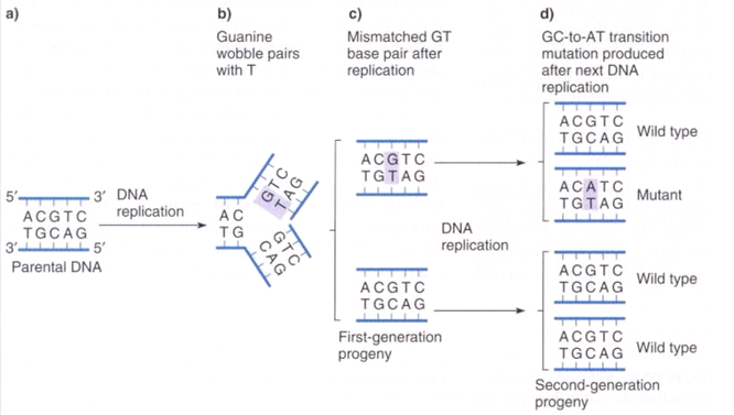
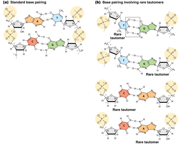
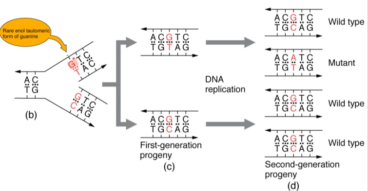
Tautomeric Shifts: DNA bases can occasionally convert into alternative structural isomers called tautomers through shifts in hydrogen placement and bonding.
Adenine and Cytosine: Normal amino form () vs. rare imino form ().
Thymine and Guanine: Normal keto form () vs. rare enol form ().
Consequence: Tautomeric shifts lead to non-standard base pairing (e.g., rare enol guanine pairing with thymine), and incorporation of incorrect bases during replication
common form of DNA lesion
Transposable Elements as Mutagenic Agents
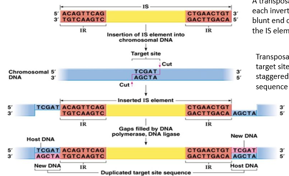
General Characteristics: Transposable elements (transposons) are integrated DNA sequences found in the genomes of all prokaryotes and eukaryotes (including humans). They cause mutations by inserting themselves into genes.
DNA Transposons: Found in both prokaryotes and eukaryotes. They move using a protein called transposase.
Bacterial IS Elements: Insertion Sequence (IS) elements in E. coli are approximately long. They contain a transposase gene bracketed by short Inverted Repeat (IR) sequences.
Mechanism: Transposase is involved in moving the IS element from one DNA location (genome or plasmid) to another. A transposase molecule binds to each IR and blunt ends cuts both sides of the IS. Transpose cuts the new target site with staggered cuts.
if transposon moves and inserts into gene it will create an insertion mutation that will cause premature termination of translation → mutant allele that can’t produce the full length protein product
Retrotransposons: Found only in eukaryotes. They move via an RNA intermediate and require the enzymes reverse transcriptase and integrase.
Mutagenic Effect: If a transposon inserts into a gene, it creates an insertion mutation, typically leading to premature termination of translation and the creation of a mutant allele that cannot produce a full-length protein.
Induced Mutations and Chemical Mutagens
Induced Mutations: These result from the interaction between DNA and environmental agents known as mutagens. The process is termed mutagenesis.
Types of Chemical Mutagens:
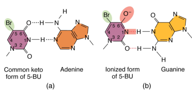
Nucleotide Base Analogs: Chemicals similar to nitrogenous bases that have altered pairing properties.
Example: -bromouracil () replaces thymine but can cause transition mutations.
Alkylating Agents: modify base structures by adding methyl or ethyl groups which can result in altered base pairing. Add methyl () or ethyl () groups to bases, altering their pairing.
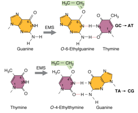
Example: Ethyl methanesulfonate (EMS).
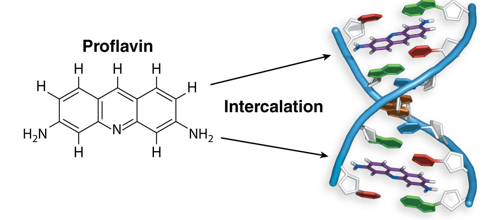
Intercalating Agents: Flat, planar molecules that wedge between base pairs, distorting the DNA helix. This leads to addition or deletion of nucleotides, causing frameshift mutations.
Examples: Proflavin, acridine orange, ethidium bromide.
Note: GelRed (used in labs) is related to ethidium bromide but is non-mutagenic because it cannot cross the membranes of living cells.
Other Agents: Deaminating agents (remove ),
hydroxylating agents (add ), oxidative agents (add oxygen or remove hydrogen), intercalating agents, and radiation induced DNA damage (UV, X-rays, gamma rays, cosmic rays).
Radiation-Induced Damage: Caused by Ultra Violet (UV) light, X-rays, gamma rays, and cosmic rays.
DNA Repair Mechanisms
Ways to repair DNA damage using correct base on the damaged DNA strand
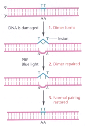
Photoreactivation Repair: Repairs damage (specifically thymine dimers) caused by UV light.
Mechanism: The Photoreactivation Enzyme (PRE), specifically photolyase in E. coli, cleaves the bond between thymine dimers (often caused by UV light).
Note: This specific mechanism is not present in humans.
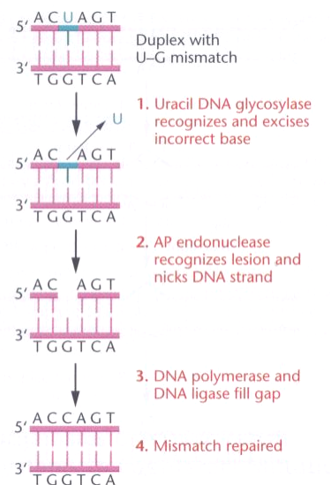
Base Excision Repair (BER): In eukaryotes, this involves:
specific DNA glycolsylase: e.g. Uracil DNA Glycosylase, Recognizes uracil as a noncomplementary base and excises it.
AP Endonuclease: recognizes lesion and nicks DNA strand.
DNA Polymerase and DNA Ligase: Fill the gap with the correct complementary base (e.g., Cytosine) and seal the nick.
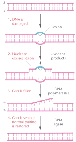
Nucleotide Excision Repair (NER): Repairs lesions that distort the DNA helix, such as thymine dimers.
In E. coli: The , , and proteins recognize the lesion and excise a segment of nucleotides. DNA polymerase I synthesizes new DNA, and DNA ligase joins the strand.
Somatic vs. Germ-line Mutations and Human Disease
Inherited Diseases of DNA Repair:
Xeroderma Pigmentosum: A condition where individuals are typically homozygous recessive for a faulty repair gene. Sufferers are extremely photosensitive; UV exposure leads to freckles, intense pigment patches, and malignant, often fatal, warty growths.
It is caused by mutant alleles in six different genes involved in human nucleotide excision repair.
Hereditary Cancer: Mutations in genes encoding DNA repair proteins significantly increase cancer risk.
BRCA2: Linked to hereditary breast cancer.
MSH2 and MLH1: Linked to hereditary colorectal cancer.
Acquired Cancer: Somatic mutations in DNA repair genes can also lead to the development of non-hereditary cancers.