Chapter 4 - Nucleic Acids & DNA Replication Objective
Nucleic Acids
Know the structure of nucleic acids; differences between nucleoside and nucleotide
Nucleic Acid: A macromolecule composed of nucleotide monomers. Generally used by cells store or transmit hereditary information. Includes ribonucleic acid and deoxyribonucleic acid.
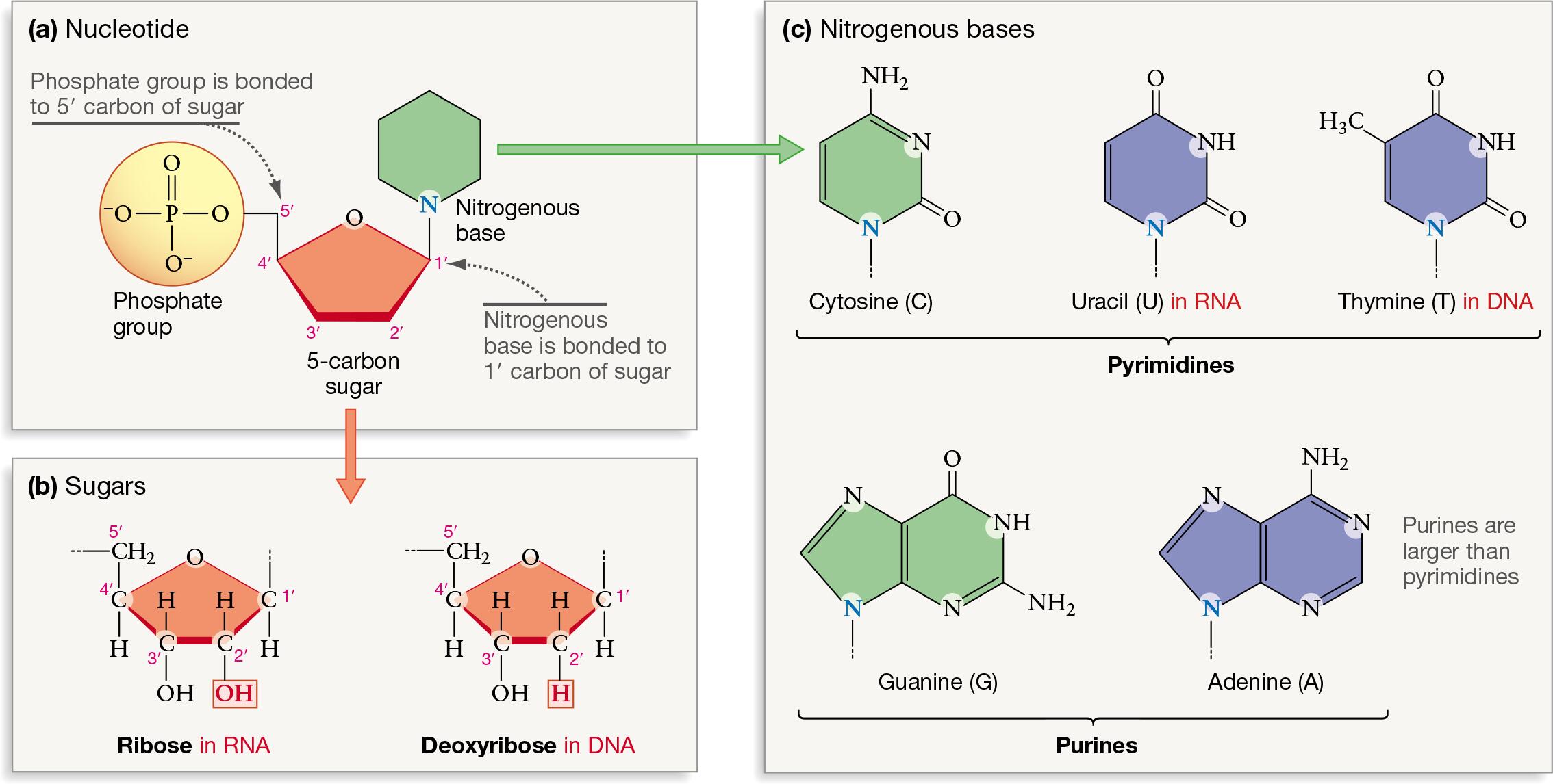
Polymers of nucleotides, each containing a sugar, phosphate group, and nitrogenous base
Sugar: give-carbon sugar, deoxyribose in DNA and ribose in RNA
Phosphate group: links sugars together
Nitrogenous base: nitrogen-containing base
DNA bases: adenine (A), guanine (G), cytosine (C), and thymine (T)
RNA bases: adenine (A), guanine (G), cytosine (C), and uracil (U)
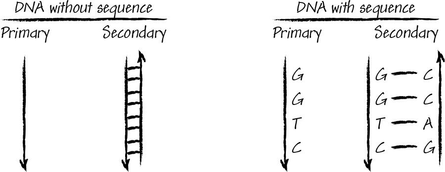
Primary structure is the sequence of these nucleotides linked by phosphodiester bonds
linear sequence of nucleotides in a single strand
phosphodiester bonds form a long chain with a sugar-phosphate backbone and a sequence of bases
sequence is read from the 5’ to 3’ direction
5’: free phosphate group, 3’ free hydroxyl group
Secondary structure: 3D shape of nucleic acid strands
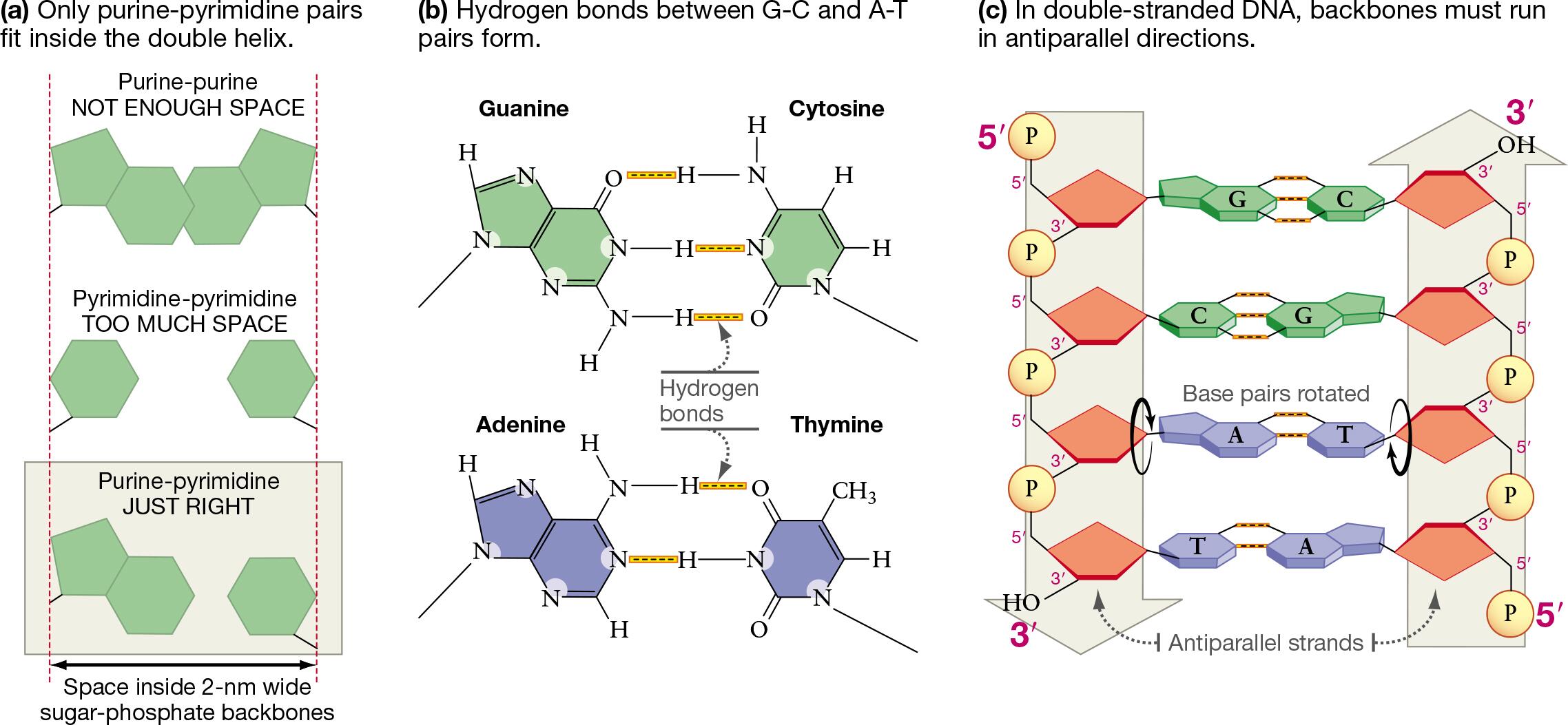
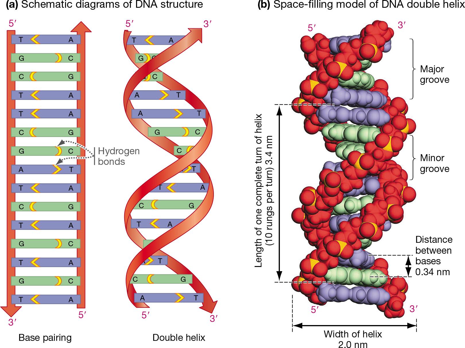
DNA’s secondary structure is a double helix of two antiparallel strands, held together by hydrogen bonds between complementary base pairs (A-T and G-C)
bases on opposite strands pair up
RNA is often a single strand that can fold into complex shapes like hairpins or loops
Nucleotide vs Nucleotide
Nucleotide: composed of a five-carbon sugar (deoxyribose/ribose)
contains a phosphate group (one or more)
components: nitrogenous base and sugar and phosphate group
function: building block for DNA and RNA, involved in energy metabolism and cell signaling
ex: ATP, GTP, cAMP
Nucleoside:
doesn’t contain a phosphate group
components: nitrogenous base and sugar
function: building block for nucleotides
ex: adenosine
Know the reaction of how they are joined and the name of the bond
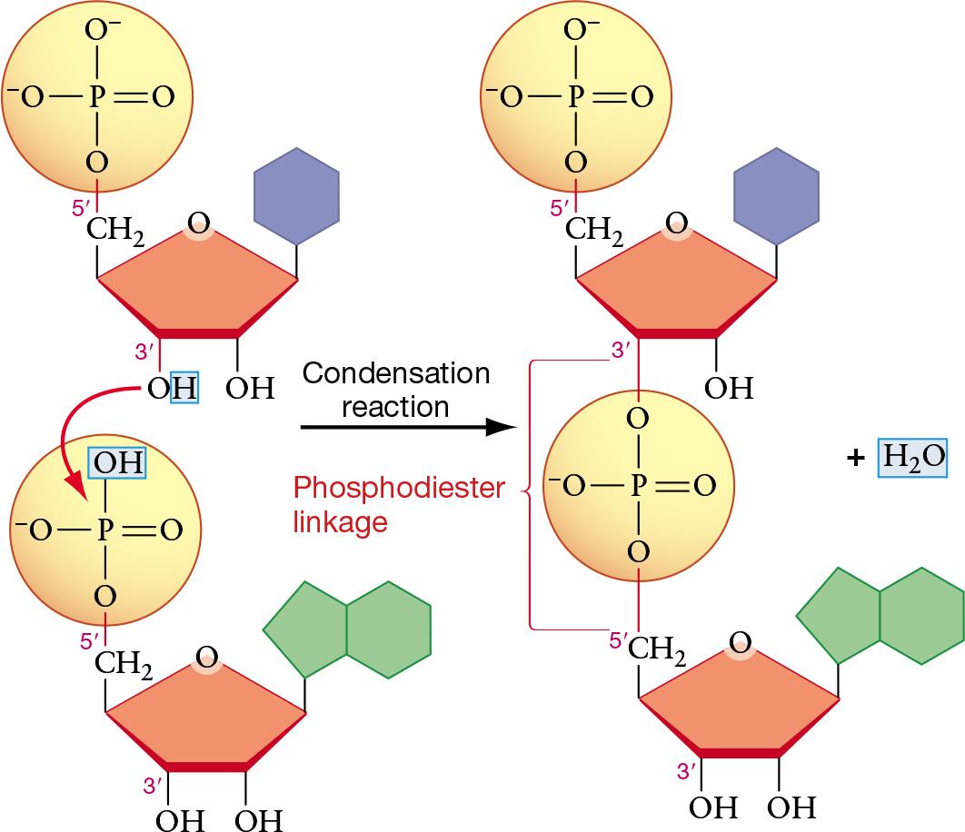
Condensation reaction that forms a covalent phosphodiester bond linking the phosphate group of one nucleotide to the sugar of the next
creates a sugar-phosphate backbone and releases a water molecule
reaction occurs between the 5’ phosphate group of one nucleotide and the 3’ hydroxyl group of another forming a long chain in the 5’ —» 3’ direction
List several functions for nucleic acids
Main functions (DNA & RNA): store and transmit genetic information, direct protein synthesis
DNA: stores genetic blueprint for an organism
RNA: Uses genetic information to build proteins which are essential for all cellular functions
Store & Transmit Genetic Information
DNA: stores the genetic blueprint of an organism, includes instructions for its development, growth, and reproduction
Heredity material: DNA is passed from parents to offspring, ensuring the transmission of heritable traits from one generation to the next
Direct Protein Synthesis:
Protein synthesis: nucleic acids are central to the process of creating proteins, which perform most the chemical processes in a cell
mRNA: messenger RNA; carries the genetic code from DNA to the ribosome
tRNA: transfer RNA; brings the correct amino acids to the ribosomes
rRNA: ribosomal RNA; a structural component of ribosomes, the site of protein synthesis
Gene expression: nucleic acids regulate which genes are turned on or off
Cellular processes: involved in processes like DNA replication, repair, and recombination to maintain genetic integrity —» highly organized
Energy transfer: modified nucleotides like ATP are crucial for energy processing in cells
Explain experiments that helped to define the structure of DNA
Erwin Chargaff - analyzed composition of DNA from different species
method: chromatography and UV spectrophotometry to measure the quantity of each of the 4 nitrogenous bases in DNA
findings: 2 rule where the amount of A=T in DNA and G=C; and ratio of (A+T) to (G+C) varies from species to species
bases are paired in a specific, non-random, way
Rosalind Franklin and Maurice Wilkins - X ray diffraction analysis
method: Franklin purified DNA fibers and sued a focused x-ray beam to produce diffraction patterns which revealed the position of atoms within crystal structure
findings: Franklin captured “photo 51” a clear diffraction image that shows a cross-shaped pattern characteristic of a helical structure —» analysis showed key measurements and confirmed that the sugar-phosphate backbone of DNA molecule was on the outside
James Watson and Francis Crick
method: used existing data from Chargaff, Franklin, and Wilkins, and knowledge of chemical bonding to assemble a physical model of DNA
findings: proposed double-helical structure for DNA and in their model the sugar-phosphate backbones form the outside the helix, nitrogenous bases (A,T,C,G) pair up in the middle forming rungs of the ladder, specific base pairing (A with T and C with G), and two strands run in opposite directions (antiparallel)
important since the structure itself suggested a mechanism for how
Meselon-Stahl Experiment
experimentally confirmed the “semi-conservative” model of DNA replication
method: grew bacteria in a medium containing “heavy” nitrogen isotope N15, causing bacteria to incorporate it into their DNA; transferred bacteria to a ‘light” nitrogen isotope N14 medium and allowed them to replicate over several generations; density of DNA at each stage was analyzed using a centrifuge
findings: after one generation, all DNA was a hybrid of heavy and light nitrogen; after two generations half the DNA was hybrid and half was entirely light; pattern perfectly patched the prediction of the semi-conservative model, where each new DNA molecule consists of one old strand and one new strand
contribution: final confirmation that the double helix model was correct and accurately explained the mechanism of genetic inheritance
Compare and contrast RNA and DNA
Both carry genetic information but differ in structure, sugar, and bases
DNA:
deoxyribonucleic acid
structure: double-stranded helix
sugar: deoxyribose
bases: adenine (A), thymine (T), guanine (G), cytosine (C)
function: long-term storage of genetic information
stability: more stable due to its structure and sugar
location: primarily in nucleus (eukaryotes)
RNA:
ribonucleic acid
structure: single-stranded (can fold into complex shapes)
sugar: ribose
bases: adenine (A), uracil (U), guanine (G), cytosine (C)
function: carries out protein synthesis, acting as a messenger (mRNA), and is a component of ribosomes (rRNA) and transfer (tRNA)
stability: less stable due to its single strand and sugar structure
location: nucleus and cytoplasm
Provide the characteristics of the W-C model of DNA
Double-helix structure, composed of two antiparallel strands with sugar-phosphate backbones on the outside and nitrogenous bases on the inside
Strands are held together by specific base pairing rules via hydrogen bonds, creating a uniform diameter and a consistent shape for the molecule
Structure: double helix, antiparallel strands, nitrogenous bases, major and minor grooves
Base pairing: complementary base pairing, hydrogen bonds
Know the Scientists involved in determining the structure of DNA
Watson and Crick proposed double helix idea
work was built on the foundational research of others including Franklin, Wilkins, and Chargaff
DNA Replication
Know the proteins involved in the process
Enzymes:
DNA polymerase: synthesizes new DNA strands by adding nucleotides to the 3’ end of the growing strand
Helicase: unwinds the DNA double helix at the replication fork
Primase: synthesizes RNA primers, which provide a starting point for DNA polymerase
Topoisomerase/Gyrase: relieves DNA supercoiling during unwinding
DNA ligase: joins Okazaki fragments on the lagging strand
Telomerase: in eukaryotic cells, synthesizes telomere sequence at the ends of linear chromosomes
Describe the process step by step with attention to orientation of the strands
1. Unwinding the DNA
origin of replication: replication begins at a specific site on the DNA helix
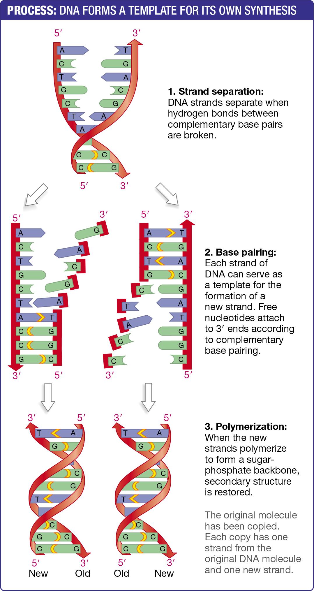
helicase: enzyme DNA helicase unwinds the double helix by breaking the hydrogen bonds between base pairs which creates a Y-shape structure called a replication fork
single-strand binding proteins: proteins bind to the separated DNA strands to prevent them from re-pairing and becoming a double-helix again
topoisomerase: ahead of the replication fork, the DNA becomes overwound or supercoiled; topoisomerase enzymes alleviate this strain by cutting, unwinding, and re-ligating the DNA
2. Building new Strands
RNA primers: DNA polymerase cannot start a new strand from scratch. An enzyme called primase must first create a short RNA primer, which provides a starting point with a free 3’-OH group for DNA polymerase to begin synthesis
3. Synthesis of the Leading Strand
the leading strand is the template strand that runs in the 3’ to 5’ direction toward the replication fork
continuous synthesis: because DNA polymerase synthesizes in the 5’ to 3’ direction, it can move along the template strand continuously following the unwinding helicase
single primer: only one RNA primer is needed for the entire leading strand
4. Synthesis of the Lagging Strand
the lagging strand in the template strand that runs in the 5’ to 3’ direction opposite to the movement of the replication fork
discontinuous synthesis: since DNA polymerase must move in the 5’ to 3’ direction, it is forced to synthesizes the lagging strand in short disconnected segments
Okazaki fragments: short segments of DNA
5. Completing the DNA Copies
primer removal: the RNA primers are eventually removed and replaced with DNA nucleotides by DNA polymerase I
ligation: after the primers are replaced, the enzyme DNA ligase seals the gaps between the Okazaki fragments by forming phosphodiester bonds
proofreading: throughout the process, DNA polymerase also proofread their work, identifying and correcting errors to ensure high yields
End Result: semiconservative replication because each new double helix consists of one original or parental strand and one newly synthesized daughter strand
Compare and contrast the process in Eukaryotes and Prokaryotes
Similarities
basic processes: semi-conservative replication model where each new DNA molecule consists of one original and one new strand
key enzymes: rely on enzymes like helicase to unwind DNA and DNA polymerase to synthesize new strands
direction: replication proceeds in a 5’ —» 3’ direction, requiring leading and lagging strands
Okazaki fragments: both create Okazaki fragments on the lagging strand
Differences
Prokaryotes:
Origin of replication: one origin
DNA structure: single, circular chromosome
replication rate: faster
DNA polymerases: DNA polymerase I and II are key; less types
primer removal: DNA polymerase I removes RNA primers
Eukaryotes:
origin of replication: multiple origins
DNA structure: multiple linear chromosomes
replication rate: slower
DNA polymerases: many types
primer removal:
Explain how telomeres are handled during the process
Linear ends of chromosomes, called telomeres, face a challenge called the “end-replication problem”
Mechanism used by DNA polymerase the lagging strand cannot be fully copied to the very end
most somatic cells don’t express the enzyme telomerase, so telomeres shorten with each division
specialized cells use telomerase to extend their telomeres and prevent shortening
End-Replication Problem
DNA polymerase can only synthesizes new DNA in the 5’ to 3’ direction, to begin replication DNA polymerase needs a short RNA primer
Leading strand: replication proceeds continuously towards the replication fork with a single primer and it copies the template strand completely
Lagging strand: replication proceeds discontinuously in short segments, called Okazaki fragments, moving away from the replication fork, and each fragment requires a new RNA primer
End-replication problem occurs specifically on the lagging strand at the chromosome’s end
1. when the final RNA primer is removed from the chromosomes end DNA polymerase cannot fill the gap
2. DNA polymerase requires a free 3’ hydroxyl group to extend the new strand, with no upstream Okazaki fragment this group is unavailable at the very tip of the chromosome
3. this leaves a non-replicated portion at the 5’ end of the new strand, leading to a net shortening of the chromosome with each cell division
How cells handle telomeres
1. Telomere shortening in most somatic cells
when telomeres become critically short, the cell enters a state of permanent growth arrest or triggers programmed cell death (apoptosis)
mechanism is a natural tumor-suppression process that prevents uncontrolled cell division
2. Telomere maintenance by telomerase in special cells
some cell types that must divide continuously throughout life express the enzyme telomerase to prevent telomere shortening
telomerase is made up of protein and RNA
Understand how the Meselon and Stahl experiment is set up and produces the results
Growing E. coli bacteria in a medium with heavy nitrogen N15 which incorporated into DNA
Bacteria were transferred to a light nitrogen N14 medium, and samples were taken after each round of replication
Found that after the first generation, all DNA was a hybrid of one heavy N15 and one light N14 strand, proving that DNA replication is semi-conservative
Results
generation 0 (before transfer): DNA was all heavy/N15 and formed a single band at the bottom of the centrifuge tube
generation 1 (after 1st replication): DNA from bacteria grown in the light medium was a single band of hybrid DNA, so each double helix contained one heavy/N15 strand and one newly synthesized light/N14 strand which results in conservative model and produces one heavy and one light DNA molecule
generation 2 (after 2nd replication): hybrid DNA replicated which results in two bands - one band of hybrid DNA N15/N14 and one band of completely light DNA N14/N14. this outcome is consistent with the semi-conservative model where the heavy parental strand separates and each template is paired with a new light strand while the previously synthesized light strands also acts as a template strand for the new light strands
subsequent generations: the ratio of light to hybrid DNA continued to increase with each generation, with the light DNA band becoming more prominent as the hybrid band becomes less so