Endocrine System
Chapter 10: The Endocrine System
Introduction
Contributors:
William C. Ober, M.D. (Art Coordinator and Illustrator)
Claire E. Ober, R.N. (Illustrator)
Kathleen Welch, M.D. (Clinical Consultant)
Kevin Petti, Ph.D. (Contributor)
Textbook: Essentials of Anatomy & Physiology, Eighth Edition, Pearson
Intercellular Communication
Purpose: Maintains homeostasis
Mechanism:
Achieved mainly through chemical messages
Long-distance communication coordinated by endocrine and nervous systems
Comparison:
Nervous System:
Faster response
Specific destinations
Short-lived effects
Endocrine System:
Slower response
Releases hormones into bloodstream
Bind to target cells
Longer-lasting effects
Nervous and Endocrine Systems Comparison
Shared features:
Release of chemicals that bind to specific receptors on target cells
Share chemical messengers (e.g., epinephrine and norepinephrine)
Both regulated by negative feedback mechanisms
Coordinate and regulate activities of other cells, tissues, organs, and systems to maintain homeostasis
The Endocrine System
Overview:
Includes all endocrine cells and tissues
Consists of glandular, secretory cells
Secretions enter extracellular fluid
Types of hormones:
Local hormones (e.g., prostaglandins) affect adjacent cells
Hormones: Released from one tissue and transported via bloodstream to target cells in other tissues
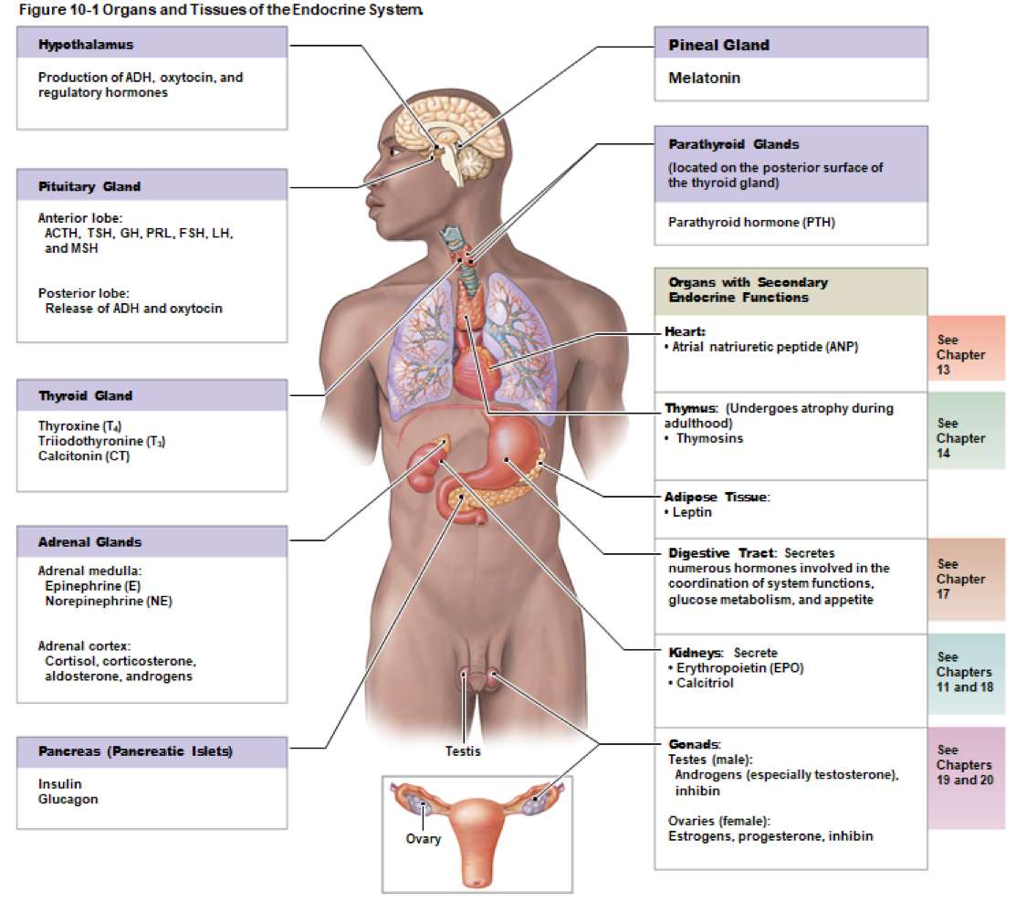
Figure 10-1: Organs and Tissues of the Endocrine System
Key organs and their functions:
Hypothalamus: Production of ADH, oxytocin, and regulatory hormones
Pineal Gland: Produces melatonin
Pituitary Gland: Releases several hormones (e.g., ACTH, TSH, GH)
Thyroid Gland: Produces thyroid hormones (T4, T3)
Adrenal Glands: (adrenal medulla, and adrenal cortex)
Medulla: Epinephrine, norepinephrine
Cortex: Cortisol, aldosterone, androgens
Pancreas: Insulin, glucagon
Ovary: Estrogens, progesterone
Parathyroid Glands: Parathyroid hormone (PTH)
Secondary Endocrine Functions: Organs such as heart (ANP), thymus (thymosins), kidneys (EPO, calcitriol), testes (androgens)
Three Groups of Hormones
Classification based on chemical structure:
Amino Acid Derivatives:
Structurally similar to amino acids
E.g., epinephrine, norepinephrine, thyroid hormones, melatonin
Peptide Hormones:
Chains of amino acids
E.g., ADH, oxytocin, hypothalamic, pituitary, and pancreatic hormones
Lipid Derivatives:
Steroid Hormones: Similar to cholesterol, bound to transport proteins
Released by reproductive organs and adrenal glands
Eicosanoids: Derived from arachidonic acid, affect local cellular functions, include prostaglandins
Hormone Action
Sensitivity: Target cell sensitivity to hormone depends on the presence of receptors
Hormones alter target cell operations including:
Types
Activities
Locations
Quantities of structural proteins and enzymes
Receptor Location:
Plasma membrane or inside the cell
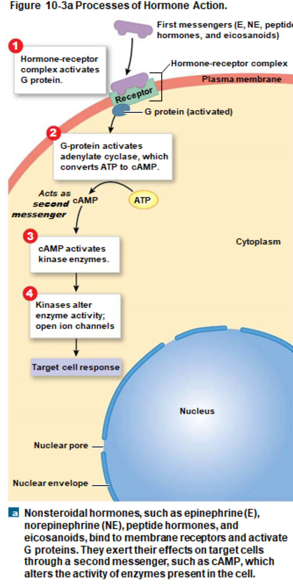
Plasma Membrane Receptors
Required for non-lipid soluble hormones:
E.g., epinephrine, norepinephrine, peptide hormones, eicosanoids
These hormones cannot diffuse through the plasma membrane
Must bind to receptors on the outside
First messengers activate second messengers in the cytoplasm, linked by a G protein
Second Messenger Systems
Amplification: A small number of hormone molecules activate thousands of second messengers
Examples of second messengers:
Cyclic-AMP
Calcium ions
Cyclic-GMP
Cyclic-AMP Second Messenger System
Mechanism:
First messenger activates G protein
G protein activates enzyme adenylate cyclase
ATP is converted to second messenger (cAMP)
cAMP activates kinase enzymes, leading to phosphorylation
Effect varies depending on the protein affected
Intracellular Receptors
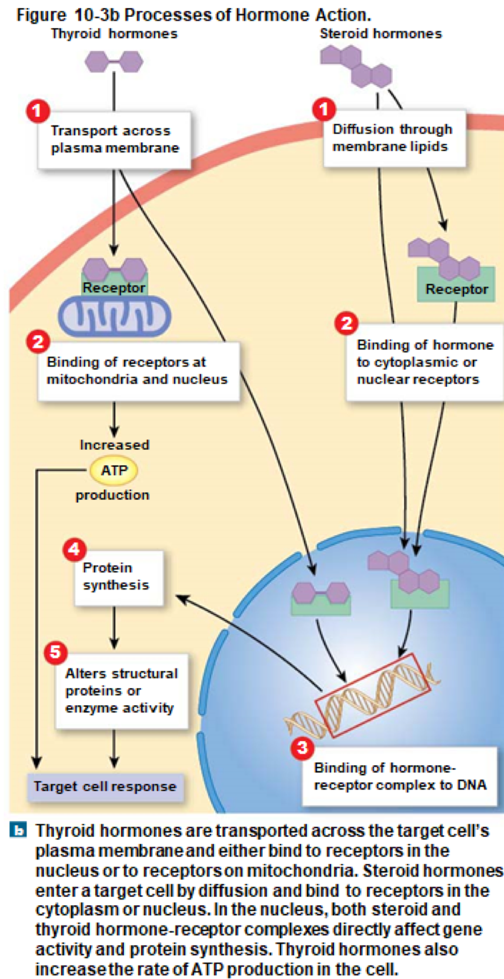
Used for lipid-soluble hormones
E.g., thyroid and steroid hormones
Mechanism:
Hormones cross plasma membrane, form hormone-receptor complex
Activates or inactivates specific genes
Alters mRNA transcription rate, changing cell structure/function
Hormone Secretion and Distribution
Hormones rapidly enter bloodstream; freely circulating hormones are short-lived
Inactivation:
Hormones bind to receptors and diffuse to target cells
Absorbed and broken down in liver and kidney
Inactivated by enzymes in plasma/interstitial fluid
Hormones bound to transport proteins stay in circulation longer
Control of Endocrine Activity
Negative Feedback Control: Hormonal secretion based on three stimuli:
Humoral Stimuli: Changes in extracellular fluid composition
Hormonal Stimuli: Changes in circulating hormone levels
Neural Stimuli: Neural stimulation of neuroglandular junctions
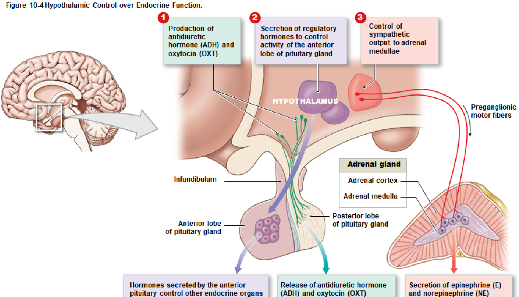
Hypothalamus and Endocrine Control
Provides the highest level of endocrine control
Regulates nervous and endocrine systems in three ways
The Pituitary Gland
Also called hypophysis
Structure: Protected by the sella turcica, hangs from the hypothalamus via infundibulum
Hormones:
Secretes nine hormones, all unique peptides or small proteins
Uses cAMP second messenger mechanism
The Anterior Lobe of the Pituitary Gland
Contains endocrine cells surrounded by a complex capillary bed (hypophyseal portal system)
Two capillary networks linked by blood vessels
Regulatory hormones diffuse onto target cells in the anterior lobe, many of which are tropic hormones (target other endocrine glands)
The Seven Anterior Lobe Hormones
Thyroid-stimulating hormone (TSH)
Adrenocorticotropic hormone (ACTH)
Follicle-stimulating hormone (FSH)
Luteinizing hormone (LH)
Prolactin (PRL)
Growth hormone (GH)
Melanocyte-stimulating hormone (MSH)
Thyroid-stimulating Hormone (TSH)
Also called thyrotropin
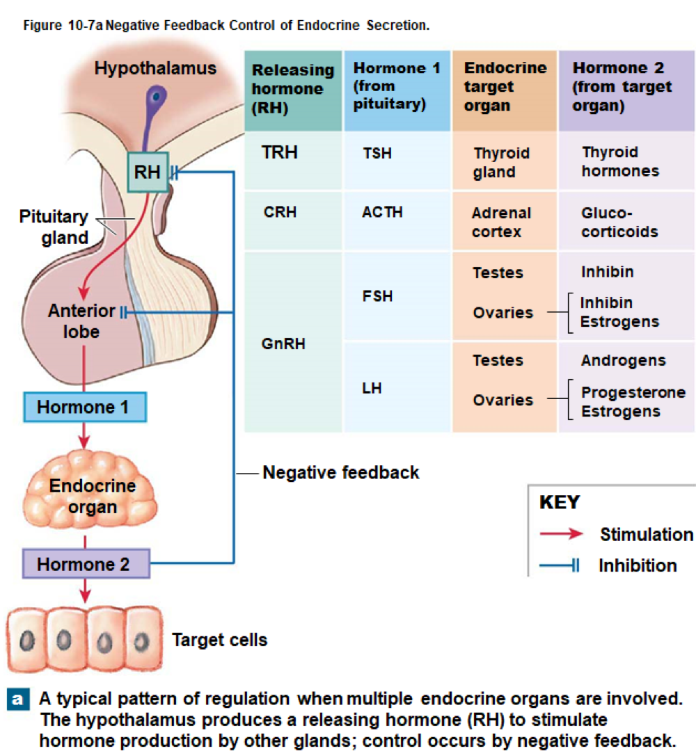
Released in response to thyrotropin-releasing hormone (TRH) from hypothalamus
Targets thyroid gland; triggers release of thyroid hormones
Increases in thyroid hormones cause decrease in TRH and TSH secretion
Adrenocorticotropic Hormone (ACTH)
Also called corticotropin
Released in response to corticotropin-releasing hormone (CRH) from hypothalamus
Targets adrenal cortex; stimulates secretion of glucocorticoids
Increases in glucocorticoids cause decrease in ACTH and CRH secretion
Gonadotropins
Released in response to gonadotropin-releasing hormone (GnRH) from hypothalamus
Target the male and female gonads: testes in males and ovaries in females
Includes two hormones:
Follicle-stimulating hormone (FSH)
Luteinizing hormone (LH)
Follicle-stimulating Hormone (FSH)
Promotes follicle development in females
Promotes sperm production in males
Cells of ovaries and testes produce inhibin which inhibits FSH production through negative feedback
Luteinizing Hormone (LH)
Induces ovulation and secretion of estrogens and progesterone in females
In males, also known as interstitial cell-stimulating hormone (ICSH) which stimulates interstitial cells in testes to produce androgens (e.g., testosterone)
Estrogens and progesterone inhibit GnRH, decreasing levels of LH
Prolactin (PRL)
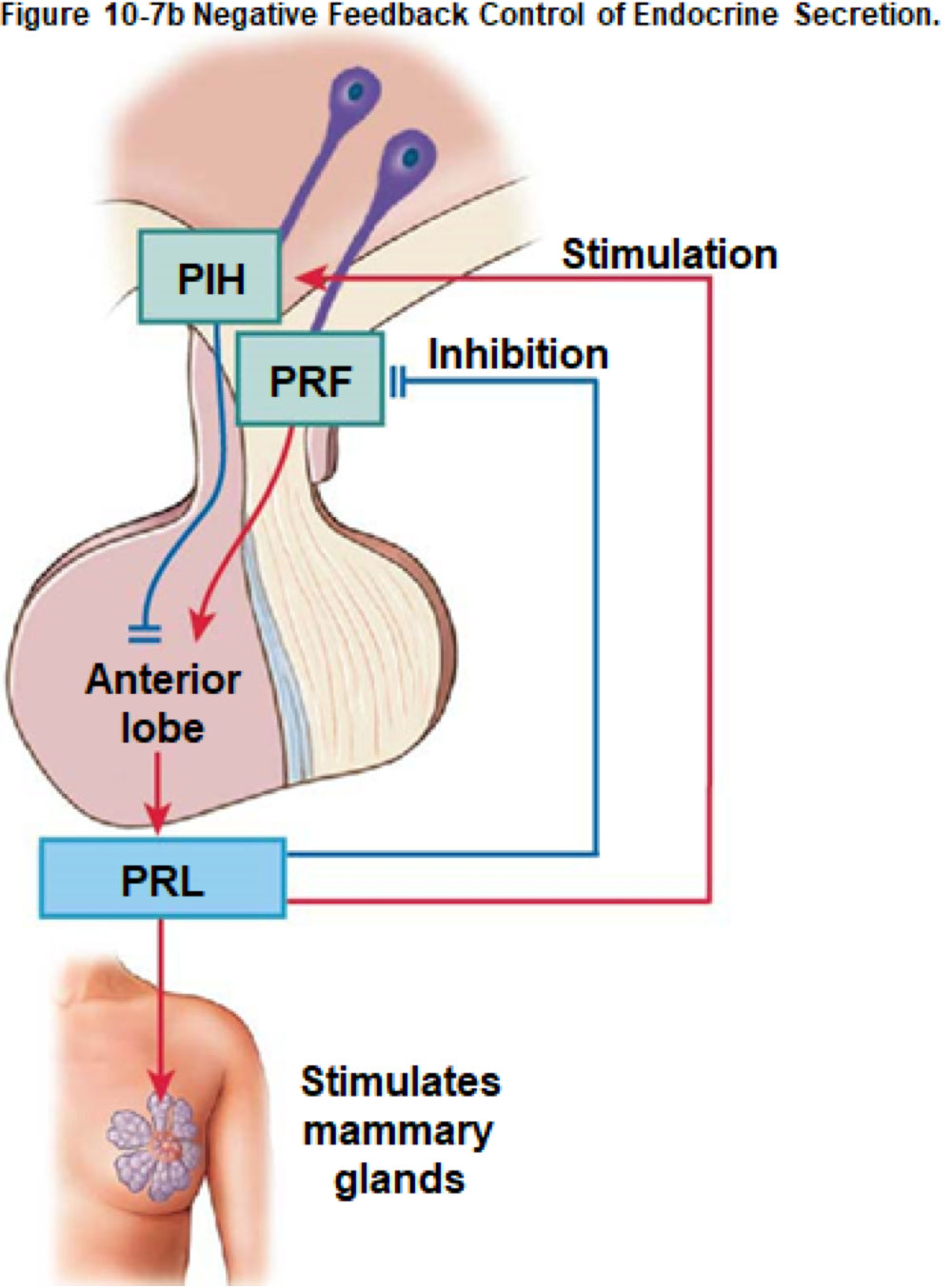
Released in response to prolactin-releasing factor (PRF) from hypothalamus
Targets mammary glands in females; stimulates development and milk production during pregnancy and nursing
Circulating PRL stimulates prolactin-inhibiting hormone (PIH) inhibiting PRF
Growth Hormone (GH)
Also called human growth hormone (hGH) or somatotropin
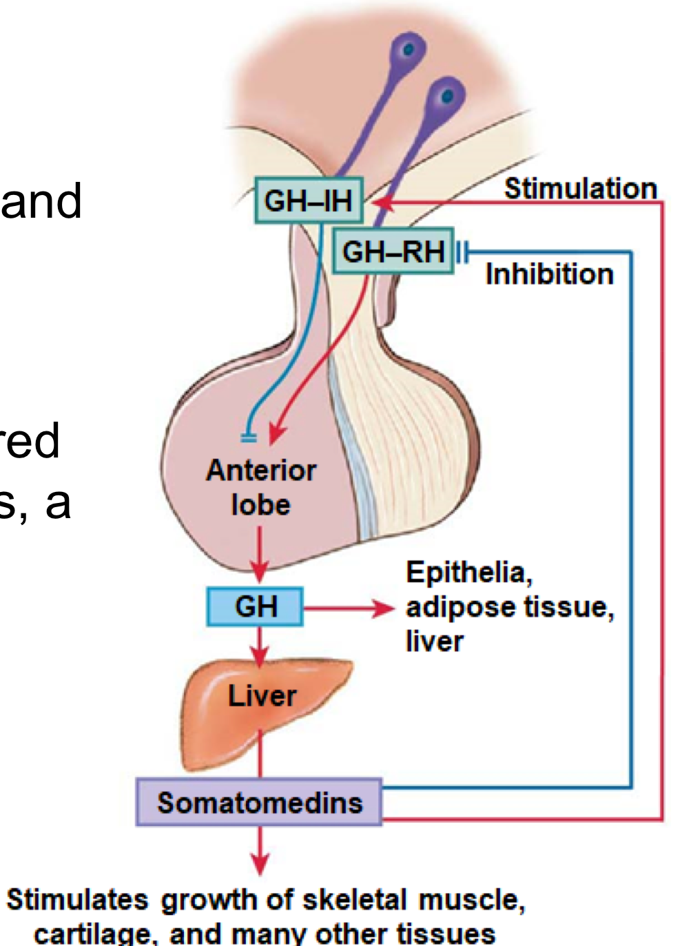
Regulated by growth hormone-releasing hormone (GH-RH) and growth hormone-inhibiting hormone (GH-IH) from the hypothalamus
Stimulates cell growth and replication, especially in skeletal muscle and chondrocytes
Indirect action: Stimulates liver to release somatomedins (insulin-like growth factors, IGFs) influencing protein synthesis
Direct Actions of Growth Hormone
In Epithelial and Connective Tissues: Stimulates stem cell division and differentiation
In Adipose Tissue: Breaks down stored fats and releases fatty acids, sparing glucose
In Liver: Breaks down glycogen reserves, releases glucose into bloodstream
Melanocyte-stimulating Hormone (MSH)
Increases activity of melanocytes in skin, increasing melanin production
Nonfunctional in adults, secreted during fetal development, early childhood, pregnancy, and certain diseases
The Posterior Lobe of the Pituitary Gland
Contains axons from two groups of hypothalamic neurons
Produces two hormones:
Antidiuretic hormone (ADH)
Oxytocin (OXT)
Hormones are transported within axons to the posterior pituitary lobe where released into capillaries
Antidiuretic Hormone (ADH)
Also called vasopressin
Released when the body is low on water (stimulated by increased solutes in blood or decreased blood volume/pressure)
Primary target: kidneys for decreasing water loss
Triggers vasoconstriction, increasing blood pressure
Oxytocin (OXT)
In women: Stimulates contraction of uterine muscles during labor
Also stimulates contraction of milk secretory cells in mammary glands
Plays an unclear role in sexual activity
In men: Stimulates smooth muscle in the walls of sperm duct, aiding in emission
The Pituitary Hormones Summary Table
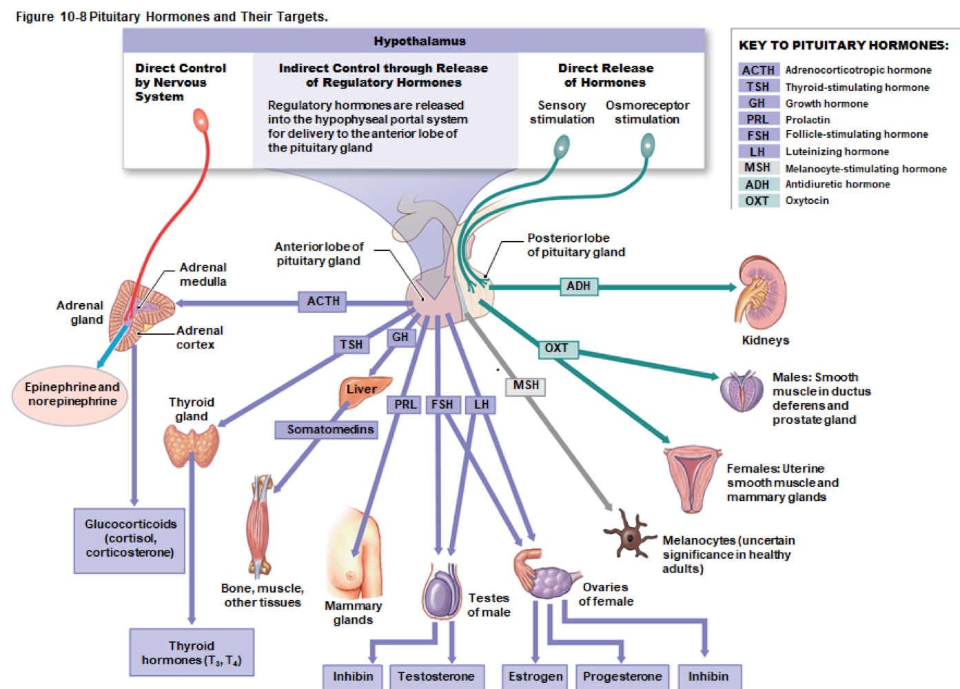
Table 10-1: The Pituitary Hormones
Pituitary Lobe/Hormone
Anterior Lobe: TSH, ACTH, FSH, LH, PRL, GH, MSH
Posterior Lobe: ADH, OXT
Target and Hormonal Effects:
TSH: Thyroid gland → thyroid hormone secretion
ACTH: Adrenal cortex → glucocorticoid secretion
FSH: Follicle cells of ovaries → estrogen secretion and follicle development
LH: Uterus/mammary glands → ovulation, progesterone secretion
PRL: Mammary glands → milk production
GH: All cells → growth, protein synthesis
MSH: Melanocytes of skin → increased melanin synthesis
ADH: Kidneys → water reabsorption
OXT: Uterus/mammary glands (females) and sperm duct/prostate (males) → labor contractions, milk ejection
The Thyroid Gland
Location: Anterior to trachea, inferior to thyroid cartilage
Structure: Two lobes connected by a narrow isthmus, contains many spherical thyroid follicles lined by simple cuboidal epithelium filled with viscous colloid
The Thyroid Follicles
Follicular cells produce thyroid hormones stored in colloid
TSH stimulates release of thyroid hormones
Thyroid hormones are transported bound to plasma proteins, providing large reserves
Thyroid hormones derived from amino acid tyrosine and iodine:
Thyroxine (T4): Four iodine atoms
Triiodothyronine (T3): Three iodine atoms, more potent than T4
The Effects of Thyroid Hormones
Affect nearly every cell in the body
Increase ATP production rates in mitochondria
Activate genes encoding enzymes for glycolysis and energy production
Calorigenic Effect: Cells use more energy, producing heat
Essential for normal skeletal, muscular, and nervous system development
The C Cells of the Thyroid Gland
Also called parafollicular cells that produce calcitonin (CT)
Triggered by increases in plasma Ca2+, inhibits osteoclasts, and stimulates calcium excretion by kidneys, essential for normal bone growth
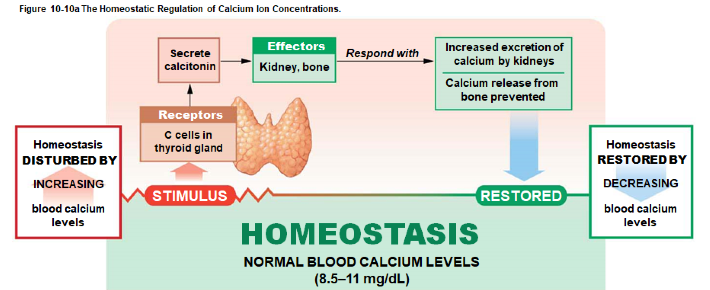
Calcium Balance and Calcitonin
Hypercalcemia (high blood calcium) reduces sodium permeability of nerves and muscles, affecting responsiveness
The Parathyroid Glands
Two pairs of small glands embedded in the posterior surface of the thyroid gland
Contains principal cells producing parathyroid hormone (PTH)
Stimulated by decreases in plasma Ca2+:
Activates osteoclasts and inhibits osteoblasts in bone
Reduces calcium excretion by kidneys
Stimulates kidney to secrete calcitriol, enhancing Ca2+ and PO43- absorption in the digestive tract
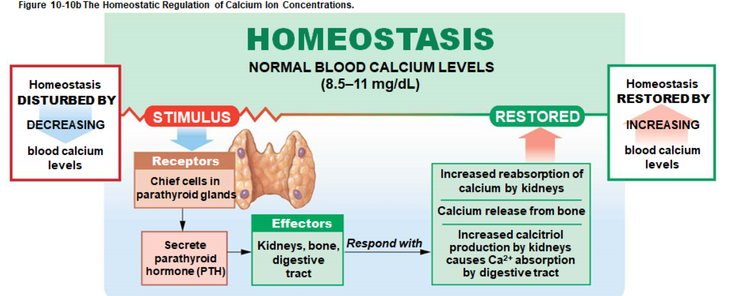
Calcium Balance and PTH
Hypocalcemia (low blood calcium) increases sodium permeability of membranes, causing excitability and spasms
PTH prevents hypocalcemia
Table 10-2: Hormones of the Thyroid Gland and Parathyroid Glands
Summary:
Gland/Cells | Hormone(s) | Targets | Effects |
|---|---|---|---|
Thyroid (Follicular epithelium) | Thyroxine (T4), Triiodothyronine (T3) | Most cells | Increased energy use, growth |
Thyroid (C cells) | Calcitonin (CT) | Bone, kidneys | Decreased calcium in body fluids |
Parathyroids (Principal cells) | PTH | Bone, kidneys | Increased calcium in body fluids |
The Adrenal Gland
Also known as the suprarenal gland, located on each kidney's superior border
Composed of two portions: adrenal cortex (outer) and adrenal medulla (inner)
The Adrenal Cortex
Contains high levels of cholesterol and fatty acids
Produces over 24 steroid hormones called corticosteroids, essential for metabolic functions and life
Divided into three zones:
Zona glomerulosa: Mineralocorticoids
Zona fasciculata: Glucocorticoids
Zona reticularis: Androgens
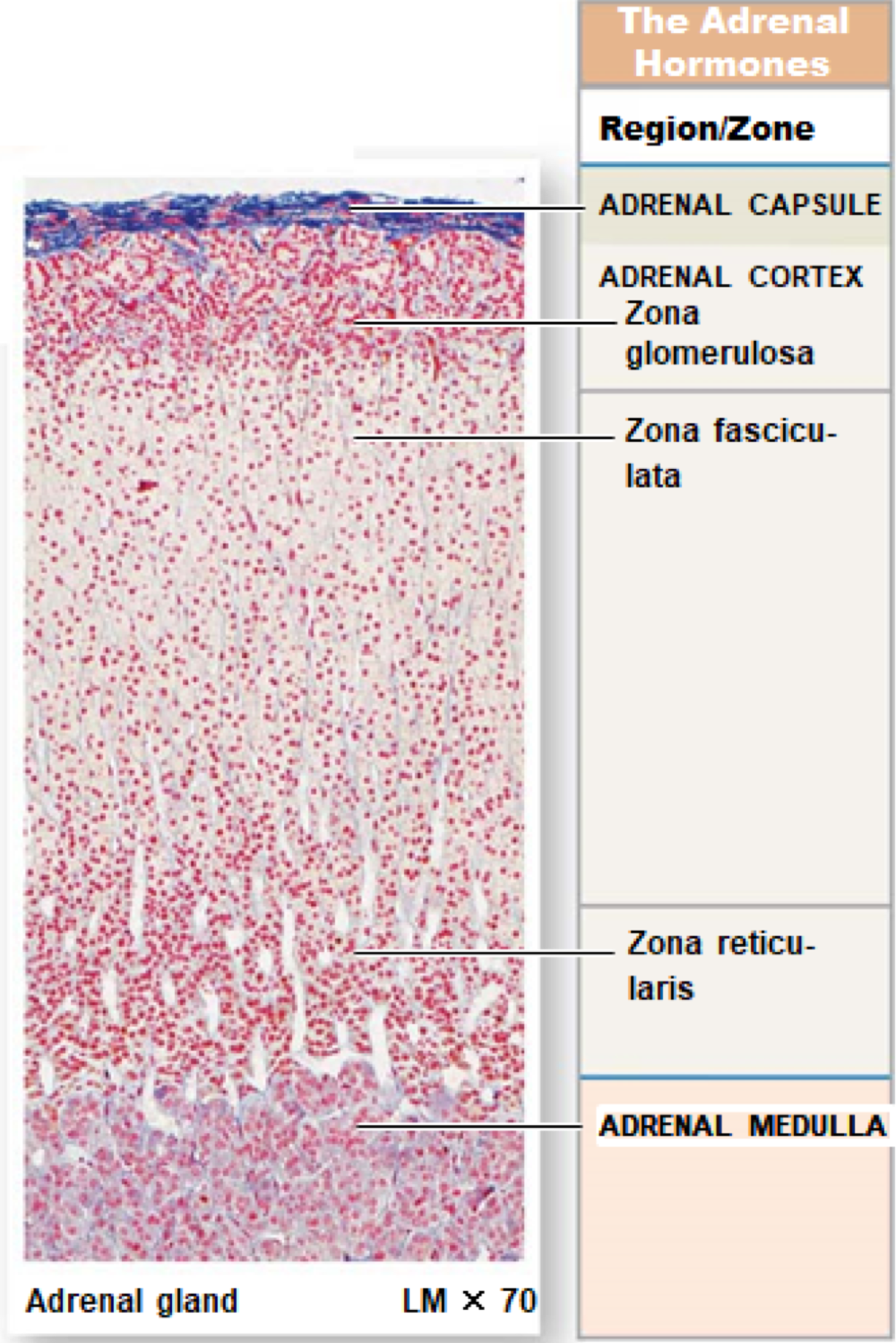
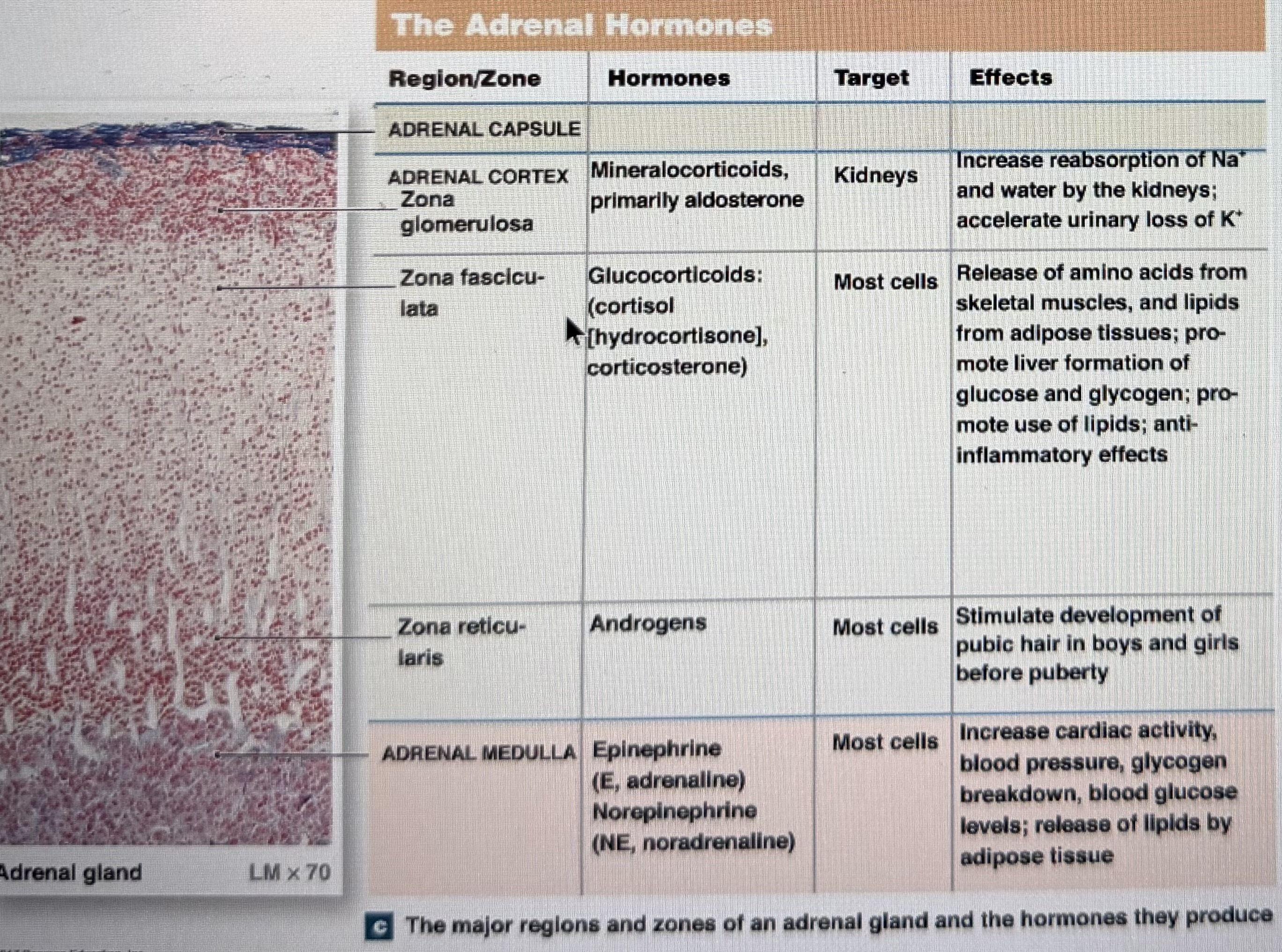
Mineralocorticoids
Main mineralocorticoid is aldosterone
Secreted in response to low Na+, low BP, high K+, or angiotensin II presence
Triggers sodium reabsorption and potassium elimination, prevents sodium loss in urine and promotes water reabsorption
Glucocorticoids
Main glucocorticoids include cortisol, corticosterone, and cortisone
Secreted in response to ACTH
Increase rates of glucose synthesis, glycogen formation, blood glucose levels
Act as anti-inflammatory agents by inhibiting components of the immune system
The Androgens
Produced in small quantities in both sexes; some convert to estrogens
Stimulate development of pubic hair before puberty
In adult women: Contribute to muscle mass, blood cell formation, and sex drive
The Adrenal Medulla
Vascular, includes cells similar to sympathetic ganglia
Secretory cells produce epinephrine (75-80%) and norepinephrine (20-25%)
Hormones trigger metabolic changes for increased energy availability, supporting the sympathetic response
The Pineal Gland
Located on the third ventricle's posterior roof
Produces melatonin, secretion influenced by light/dark cycles
May influence sexual maturation timing and protect CNS neurons with antioxidant activity
The Endocrine Pancreas
Lies between the stomach and proximal small intestine
Contains both exocrine (digestive enzymes) and endocrine cells
Endocrine Cells in Pancreas
Located in islets of Langerhans, accounting for 1% of pancreatic cells
Alpha cells: Secrete glucagon
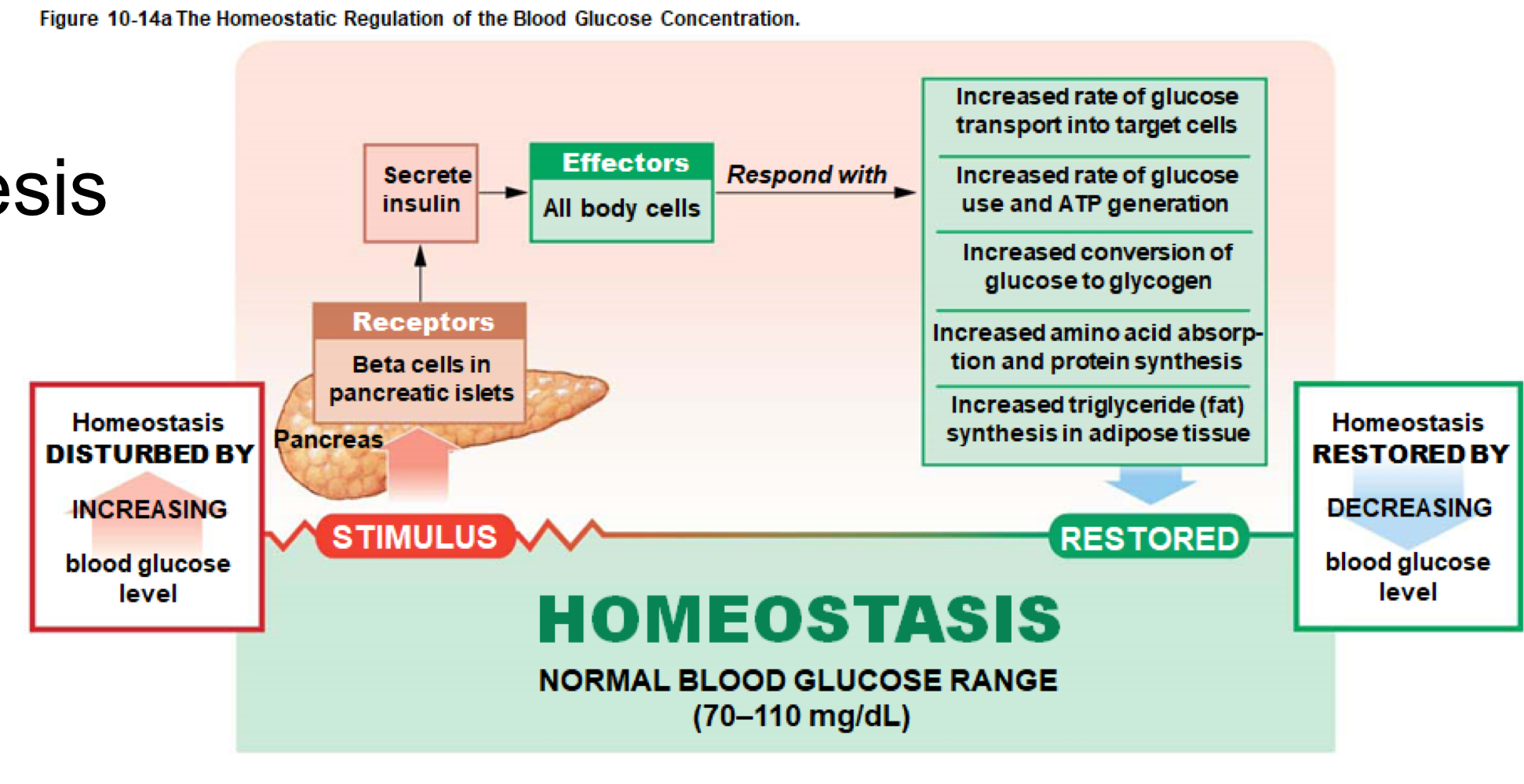
Beta cells: Secrete insulin, regulating blood glucose levels
Blood Glucose and Insulin
Increases in blood glucose stimulate beta cells to release more insulin
Insulin triggers glucose uptake by almost all cells (exceptions: neurons, RBCs, intestinal epithelium)
Increases protein synthesis and fat storage, leading to lower blood glucose after meals
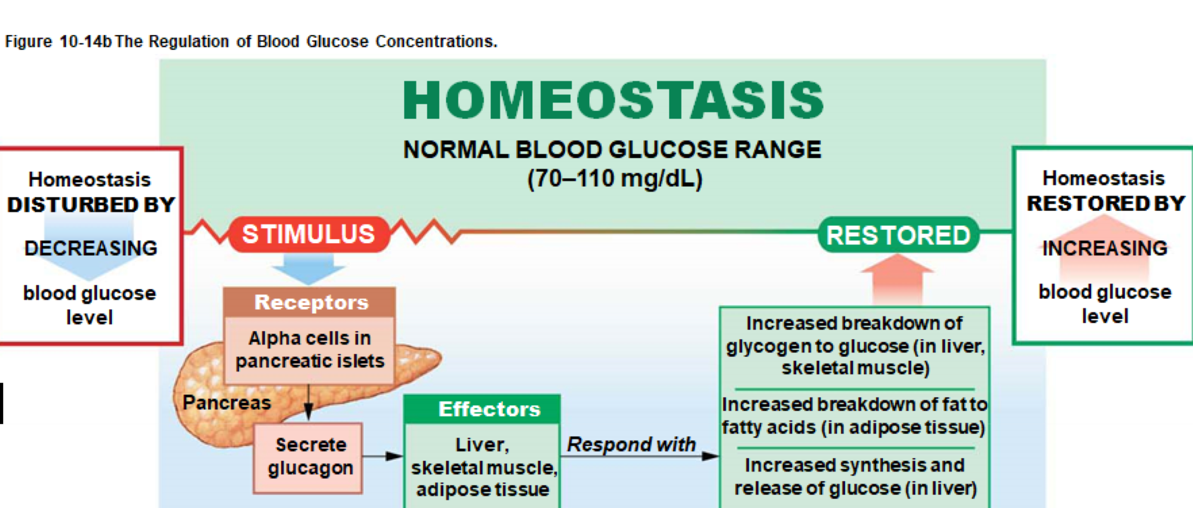
Blood Glucose and Glucagon
Decreases in blood glucose activate alpha cells to release glucagon
Mobilizes energy reserves by breaking down glycogen and releasing fatty acids
Pancreatic Regulation of Blood Glucose
Secretion of insulin and glucagon not directly stimulated by nerves
Affected by hormones influencing blood glucose e.g., cortisol
ANS activity: Parasympathetic stimulates insulin release, sympathetic inhibits it
Diabetes Mellitus
Faulty glucose metabolism causing hyperglycemia
Type 1 Diabetes: Inadequate insulin production
Type 2 Diabetes: Insulin resistance
Symptoms include hyperglycemia, glycosuria, polyuria
Complications of Diabetes Mellitus
Retinal Damage: Diabetic retinopathy can lead to blindness
Kidney Degeneration: Diabetic nephropathy can cause kidney failure
Cardiac Issues: Early heart attacks are more likely
Peripheral Nerve Problems: Abnormal blood flow causing various neural issues
Tissue Damage: Reduced blood flow leading to ulceration and infection
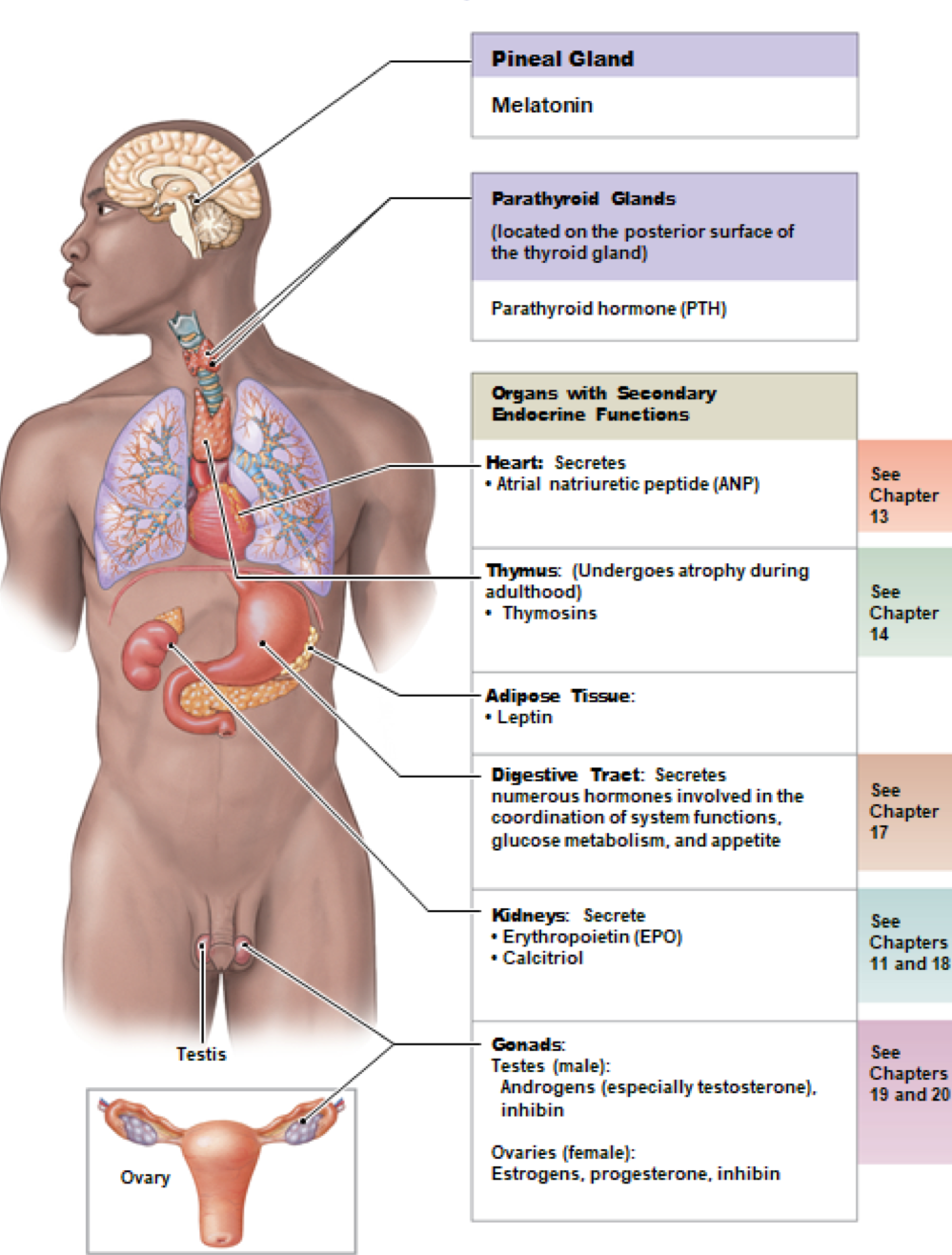
Secondary Endocrine Organs
Various organs produce hormones but primarily function in other systems
Examples:
Intestines: Release local hormones coordinating digestive activities
Kidneys:
Calcitriol: Stimulated by PTH, increases calcium absorption
Erythropoietin (EPO): Stimulated by low oxygen levels, increases RBC production
Renin: Triggers hormonal reactions to increase blood pressure
Heart: Atrial natriuretic peptide (ANP) released in response to increased blood volume
Thymus: Releases thymosins for immune defense development
Gonads and Adipose Tissue: Produce hormones impacting reproductive functions
The Intestines
Release hormones to coordinate digestive activities, major control over digestion rate influenced by ANS
Hormones Released by the Kidneys
Calcitriol: Increases calcium absorption
Erythropoietin: Increases RBC production
Renin: Triggers the RAAS for blood pressure regulation
The Heart
Endocrine cells in right atrium release ANP in response to increased blood volume, promoting sodium and water loss
The Thymus
Deep to sternum, active in childhood, atrophies in adulthood
Releases thymosins to support immune defenses
The Gonads: The Testes
Produce androgens (mainly testosterone)
Promote sperm production and maintain male reproductive structures
Inhibin secreted by nurse cells regulates FSH production
The Gonads: The Ovaries
Follicular cells produce estrogens and inhibins
Estrogens support egg maturation and uterine lining growth
After ovulation, corpus luteum secretes estrogen and progesterone preparing for embryo implantation
Hormones of the Reproductive System
Table 10-3: Summary of Reproductive Hormones
Hormone | Primary Target | Effects | Structure/Cells |
|---|---|---|---|
Androgens (testes) | Most cells | Supports sperm maturation, secondary sex characteristics | Interstitial cells |
Inhibin (testes) | Anterior pituitary | Inhibits FSH secretion | Nurse cells |
Estrogens (ovaries) | Most cells | Supports follicle maturation, secondary sex characteristics | Follicular cells |
Progesterone (ovaries) | Uterus, mammary glands | Prepares for embryo implantation, milk production | Corpus luteum |
Adipose Tissue
Produces leptin, regulating appetite
Triggers satiation, suppressing appetite
Vital for normal GnRH and gonadotropin synthesis
Low body fat can delay puberty; increased fat may enhance fertility
Hormonal Interactions
Hormones in extracellular fluid can produce various effects:
Antagonistic Effects: Opposing (e.g., PTH and calcitonin)
Synergistic Effects: Greater combined effect
Permissive Effects: One hormone's presence is necessary for another's function
Integrative Effects: Different but complementary effects
Hormones and Growth
Six hormones essential for normal growth:
Growth Hormone: Supports muscle and skeletal growth
Thyroid Hormones: Required for nervous system development
Insulin: Supplies energy to growing cells
PTH: Promotes calcium availability for bone growth
Calcitriol: Calcium absorption support
Reproductive Hormones: Affect osteoblast activity and secondary characteristics
Hormones and Stress
Stress triggers:
Physical (injury/disease), emotional (anxiety), environmental (extremes), metabolic (starvation)
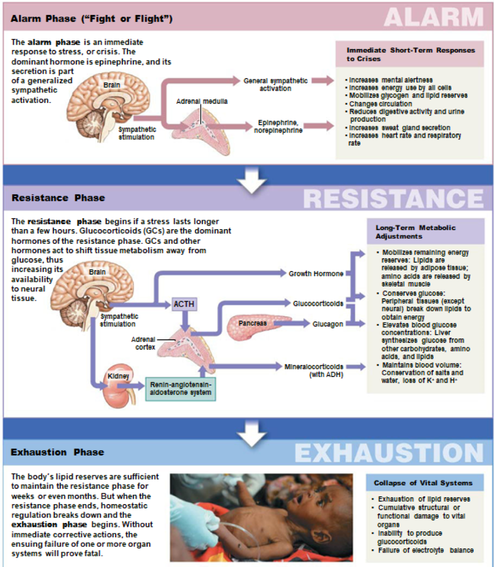
Triggers the general adaptation syndrome (GAS) which has three phases:
Alarm Phase: Immediate fight or flight response, dominated by epinephrine
Resistance Phase: Lasts several hours, characterized by glucocorticoids
Exhaustion Phase: Homeostatic breakdown leads to organ failure possibility
Hormones and Behavior
Regulated by the hypothalamus
Hormonal imbalances can alter behavior (e.g., precocious puberty) affecting aggression and assertiveness
Hormones and Aging
Usually most hormones remain the same throughout adulthood, except reproductive hormones.
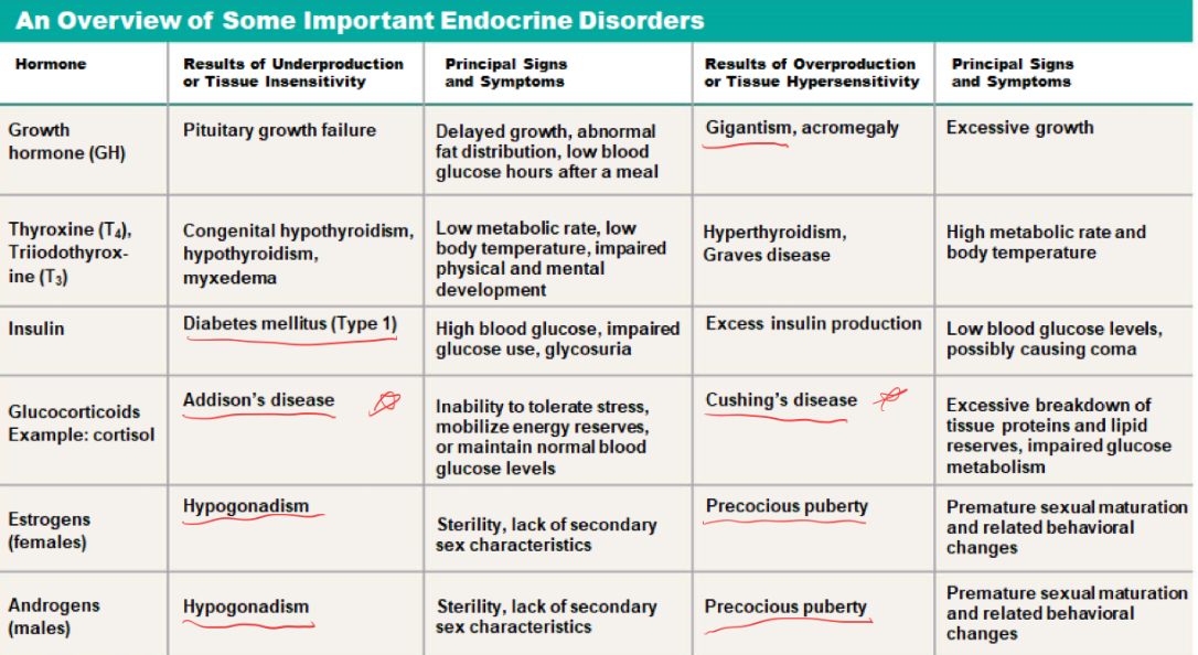
Endocrine Disorders