Prenatal G&D 1
Prenatal Development
Introduction
When does development start?
stages of prenatal development
ovum
from fertilization to end of cleavage
embryonic
from implantation to fetal stage
fetal
from beginning of fetal stage to parturition
stages of fertilization
oocyte → ovulated
oocyte surrounded by the zona pellucida
a protective layer made of glycoproteins
thickens after ovulation to protect oocyte
sperm makes contact with egg
acrosome in the sperm head reacts to ZP (acrosome reaction)
sperm fuses with cell membrane of egg and releases its contents
ZP hardens
prevents polyspermy
pronuclei of male and female fuse and egg completes Meiosis II
results in a second polar body
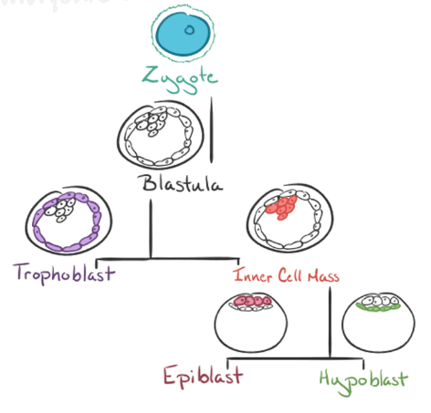
now a fertilized egg = zygote
fertilized ovum + sperm = zygote
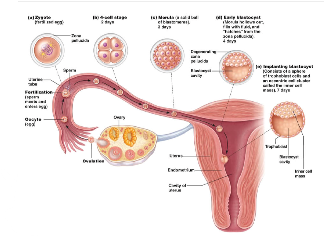
Ovum Phase
begins at fertilization
11-14 days, ending at the end of cleavage
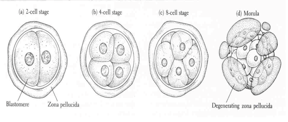
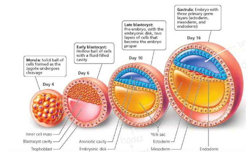
cleavage
rapid cell division = hyperplasia
no protein synthesis
>DNA: protein
ratio
increasing in cell number → increasing DNA, stagnant protein
ends with implantation
no morphogenesis or differentiation
stages:
early cleavage
two cells → four cells → eight cells
late cleavage
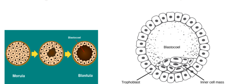
sixteen cells→ thirty-two cells (Morula)
increase in cell number, decrease in cell size
zona pellucida prevents cells from growing in size too much
in the morula phase, the zona pellucida begins to degenerate/crack
“hatching”
blastulation
conceptus now referred to as a blastocyst
center of morula becomes hollow
lumen (blastocoel)
cells begin to differentiate (blastula)
trophoblasts
allows for implantation and becomes the fetal portion of the placenta
around the perimeter
inner cell mass
becomes the embryo
elongation
lengthening of the fetal tissues
oval shape → tubule → filamentous shape
horses and humans:
conceptus remains in egg shape
do not go through elongation
increased placental surface area
increase absorption of nutrients from placenta
Embryonic Phase
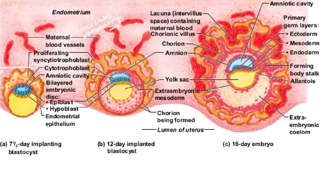
Implantation
conceptus imbeds itself into the endometrial wall of uterus and into maternal side
occurs 1-5 weeks after fertilization
time frame depends on species
still referred to as a conceptus, or fetal tissue
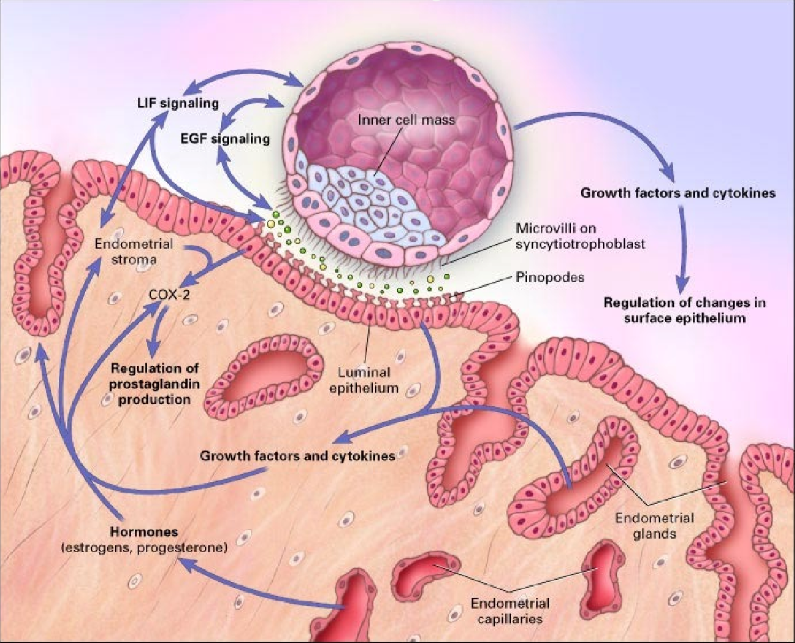
Key player: Progesterone
maintain pregnancy, hormone of pregnancy
increase vascularity and glycogen secretions
glycogen - stored glucose (sugar)
fetal nutrients/energy
fetal tissue receives nutrients from blood
inhibit muscular contractions
Special note:
Leukemia Inhibitory Factor
produced by endometrial glands
in humans and mice
preps uterus and conceptus
without this, implantation can’t occur
in cattle, it is not believed to be involved in implantation
rather cell differentiation
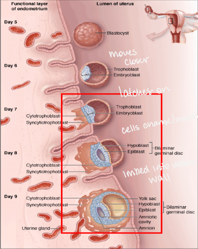
inner cell mass pushes to one side of blasocyst to form embryonic disc
zygote → blastula → trophoblast & inner cell mass → epiblast & hypoblast
Factors of maternal recognition of pregnancy (limiting maternal immunity)
FAS Ligand
binds to Fas
receptor on the maternal cytotoxic T cells
causes apoptosis of cytotoxic T cells
Indoleamine 2,3 dioxygenase (IDO)
trophoblast produces IDO which destroys tryptophan
tryptophan (amino acid) activates maternal cytotoxic T cells
Interferon Tau
ruminants
effects hormone cycles
acts on uterine peithelium
decrease estrogen and oxytocin
in result, decrease PGF2a
Synctiotrophoblast/Synctial Plaques
endogenous retroviruses
form feto-maternal interface
nutrient exchange, hormone secretion, immune modulator (decrease dam’s immune system)
non-ruminant: Syncytiotrophoblast
ruminant: syncytial plaques
Embryonic Phase
formation of embryo
hyperplastic growth
multipotent cells → different cell lineages
eventually form different tissues
driven by gene expression
stages:
(implantation), gastrulation, neurulation, embryonic folding, organogenesis
gastrulation
formation and development of the gut
epiblast cells replicate to form the primitive streak
depression forms → primitive groove
epiblast cells migrate toward the hypoblast to form germ layers, fold themselves to center and in result, push hypoblast down to form third layer
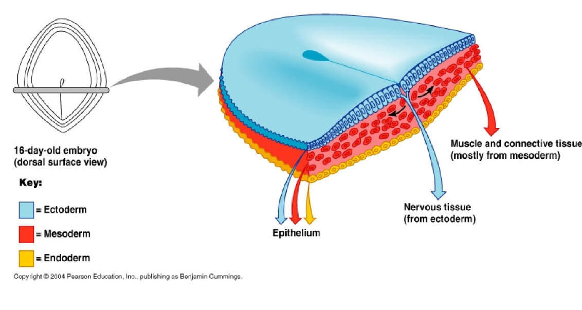
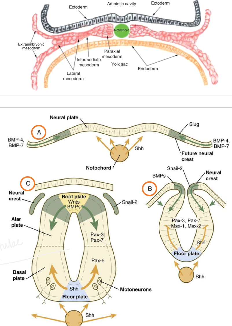
ectoderm, mesoderm, endoderm

three germ layers
ectoderm
outer layer
integument, sensory organs, oral cavity, nervous system, mammary and sweat glands
mesoderm
middle layer
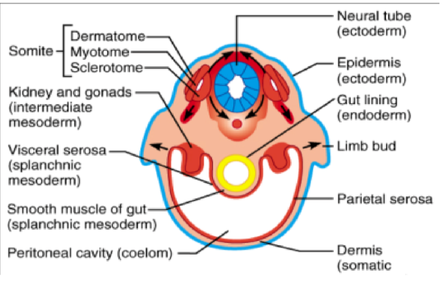
musculoskeletal, excretory, reproductive (except germ cells), cardiovascular and circular systems, visceral and parietal peritoneum, and mesenchyme
endoderm
inner layer
digestive tract, liver, lungs, pancreas, thyroid, respiratory tract, germ cells
neurulation
notochord
formation of spinal cord precursors
influences and initiates folding of the embryo
sonic hedge hog (Shh)
induces neural fold elevation
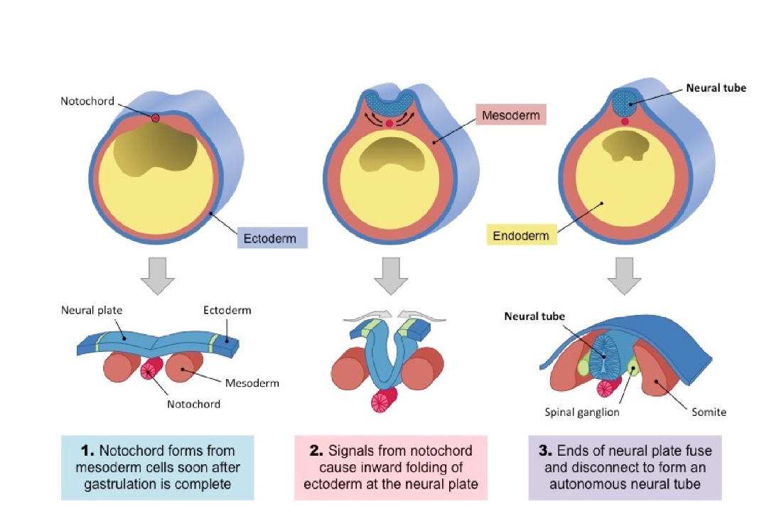
embryonic folding
lateral body folds across median and horizontal planes
involves ectoderm, mesoderm and endoderm
endoderm moves towards midline to form primitive gut tube
foregut, midgut, hindgut
ectoderm moves to cover the outside of the embryo
body plan developed
germ layers continue to differentiate to form organ systems
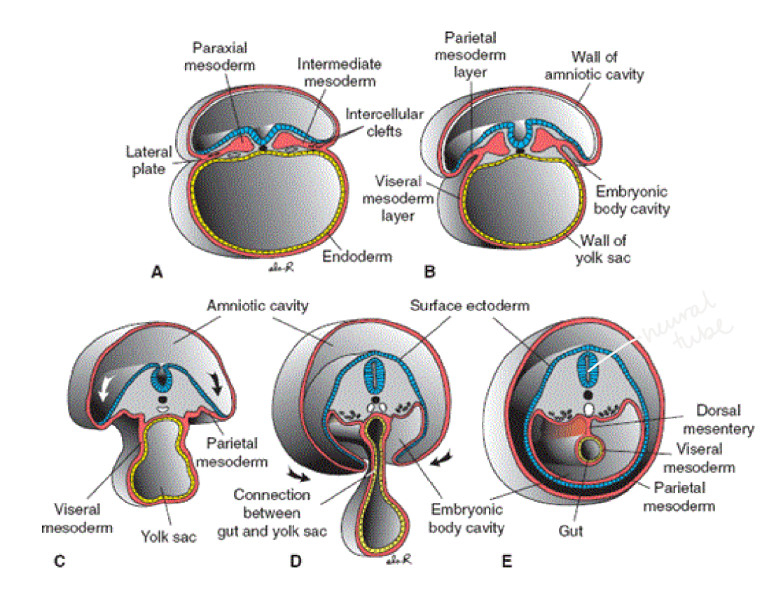
somites
block of mesoderm tissue, lateral to notochord
line the vertebrae
one pair of somites for every vertebrae
each somite differentiates into three regions
sclerotome (skeleton)
vertebrae, ribs, endochondral bones of skull
dermatome
dermis of skin
myotome
skeletal muscle
organogenesis
the formation of organs and organ systems
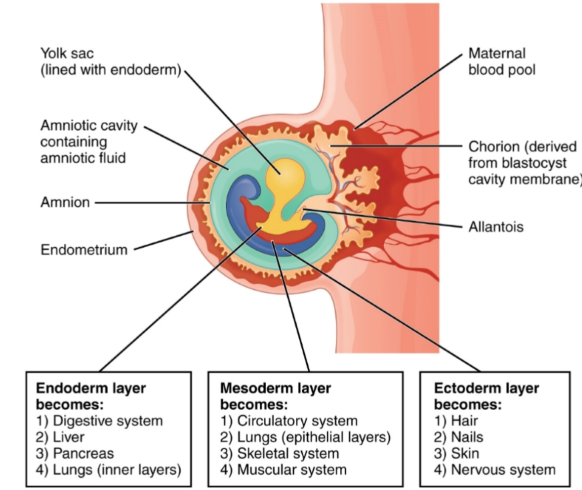
endoderm layer becomes:
digestive system, liver, pancreas, lungs (inner layers)
mesoderm layer becomes:
circulatory system, lungs (epithelial layers), skeletal system, muscular system
ectoderm layer becomes:
hair, nails, skin, nervous system
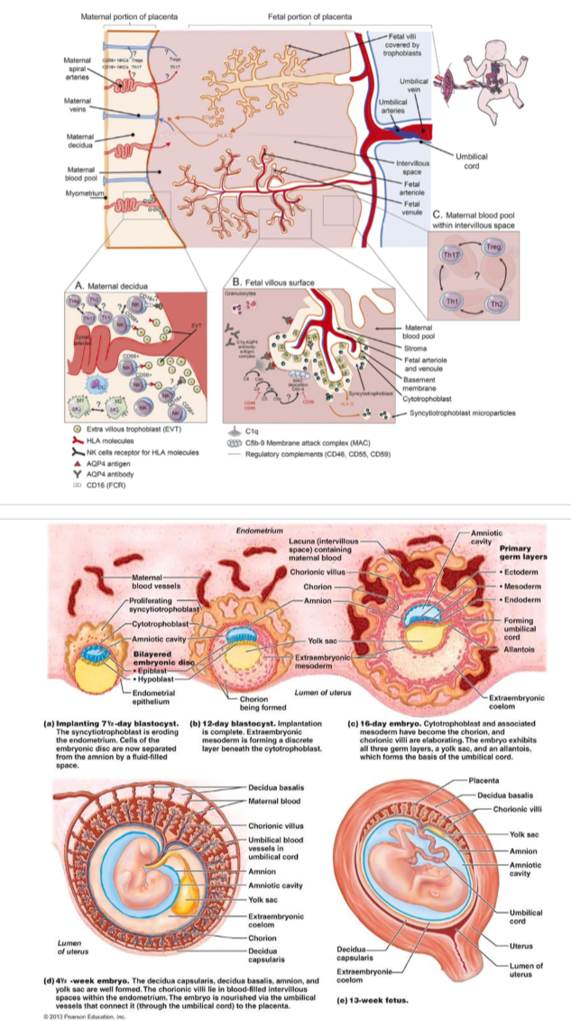
Placenta
Introduction
How does the embryo grow and develop?
by way of the placenta
nutrient transfer
Anatomy
Basics
trophoblast cells
implantation
increased size of conceptus = increased size of placental tissue
why does a cow eat her placenta?
to deter predators
placental variation based on species
placentas are classified by:
layers between fetal and maternal blood supply
shape and contact of chorionic villi
less layers = less connection
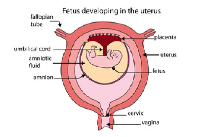
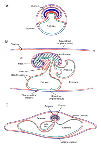
Umbilical Cord
umbilical artery and umbilical vein
wrapped in connective tissue
Layers Surrounding Fetal Membrane
chorion
trophoblastic layer
avascular
2 layers thick
encloses the embryo and fetal membrane
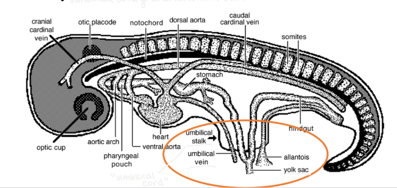
allantois
precursor of the umbilical cord
outgrowth of the hindgut
amnion
formed by folding of membranes around the Internal Cell Mass (ICM)
encloses the fetus in a fluid-filled cavity
yolk sac
formed by the endoderm spreading over the surface of the trophoblast
important for placental development
part of the primitive gut
early nutrition for the embryo
prior to placental development
aka no longer getting energy from glycogen derived from uterus
in between glycogen secretions between implantation and formation of placenta
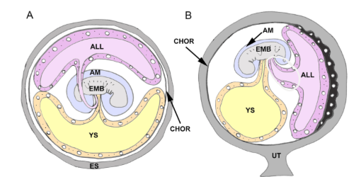
Chorioallantois Layer/Membrane
allantois and chorion fuse to form the allanto-chorion
in this process the yolk sac has been displaced
surrounds entire fetus
expansion of allantois
formation of chorioallantoic placenta
the chorionic villi
interdigitate with the endometrium
nutrient, gas, and waste exchange
blood supply changes depending on species
location of these villi changes
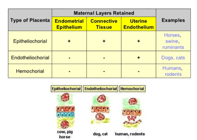
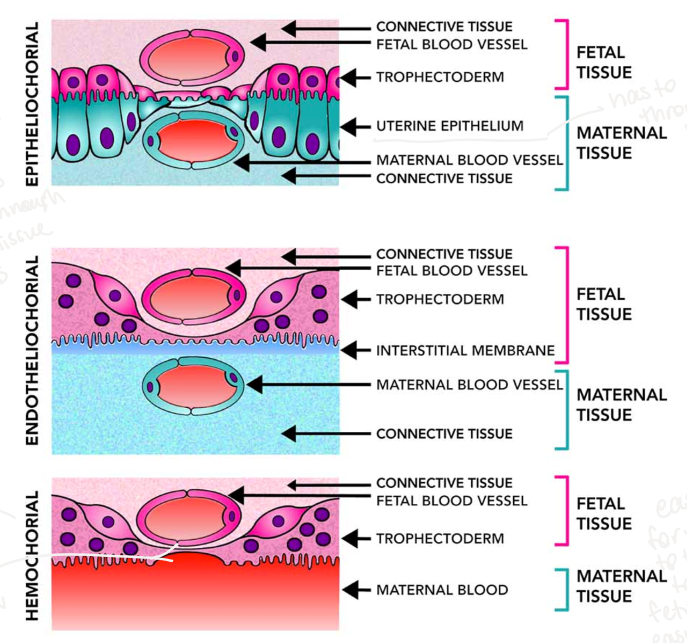
Classification by Layers
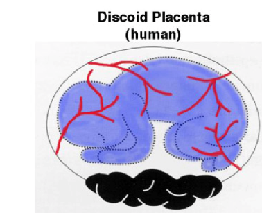
Hemochorial
most fetal-maternal interface
closest connection between mom/fetus
blood escapes maternal capillary and circulates unimpeded against placenta
example: primates (humans) and rodents
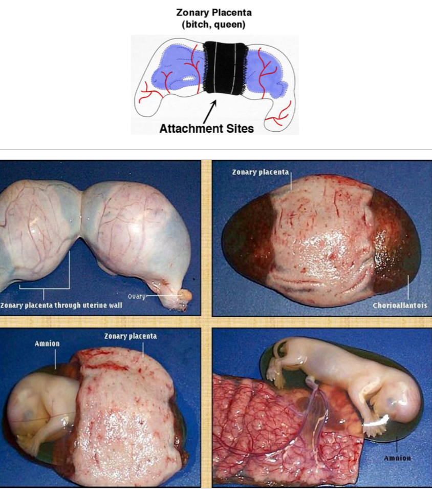
Endotheliochorial
placental epithelium invades maternal epithelium
immediately adjacent to maternal blood supply
example: carnivores (cats and dogs)
Epitheliochorial
placenta + maternal epithelium
2 membranes
placental epithelium + maternal epithelium
separate maternal and fetal blood
example: pigs, horses
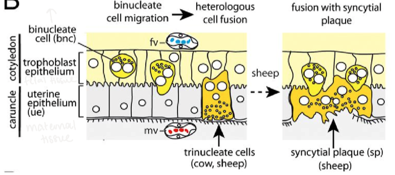
Synepitheliochorial
similar to epitheliochorial
connective tissue layer between placental epithelium and maternal vasculature
Binucleate Cells (BNC) of trophectoderm produce syncytium
produce syncytial plaques
example: cattle, sheep & goats
Classification by Chorionic Villi
Diffuse
chorionic villi are distributed over almost the entire surface of the chorionic sac
almost entirety of the chorioallantoic membrane is attached
villi projections/velvety over entirety of placenta
many layers, lots of connection
nutrients to baby
Example: swine, horses, camel
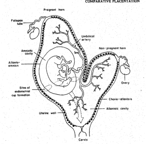
Horses:
endometrial cups form early until day 120 (then die)
produce equine choriogonadotropin
helps with placenta formation

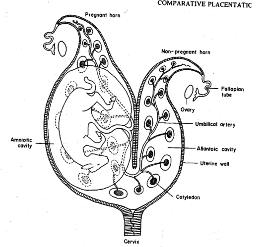
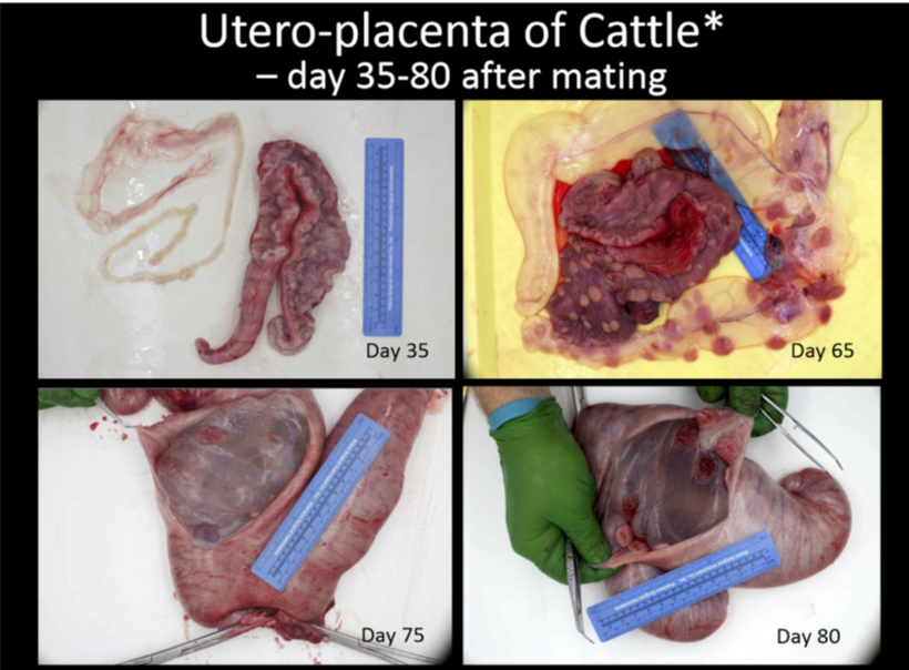
Cotyledonary (multiplex)
chorionic villi are normally restricted villi are normally restricted to circular or oval areas of the chorionic sac
number of cotyledons varies in species
multiple discrete sites of attachment
a lot of layers
placentome
fetal portion = cotyledon
maternal portion = caruncles
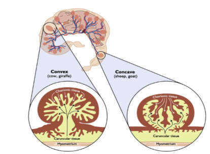
Example: ruminants
sheep:
no connections, but rather discrete pockets
start out small, grow to softball width
grows as placenta and baby grows
differ from mare and sow because of synepitheliochoriol
BNC produce syncytial plaques make this connection
BNC cluster in placentome-like pattern
maternal epithelium and capillaries push back towards syncytium and chorion (convex)
like inflating rubber glove fingers (fetal villi) into a swelling mound of jelly
once pattern is established, growth (IGF -make placenta grow) and angiogenic (VEGF-increase blood supply, make new blood vessels) drive mutual growth and interaction
Zonary Attachment
chorionic villi are restricted to an equatorial girdle
site of attachment is around a band of tissue that surrounds the fetus
example: carnivores (dogs, cats, seals, bears, elephants)
band around each offspring
nutrients have few layers to get through versus previous species
incomplete zonary placenta
resembles single or double discoidal condition
can be distinguished by the presence of central or marginal effusions of maternal blood
example: mink
Discoid
chorionic villi are arranged in a circular plate
a single placenta is formed is a discoid shape
chorionic villi distributed in a circular plate
example: primates (humans), rodents
only ONE site of attachment
primarily at the bottom
not necessary to have a lot of attachment because of hemochorial layers
very easy to diffuse nutrients
double discoid
certain monkeys, occasionally an abnormality in humans
Classification of Placenta by Species
Type of Placenta → Common Examples
Diffuse, epitheliochorial → horses and pigs
Cotyledonary, synepitheliochorial →ruminants
Zonary, endotheliochorial → carnivores
Discoid, hemochorial → humans, primates, rodents
Function
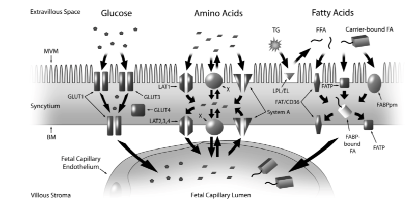
Transfer of Nutrients
maternal organs work for fetus
respiratory tract, digestive tract, kidneys
selective permeability
only some nutrients can pass
this is a good thing as dam can ingest something toxic
syncytiotrophoblast/syncytial plaques immune functions
increase dam immune function
placental fuel
all glucose delivered to uterine circulation not umbilical circulation
whatever is in maternal circulation does not mean it will pass into fetal blood
Types of Diffusion:
Simple Diffusion
maternal heart and lungs work extra hard
increased: BP, HR, RR
O2
partial pressure
hemoglobin concentration >50% than maternal blood
higher oxygen carrying capacity
CO2
byproduct of biochemical processes
from fetal blood to maternal circulation → expiration
fetus intakes O2, expels CO2 to maternal circulation for dam to excrete
Na+, K+, Cl-, Ca, P, H2O
necessary in small amounts
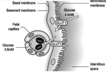
Facilitated Diffusion
glucose
60% of energy
GLUT 1 (primary), GLUT 3, and GLUT 4 transporters during early pregnancy
fetal circulation dependent on maternal circulation
fetal circulation does depend on maternal circulation
strange phenomenon:
maternal stores will be given to fetus in desperate times
Active Transport
amino acids
30% of energy
fetal circulation greater than maternal
requirement determined by growth rate, protein deposition & energy demands
placenta expresses over 15 transporters
fatty acids
triglycerides too large for transport
must become free fatty acids (lipases)
placenta metabolizes long chain NEFAs and supplies fetus with long chain metabolites
very low concentrations of fat soluble vitamins
Endocrine Organ of Pregnancy
Chorionic Gonadotropin
maintains CL during pregnancy
estrogen
stimulates growth of myometrium
aids in preparation of mammary glands for lactation
progesterone
suppresses uterine contractions
aids in preparation of mammary glands for lactation
promotes the formation of the cervical plug to prevent uterine contamination
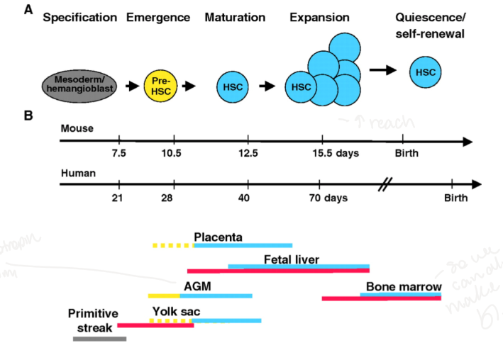
Blood Supply
fetal and maternal blood are separate
fetal blood is formed in the yolk sac
hematopoietic stem cells from mesoderm
hematopoietic → blood generation cells
runx1 necessary for HSC to become endothelium
mesoderm→hemogenic endothelial cells→pre-hematopoietic stem cells→hematopoietic stem cells
Steps of hematopoietic development:
Specification: mesoderm/hemangioblast
primitive streak
Emergence: Pre-HSC
aortic gonadotropin mesoderm (AGM), placenta, yolk sac
Maturation: HSC
placenta, AGM, yolk sac, fetal liver
Expansion: multiplication of HSC
increase reach
placenta, fetal liver, AGM, yolk sac
Quiescence/Self-Renewal
bone marrow
ability to make more blood