Nov 10 - Lecture 29 ~ GPCR
alpha and beta gamma protein subunits have distinct functions
when bound together, the alpha subunit and beta gamma subunit complex mutually inhibit each other
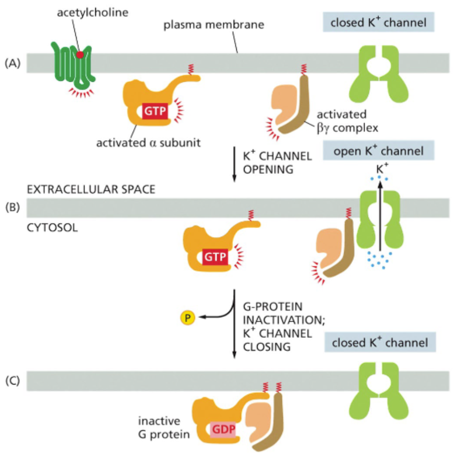
upon separation, alpha and beta gamma can interact with their own target proteins
e.g. — the separation of G protein associated with an Ach receptor allows the beta gamma complex to bind and stimulate the opening of a K+ channel
the outflow of K ions alters the membrane potential
neuron signaling that opens ion channels indirectly via intracellular signaling is metabotrophic
the alpha subunit in this pathway has its own interaction partner
specifically, it activates or inhibits an enzyme called adenylyl cyclase
alpha subunits signal through second messengers
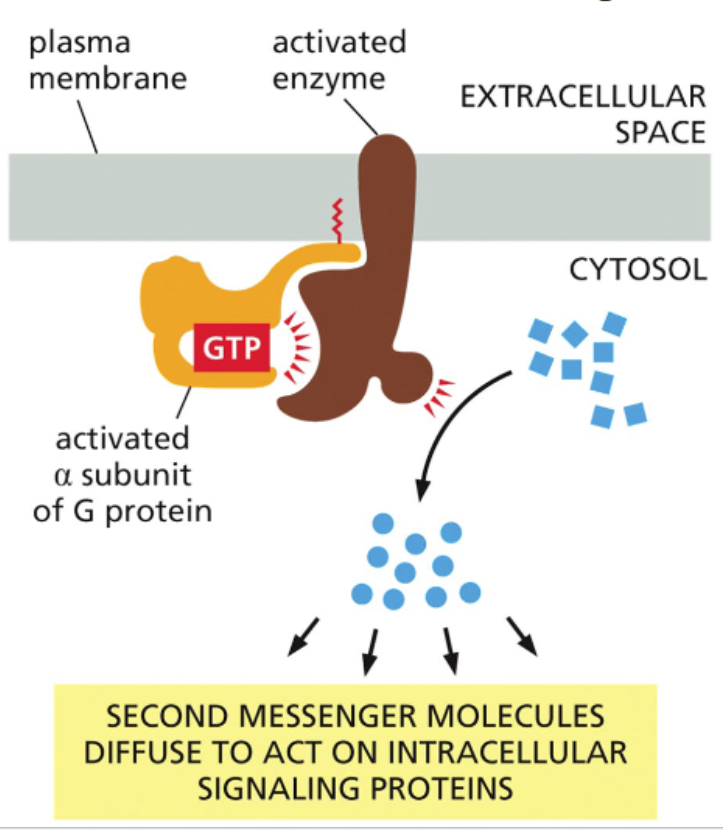
alpha subunits signal through their association with enzymes
the rxn catalyzed by the enzyme and the effect of the association depends on the alpha subunit itself
signal molecules produced (or released) via enzymes are referred to as second messengers
examples include: cAMP, IP3, DAG, and Ca2+
a single activated enzyme can produce many second messenger molecules — more signal amplification
Gas and Gai subunits alter cAMP concentrations
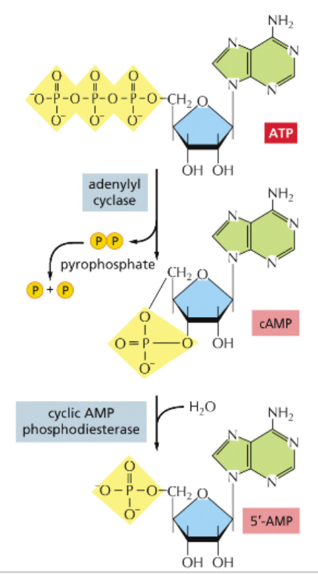
cyclic AMP is a metabolite of ATP
Adenylyl cyclase catalyzes phosphodiester bond formation between a 5’ phosphate and 3’ hydroxyl of the same ribose
cyclic AMP phosphodiesterase catalyzes hydrolysis of the 3’ phosphoester bond and conversion of cAMP to AMP
cAMP is an activator of protein kinase A (PKA)
cAMP has multiple potential target that vary according to cell type
cAMP-gated channels open when bound by cAMP
cAMP-sensitive channels close when bound by cAM
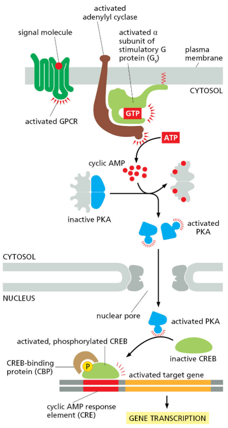
PKA depends on cAMP binding for full activity
cAMP binding to a regulatory protein releases active PKA
once bound to cAMP, PKA phosphorylates its protein target using ATP as a donor
PKA can regulate the function of cytosolic proteins via phosphorylation
cholera and pertussis toxins increase cAMP through different mechanisms
Cholera and whooping cough are caused by two different bacterial pathogens that colonize different tissues
Cholera – Vibrio cholerae in the intestine
Whooping cough – Bordetella pertussis in the airways
Both toxins covalently modify α subunits
Cholera toxin enhances cAMP signaling in enterocytes by locking Gαs in an active state – this leads to increased expression of the chloride channel CFTR on the apical membrane, water secretion, and diarrhea
Pertussis toxin enhances cAMP signaling in immune cells (maybe) by preventing activation of Gαi – this is hypothesized to inactivate the immune cells
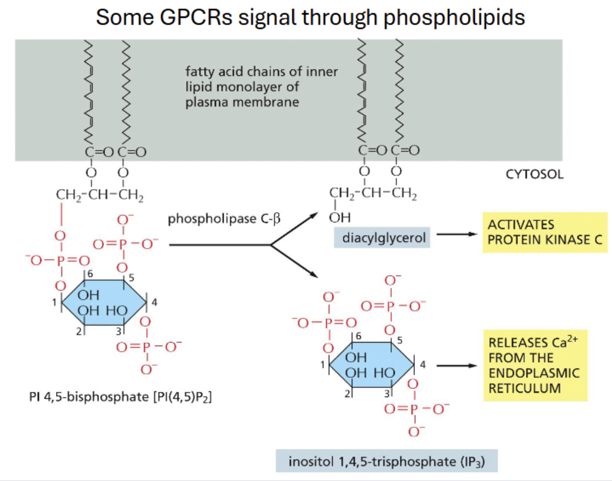
Gq signals through phospholipase C (PLC) and protein kinase C (PKC)
Gaq and GBy activate phospholipase C-B (PLC-B), a lipid-hydrolyzing enzyme
the main substrate for PLC PI(4,5)P2 which when cleaved yields IP3 and DAG
IP3 opens a Ca2+ channel on the ER membrane
Ca2+ and DAG activate protein kinase C (PKC) which phosphorylates its own target
signaling can result in rapid rises in cytosolic calcium concentration
opening of IP3 receptors yields an increase in cytosolic calcium
increased calcium conc leads to opening of calcium-dependent ryanodine channels that increase the rate of calcium uptake
positive and negative feedback contributes to rapid Ca2+ oscillations
opening of ryanodine calcium channels by calcium from IP3 receptors produces a positive feedback process that opens more ryanodine channels
as calcium rises, it causes the channels to close
ATPases return Ca2+back to the ER causing a drop in calcium concentration, which allows channels to open again
this cycle produces oscillations of Ca2+ concentration with frequencies proportional to signal strength
calcium-binding regulatory proteins turn calcium mobilization into responses
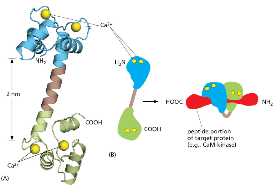
calmodulin is a dumbbell-shaped protein with a pair of calcium binding sites on each head
without Ca2+, it has a linear structure; upon Ca2+ binding, the protein jackknifes placing the head groups side-by-side
this creates a protein interaction domain that allows calmodulin to bind target proteins
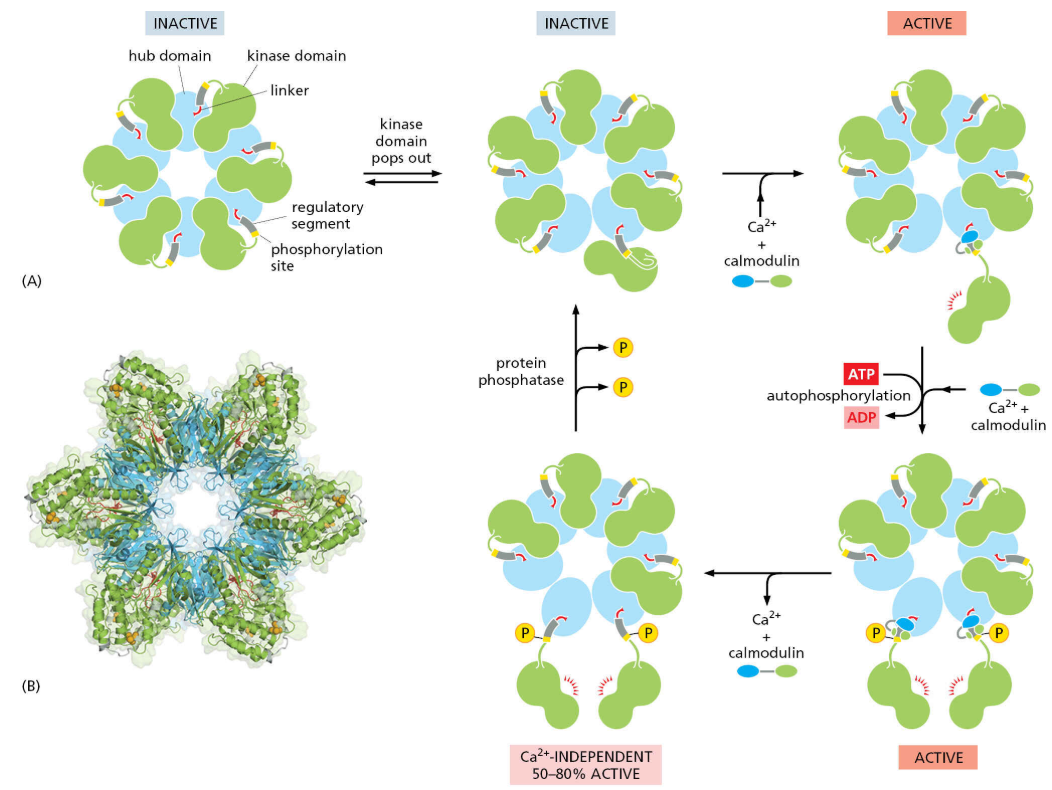
calmodulin-dependent protein kinase II is one of many targets of calmodulin
the transcription factor NFAT can enter the nucleus only when it isn’t phosphorylated; calcineurin is a calmodulin-dependent phosphatase that dephosphorylates NFAT
calmodulin stabilizes CAMK II in an active conformation
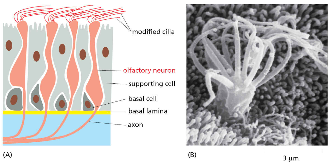
examples of GPCR signalling — olfactory neurons signal through cAMP
when an odorant binds to the olfactory receptors in the olfactory neurons that line the nose, it activates Golf
Golf then activates adenylyl cyclase → cAMP → opens cation channel to initiate action potential
Na+ influx causes depolarization of the membrane which sends a nerve impulse that travels to the brain
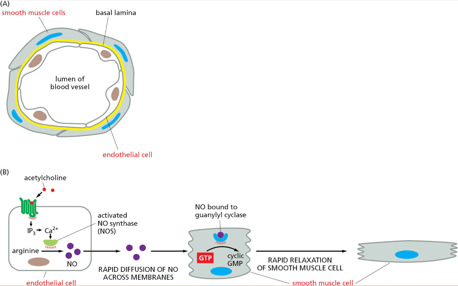
Gaq signaling in endothelial cells leads to nitric oxide production which relaxes smooth muscle
vasorelaxation can be initiated via endothelial cells that line the inner surface of the vessel
Ach activates Gaq couple receptor in endothelial cells — the increase of Ca2+ activates the enzyme nitric oxide synthase (NOS)
NO gas binds and activates guanylyl cyclase which produces cGMP
cGMP hyperpolarizes the cell and closes Ca2+ channels
this results in the cell relaxing and cGMP broken down by cGMP phosphodiesterase
sensory neuron — eyesight
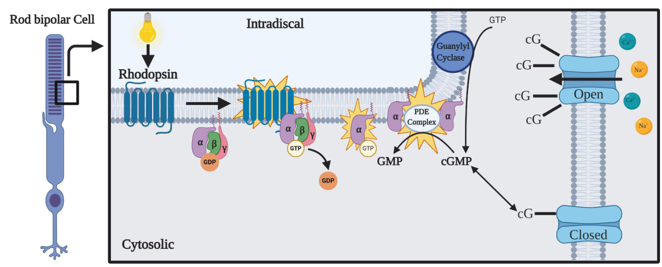
rhodopsin is a GPCR that is activated by light
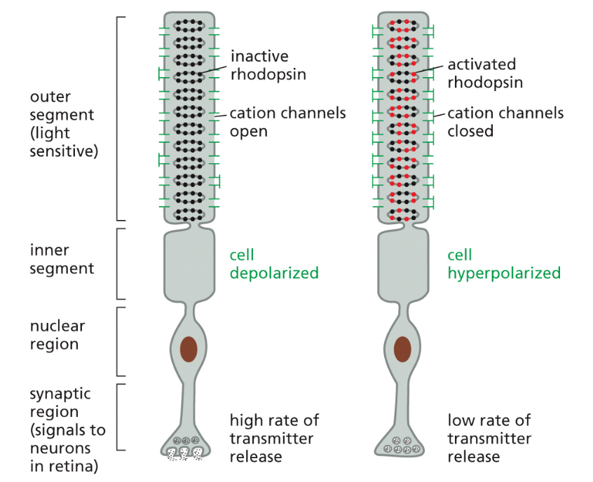
Photodetection is an unusual sense in that the sensing cells are depolarized when they are not stimulated
Rhodopsin is a GPCR that associates with a special α subunit called transducin (GαT )
GαT activates cGMP phosphodiesterase, which degrades cGMP
Falling levels of cGMP cause a cGMP-gated cation channel to close, which hyperpolarizes the cell
Because of this, photoreceptors release neurotransmitter in the dark and stop releasing when hit by a photon
light inhibits inhibitory nT release
nT are inhibitory — they inhibit nerve impulse from travelling to the brain
when inhibition is removed, nerve impulse is able to travel to the brain
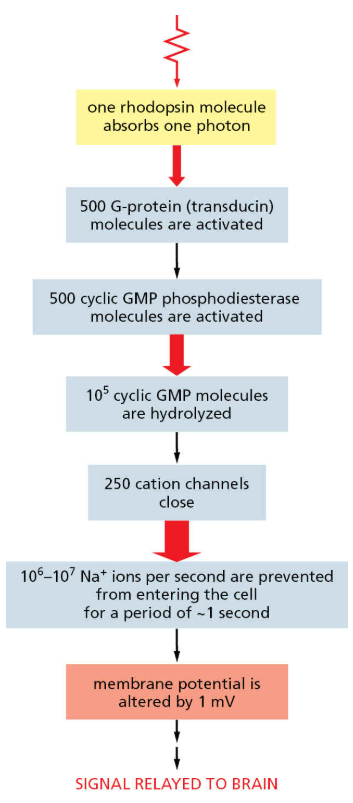
amplification of GPCR signals produce strong responses from few receptors
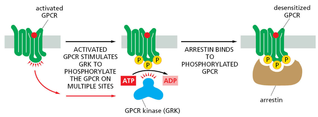
GPCR kinases and arrestins decrease sensitivity of GPCRs
Summary / Key Concepts
Gαq signaling acts first through phospholipase C (PLC). This enzyme cleaves PI(4,5)P2 to release IP3 & DAG. IP3 increases cytosolic calcium concentration and DAG cooperates with calcium to activate protein kinase C (PKC).
Like PKA, PKC has it’s own phosphorylation targets.
Calcium can also alter protein function via binding to regulatory proteins like calmodulin. Calcium oscillations are translatable by CAMK II
Photoreceptors use GPCRs that signal through transducin, which activates phosphodiesterase that breaks down cyclic GMP – this closes cGMP-gated cation channels and hyperpolarizes the photoreceptor cell.
GPCR activation can lead to phosphorylation by GPCR kinases, followed by binding of arrestin and desensitization