Molecular Biology Techniques: Hybridization, Sequencing, and PCR
Northern Blotting Techniques
Definition and Purpose: Northern blotting is a technique closely related to Southern blotting but specifically designed for the detection of RNA rather than DNA.
It identifies specific RNA sequences using complementary probes.
The most frequent application is determining gene expression levels, specifically whether a gene is being transcribed into mRNA within a particular sample or tissue.
Key Principles of Gene Expression:
Gene expression is spatio-temporally regulated; not all genes are expressed in every tissue or at all times.
A single gene may produce multiple mRNA transcripts of varying sizes.
The Northern Blotting Workflow:
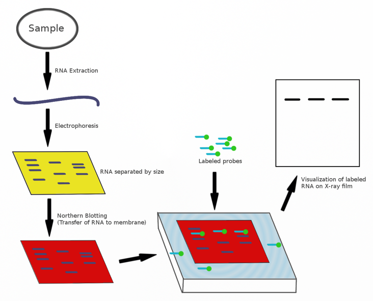
RNA Extraction: Single-stranded RNA is extracted from the sample.
Electrophoresis: The extracted RNAs are applied to an agarose gel to be separated.
Transfer (Blotting): The separated RNA molecules are transferred (blot) onto a nitrocellulose membrane.
Hybridization: hybridise with ssDNA probe that is complementary to the gene of interest.
Detection: wash and detect probe to show if the RNA of the gene of interest was present (and estimate its size)
Applications of Northern Blotting:
Determining tissue-specific gene expression.
Studying the regulation of gene expression levels over time, such as during different stages of biological development.
In Situ Hybridization (ISH)
Methodological Overview:
In situ hybridization is a technique used to detect specific RNA molecules directly within cells or tissues, without blotting.
Probes (either DNA or RNA) hybridize to the target mRNA at its functional site within the cell (in situ).
The procedure can be performed on thin tissue sections or on whole organisms.
Significance in Developmental Genetics:
The technique is widely used to determine "where and when" genes are expressed.
It reveals the precise cellular location and tissue distribution of specific mRNAs when viewed under a microscope.
It can also be utilized to locate specific genes or sequences on whole chromosomes.
Labeling and Detection Methods: Colorimetric In Situ Hybridization (CISH):
Uses a color-generating affinity probe.
Example: Digoxigenin-labeled probes detected by an antibody conjugated to a colorimetric enzyme, such as Alkaline Phosphatase.
Case Study: Detection of myogenin mRNA in mouse embryos. is essential for muscle development.
Fluorescent In Situ Hybridization (FISH):
Uses fluorescently labeled probes for visualization.
Tissue Localization Example: Simultaneous use of five different probes in a Drosophila embryo, each labeled with a distinct fluorescent molecule.
Revealing chromosome location of genes/sequences, example: Identifying deletions in the Duchenne muscular dystrophy (DMD) gene. In a heterozygous female, the DMD gene probe appears red while the X chromosome centromeres appear green. A deleted gene is indicated by an X chromosome lacking the red signal.
Summary of Gene Expression Hybridization Techniques
Northern blots separate and detect RNA (gene products) using DNA probes.
These techniques reveal the timing (developmental stage) and location (tissue type) of gene expression.
DNA probes used in high-resolution microscopy on "chromosome spreads" can detect gene locations, mutations, or chromosomal count abnormalities (ploidy).
DNA Sequencing Principles and Sanger Method
Definition and Utility:
DNA sequencing provides a base-by-base sequence of genes within a genome.
Sequence data allows for the prediction of amino acid sequences and the properties of encoded proteins.
It reveals genome organization, including regulatory elements like promoters and enhancers, as well as structural features like telomeres and centromeres.
The Sanger Method (Dideoxy/Chain-Termination Sequencing):
Developed by Fred Sanger, who was awarded the Nobel Prize in Chemistry in .
Core Mechanism: Uses DNA polymerase to copy a single-stranded DNA template.
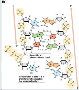
incorporation of special dideoxy-nucleotides (ddNTPs) to terminate DNA strand extension - dideoxy sequencing
can be automated, enabling rapid sequencing of large numbers of DNA frags.
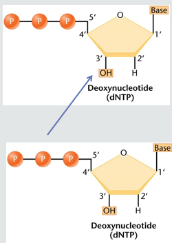
Standard Nucleotides (dNTPs): DNA is typically replicated using four deoxynucleotides: dATP, dCTP, dGTP, dTTP. These form phosphodiester bonds between the 5’ -PO4 group of the incoming nucleotide and the 3’ -OH group of the previous nucleotide.
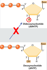
Dideoxynucleotides (ddNTPs): These special nucleotides lack a hydroxyl -OH group on the 3’ carbon. Once a ddNTP is incorporated, the DNA polymerase cannot add further nucleotides because there is no 3’ OH to form the next phosphodiester bond, resulting in chain termination.
Ideoxynucleotide sequencing:
4 reactions are needed to sequence the entire DNA fragment - one for each nucleotide A,C,G,T
dsDNA is seperated (denatured) into single strands
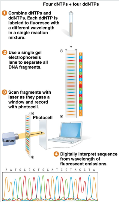
all dNTPs plus a small amt of each of the 4 ddNTPs are fluorescently labelled with a diff colour and are added together with DNA polymerase and primer
in each reation the primer is extended to copu the DNA frag, the rxn will terminate at diff points if a ddNTP is incorporated
rxn are repeated in many cycles until a fragment of every possible length is made
Automated DNA Sequencing:
The resulting fragments are separated by capillary gel electrophoresis.
each ddNTP is fluorescently labelled with a diff colour determined by nucleotide at the end of it that leads to termination
As fragments pass a laser, their fluorescent color (representing the terminal nucleotide) is recorded.
The sequence of recorded colors reveals the sequence of the DNA.
increased throughput compared the the Sanger method
Genomic Sequencing and Evolution of Technology
Whole-Genome Shotgun Sequencing:
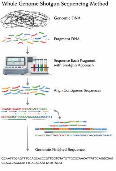
Sanger sequencing typically reads fragments of 30-1000bp
In the shotgun approach, the entire genome is broken into small fragments, each fragment is sequences and the complete genome is reconstructed by computationally assembling overlapping sequences
Next-Generation Sequencing (NGS):
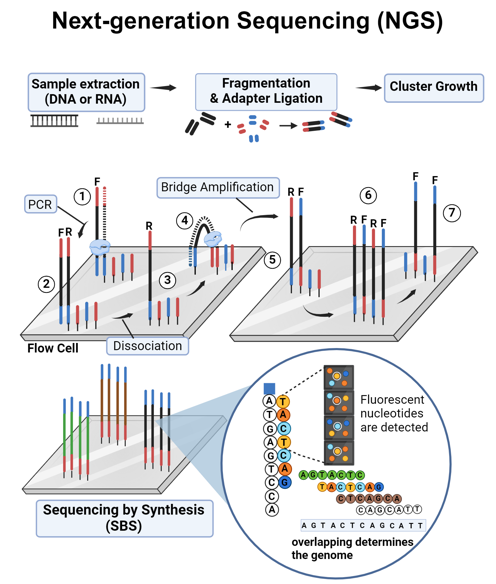
Also known as massively parallel sequencing.
Sequences millions of short fragments in parallel vy the sequential addition of nucleotides to DNA fragments on a solid surfae
enables rapid, high-throuput genome cale analysis
sequencing technologies
original sequencing too 10 years and $2 billion, now possible to sequence human genome for less than $1000
third generation sequencing allows faster, cheaper sequencing and longer reads
companies exist to provide personal genome sequences
The Human Genome Projects:
reference genome, built from a small number of individuals
one thousand genomes project
sequenced 1092 human genomes from across the globe
Catalogues the basis of genetic variation in humans to explain individual differences in susceptibility to disease, response to drugs or reaction to environmental factors
humans are 99.9% identical
single base pair differences between individuals can explain differences in disease risk, drug response
Genomic sequence in other organisms
genomes of more than 340 eukaryotes have been sequenced
+ 30000 bacteria
Polymerase Chain Reaction (PCR)
Fundamental Concept:
Invented by Kary Mullis (Nobel Prize ).
A method for selectively amplifying a specific DNA sequence.
The Enzyme: Uses Taq polymerase, a thermostable enzyme derived from the thermophilic bacterium Thermus aquaticus, to copy DNA. This enzyme functions at high temperatures.
rapidly amplifies specific DNA fragments without the need for cloning vectors or restriction enzymes
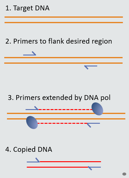
requires primers that bind to known DNA sequences that flank the target region
extremely sensitive, can amplify DNA from pico to femtogram quantities
widely used in diagnostic and forensic science
The PCR Cycle (Three Steps):
uses repeated cycles of heating and cooling to copy a specific DNA fragment
Denaturation (): temperature is increased for ard 30s to seperate DNA strands into single strands.
Primer Annealing (-): Temp is decreased for ard 30s to allow primers to anneal to base pair to complementary sequences in target DNA template
Extension (): Taq polymerase extends primer and synthesize nascent DNA strand for ard 1min.
PCR cycle 2: bi increasing reaction temp, the process begins again
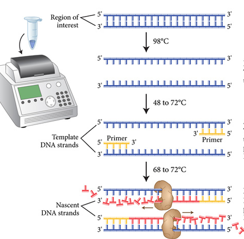
Amplification Dynamics:
Amplification is exponential (), doubling the number of copies every cycle ().
Newly made DNA strands become templates in the next cycle
After cycles, one fragment can become approximately 1bil copies ().
amplified DNA frags can be separated by gel electrophoresis
PCR product size should be equal to the amount of DNA between primers
Advantages and Clinical Utility:
makes millions of DNA copies without cloning
Sensitivity: Can amplify DNA from picogram () or femtogram () quantities.
Applications:
Diagnostic testing for bacteria, viruses, and genetic mutations.
Forensic identification from trace samples (e.g., a single hair).
Food testing for pathogens (e.g., ) or species contamination (horse or whale DNA).
Paternity testing.
Limitations:
Requires prior knowledge of the DNA sequence to design appropriate primers.
Extremely sensitive to contamination.
Typically limited to fragments between and .
Advanced PCR Adaptations and Genotyping
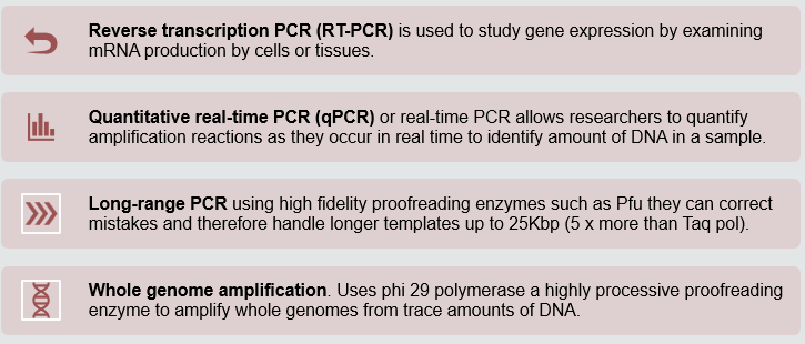
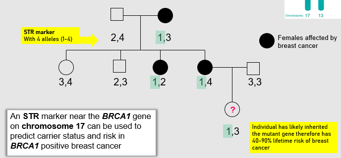
Genotyping with Short Tandem Repeats (STRs): * Used to distinguish alleles by size using PCR followed by electrophoresis. * Clinical Example: An STR marker on a chromosome near the gene can predict carrier status for hereditary breast cancer. Females with specific inherited alleles may have a - lifetime risk of the disease.
o