Chapter 10: Cell Divison
The ability of organisms to produce more of their own kind is the one characteristic that distinguishes living things from nonliving matter
The continuity of life is based on the reproduction of cells, or cell division
Key Roles of Cell Division
Cell division plays several important roles in life
Single-celled organisms given rise to new organisms through cell division
Multicellular eukaryotes undergo embryonic development through cell division
Cell division continues to function in renewal and repair in fully grown multicellular eukaryotes
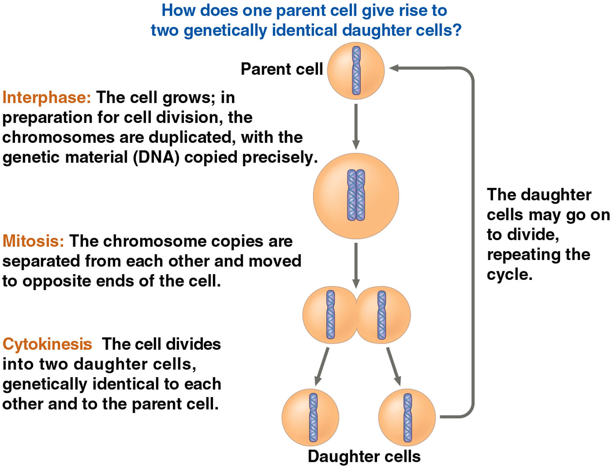
A crucial function of most cell division is the distribution of identical genetic material to the two daughter cells
Cell division is remarkably accurate in passing DNA from one generation to the next
Cellular Organization of the Genetic Material
All the DNA in a cell constitutes the cell’s genome
A genome can consist of a single DNA molecule (common in prokaryotic cells) or a number of DNA molecules (common in eukaryotic cells)
DNA molecules in a cell are packaged into chromosomes
The DNA molecule of a chromosome carries several hundred to a few thousand genes
Eukaryotic chromosomes consist of chromatin, a complex of DNA and protein that condenses during cell division
Every eukaryotic species has a characteristic number of chromosomes in each cell nucleus
Somatic Cells (nonreproductive cells) have two sets of chromosomes
Gametes (reproductive cells: sperm and eggs) have half as many chromosomes as somatic cells
Distribution of Chromosomes during eukaryotic cell division
In preparation for cell division, DNA is replicated and the chromosomes condense
Each duplicated chromosome has two sister chromatids (joined copies of the original chromosome), attached along their lengths by cohesins
The centrome is the narrow waist of the duplicated chromosome, where the rwo chromatids are most closely attaches
During cell division, the two sister chromatids of each duplicated chromosome separate and move into two nuclei
Once separate, the chromatids are called chromosomes
Eukaryotic cell division consists of
mitosis, the division of the genetic material the nucleus
cytokinesis, the division of the cytoplasm
Gametes are produced by a variation of cell division called meiosis
Meiosis yields nonidentical daughter cells that have half as many chromosomes as the parent cell
Phases of the Cell Cycle
The cell cycle consists of
mitotic (M) phase (mitosis and cytokinesis)
interphase (cell growth and copying of chromosomes in preparation for cell division)
Interphase (about 90% of the cell cycle) can be divided into three phases
G1 phase (first gap)
S Phase (synthesis)
G2 phase (second gap)
The cell grows during all three phases, but chromosomes are duplicated only during the S phase
Mitosis is conventionally broken down into 5 stages
prophase
prometaphase
metaphase
anaphase
telophase
The mitotic spindle:
The mitotic spindle is a structure made of microtubules that controls chromosome movement during mitosis
In animal cells, assembly of spindle microtubules begins in the centrosome, a type of microtubule organizing center
The centrosome replicates during interphase, forming two centrosomes that migrate to opposite ends of the cell during prophase and prometaphase
By the end of prometaphase, the two centrosomes are at opposite end of the cell
An aster (a radial array of short microtubules) extends from each centrosome
The spindle includes the centrosomes, the spindle microtubules, and the asters
Each sister chromatid has a kinetochore
A kinetochore is a protein complex associated with centromeres
During prometaphase, some spindle microtubules (kinetochore microtubules) attach to the kinetochores
At metaphase, the chromosomes are all lined up at the metaphase plate, an imaginary plane midway between the spindles two poles
In anaphase the cohesins are cleaved by an enzyme called separase
Sister chromatids separate and move along the kinetochore microtubules toward opposite ends of the cell
The microtubules shorten by depolymerizing at their kinetochore ends
Results of a clever experiment suggest that motor proteins on kinetochores “walk” the chromosomes along the microtubules during anaphase
The depolymerization of the microtubules at the kinetochore ends occurs after the motor proteins have passed
this is called the pac-man mechanism
Other research shows that chromosomes are reeled in by motor proteins at the spindle poles
Microtubules depolymerize after they pass by the motor proteins at the poles
The general consensus is that both mechanisms are used
Nonkinetochore microtubules from opposite poles overlap and push against each other, elongating the cell
At the end of anaphase, duplicate groups of chromosomes have arrived at opposite ends of the elongated cell
Cytokinesis begins during anaphase or telophase and the spindle eventually disassembles
Cytokinesis: A closer look
In animal cells, cytokinesis occurs by a process known as cleavasge
The first sign of cleavage is the appearance of a cleavage furrow, a shallow groove in the cell surface near the old metaphase plate
In plant cells, a cell plate forms during cytokinesis
Binary Fission in Bacteria
Prokaryotes (bacteria and archaea) reproduce by a type of cell division called binary fission
In binary fission, the chromosome replicated (beginning at the origin of replication) and the two daughter chromosomes actively move apart
The plasma membrane pinches inward, dividing the cell into two
How bacterial chromosomes move and their location established are active areas of research
The evolution of Mitosis
Because prokaryotes evolved before eukaryotes, mitosis probably evolved from binary fission
Certain unicellular eukaryotes exhibit types of cell division that seem intermediate between binary fission and mitosis
The frequency of cell division varies with the type of cell
These difference results from regulation at the molecular level
Cancer cells manage to escape the usual controls on the cell cycle
The frequency of cell division varies with the type of cell
These differences result from regulation at the molecular level
Cancer cells manage to escape the usual controls on the cell cycle
The cell cycle appears to be driven by specific signaling molecules present in the cytoplasm
Some evidence for this hypothesis comes from experiments in which culture mammalian cells at different phases of the cell cycle were fused to form a single cell with two nuclei
Signals in the cytoplasm of the fused cell caused both nuclei to enter the same phase of the cell cycle
The sequential events of the cell cycle are directed by a distinct cell cycle control system
The cell cycle control system is regulated by both internal and external controls
The clock has specific checkpoints where the cell cycle stops until a go-ahead signal is received
Two types of regulatory proteins are involved in cell cycle control: cyclins and cyclin-dependent kinases (Cdks)
Cyclins are named for their cyclically fluctuating concentrations in the cell
The activity of a Cdk rises and falls with changes in concentration of its cyclin partner
Cdks must be attached to a cyclin to be active
MPF (maturation-promoting factor) is a cyclin-Cdk complex that triggers a cell’s passage past the G2 checkpoint into the M phase
Peaks of MPF activity correspond to the peaks of cyclin concentration
MPF acts both as a kinase and indirectly through activating other kinases